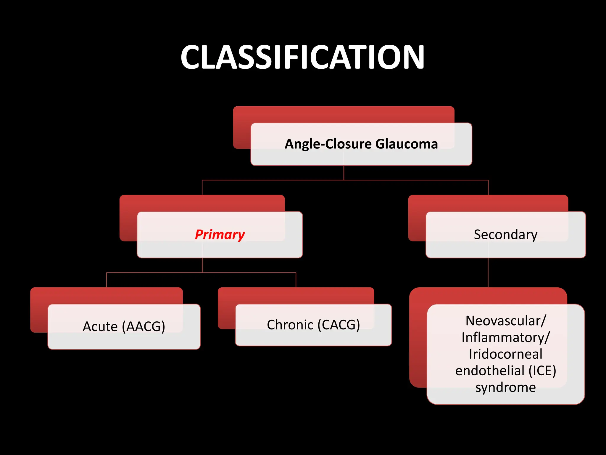
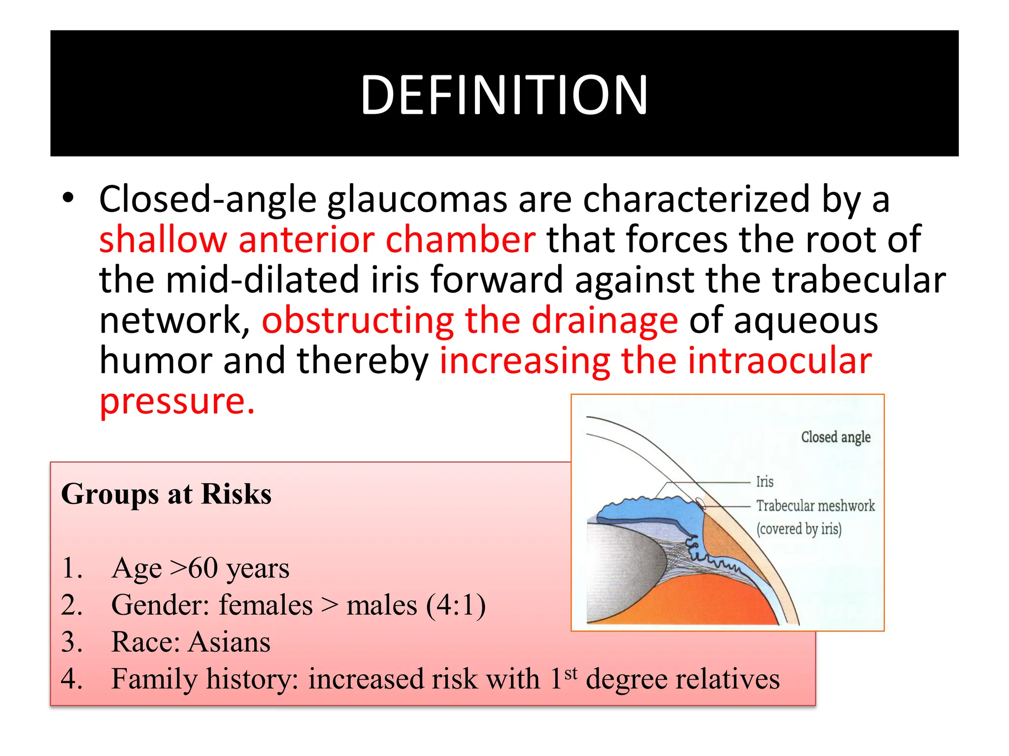
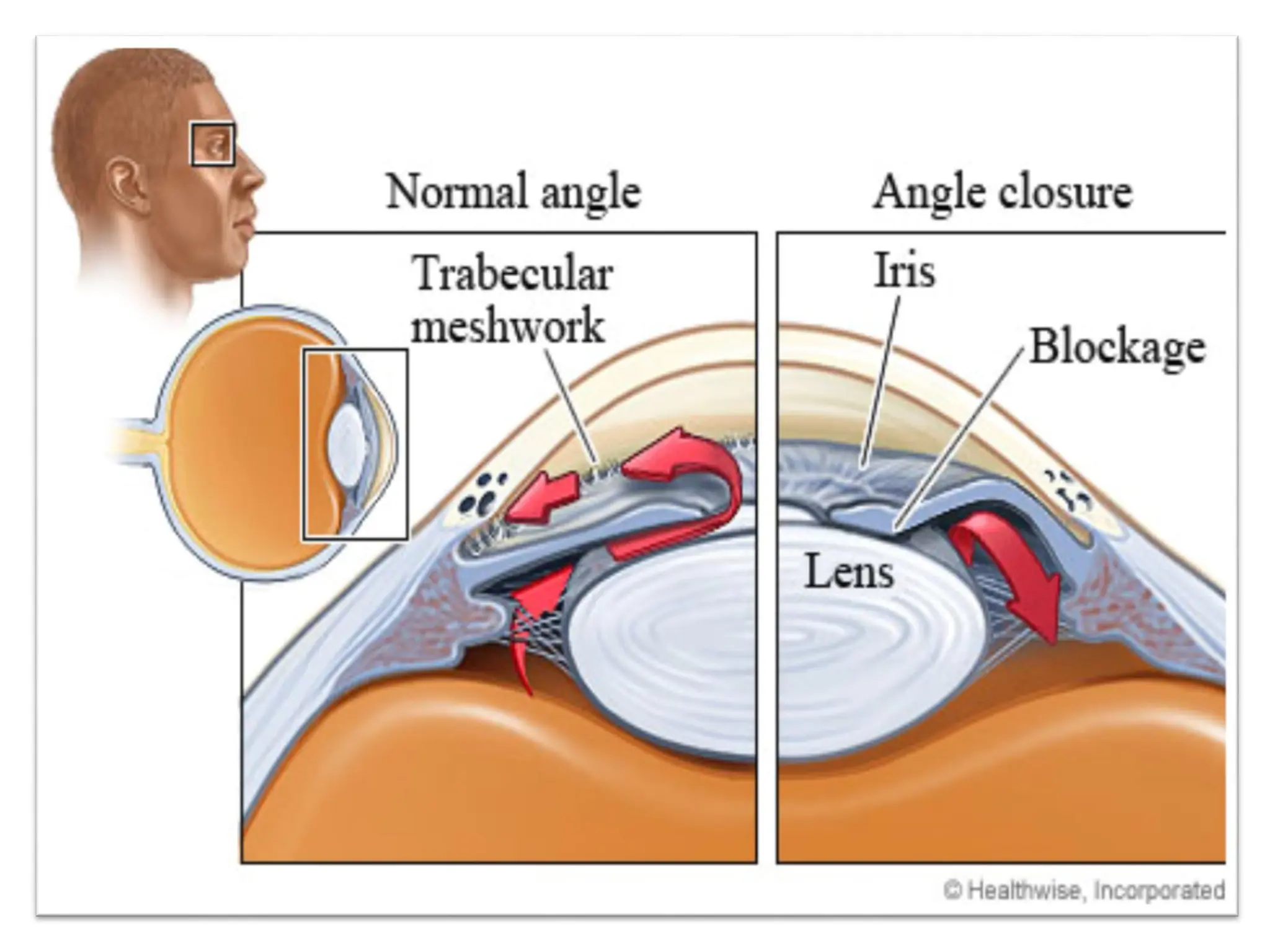
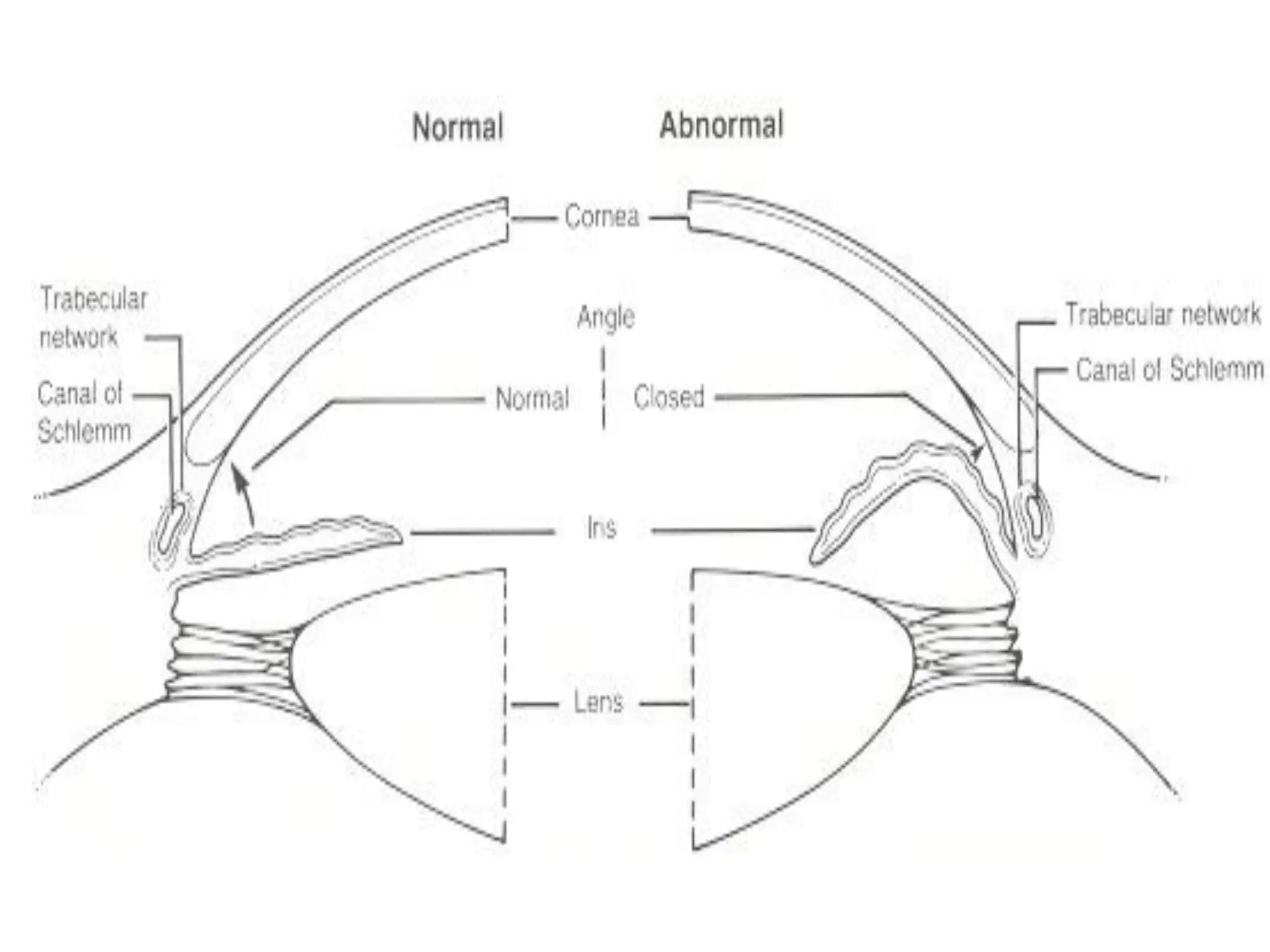
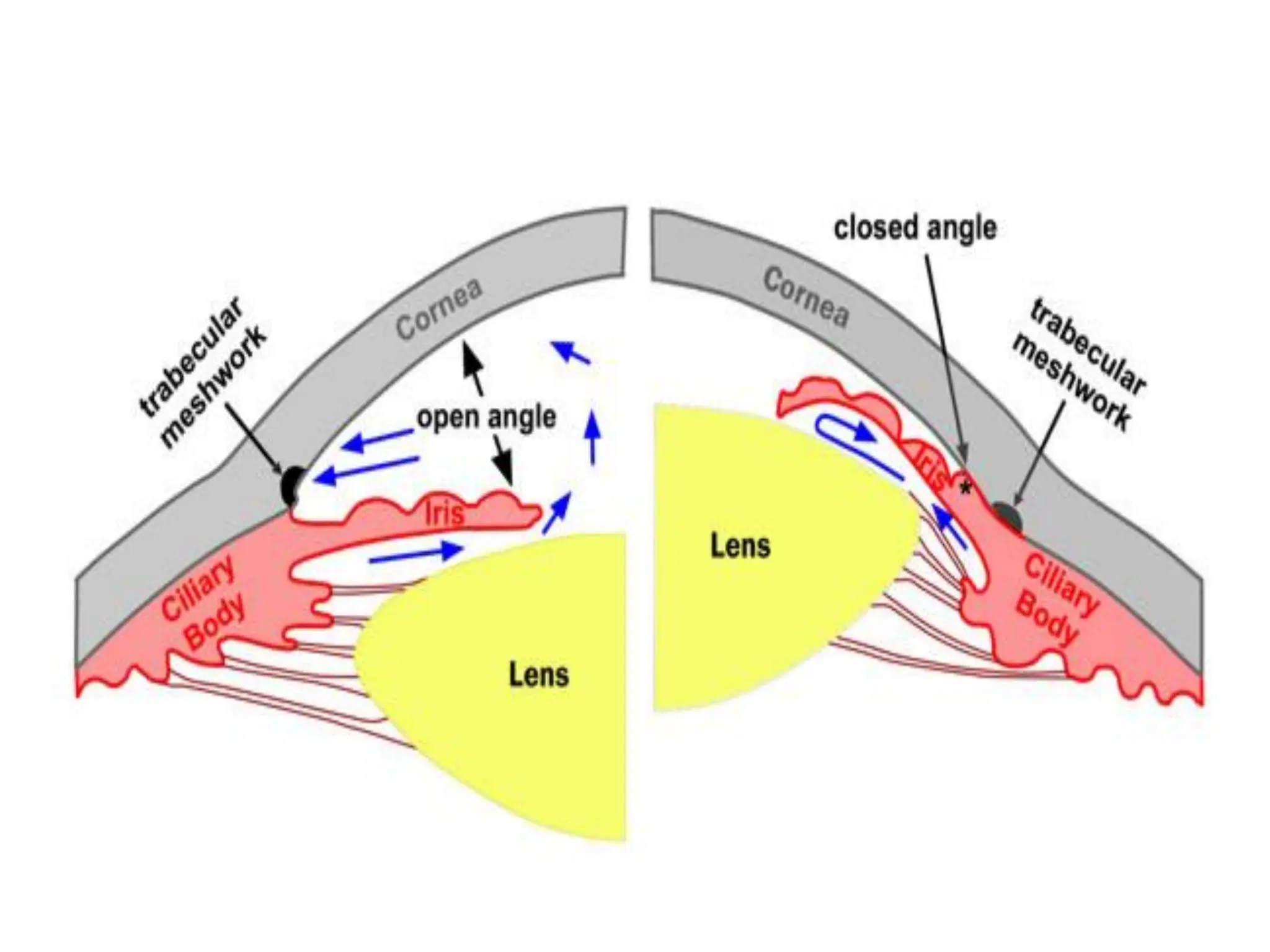
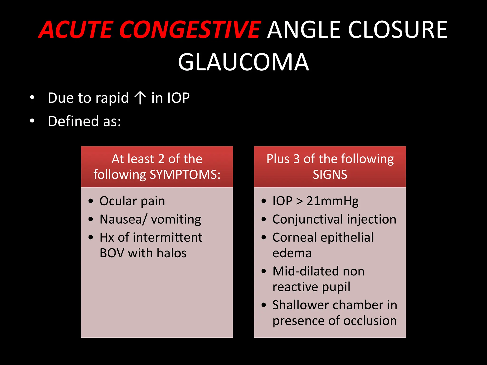
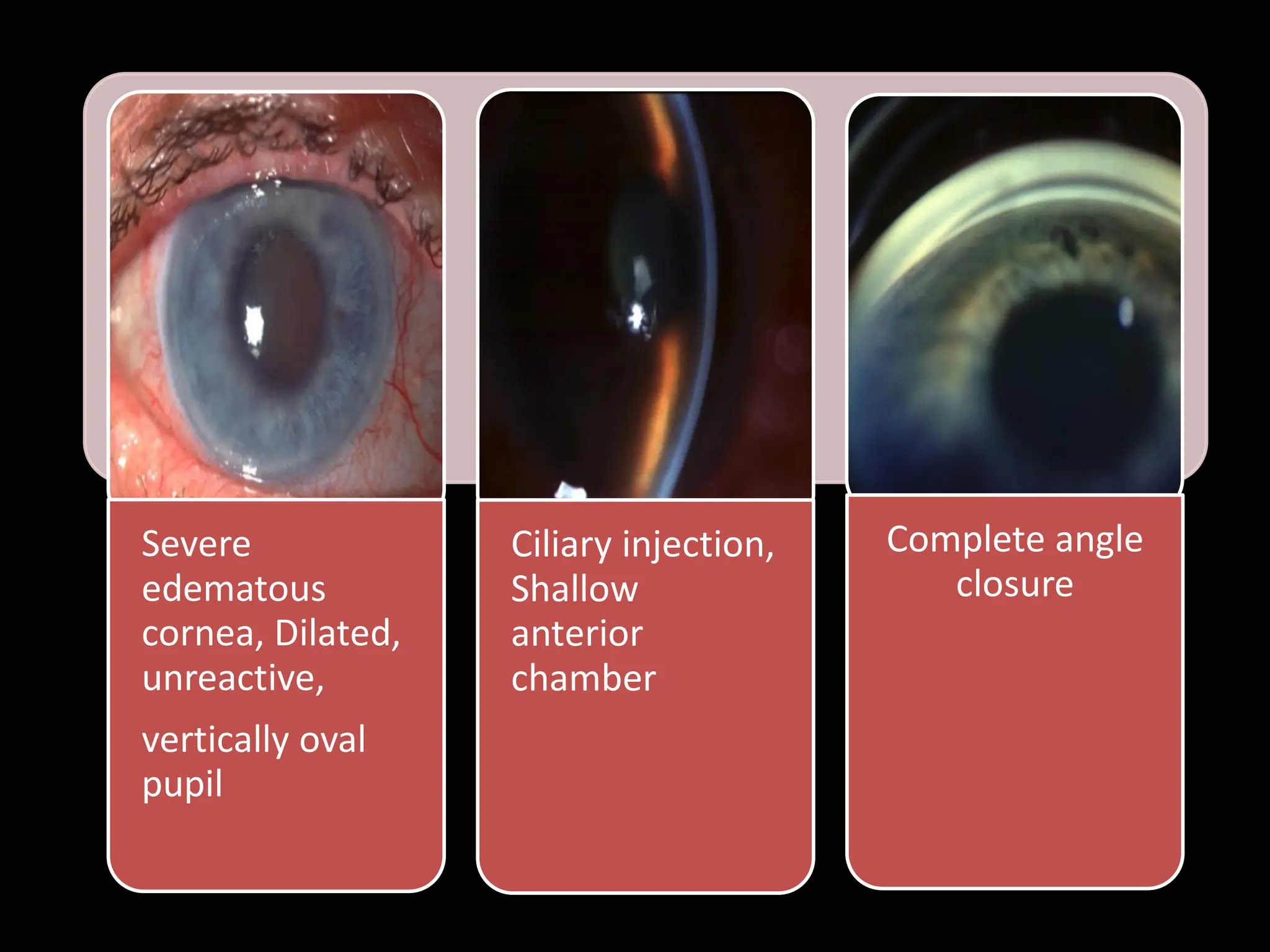
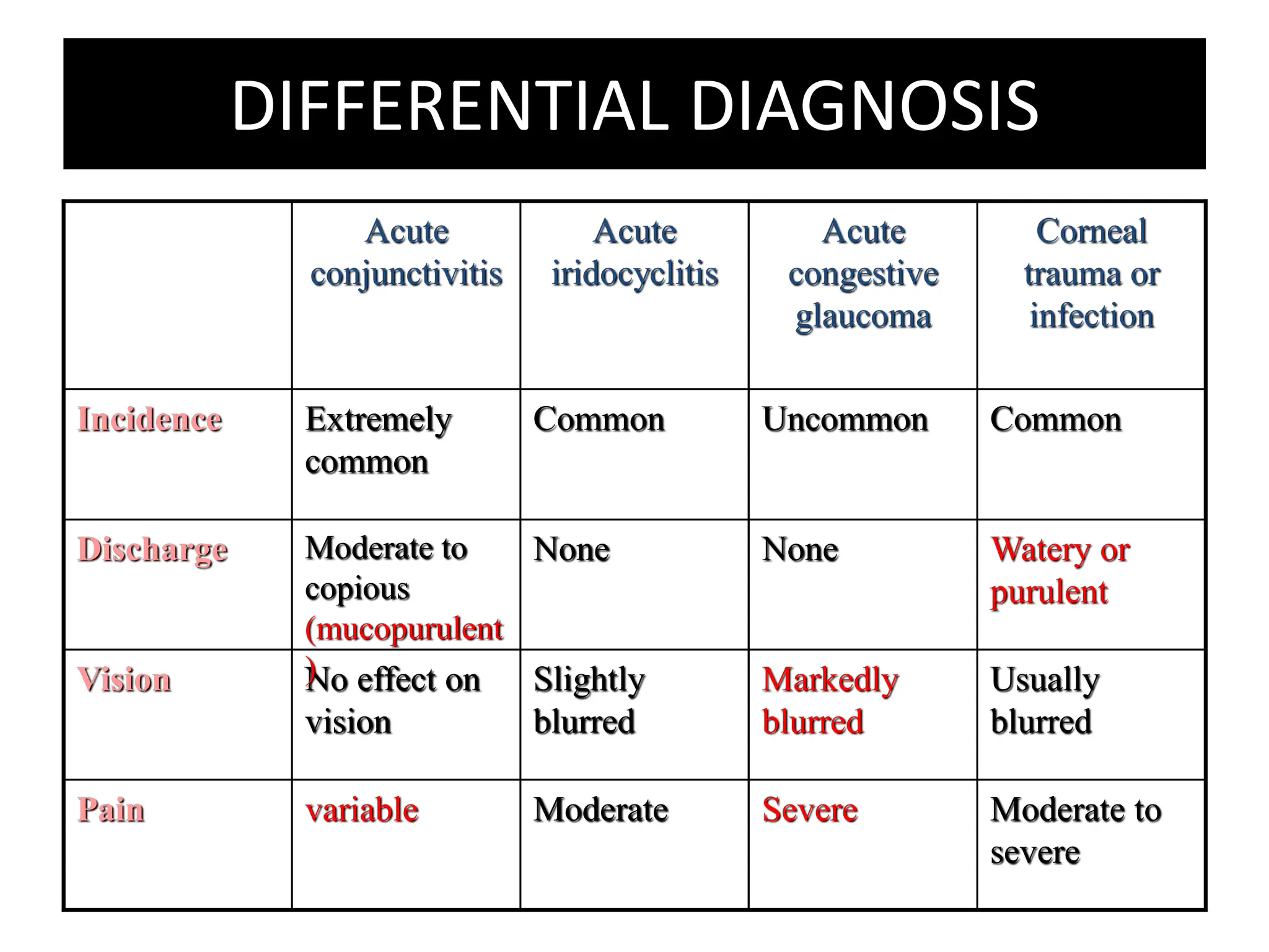
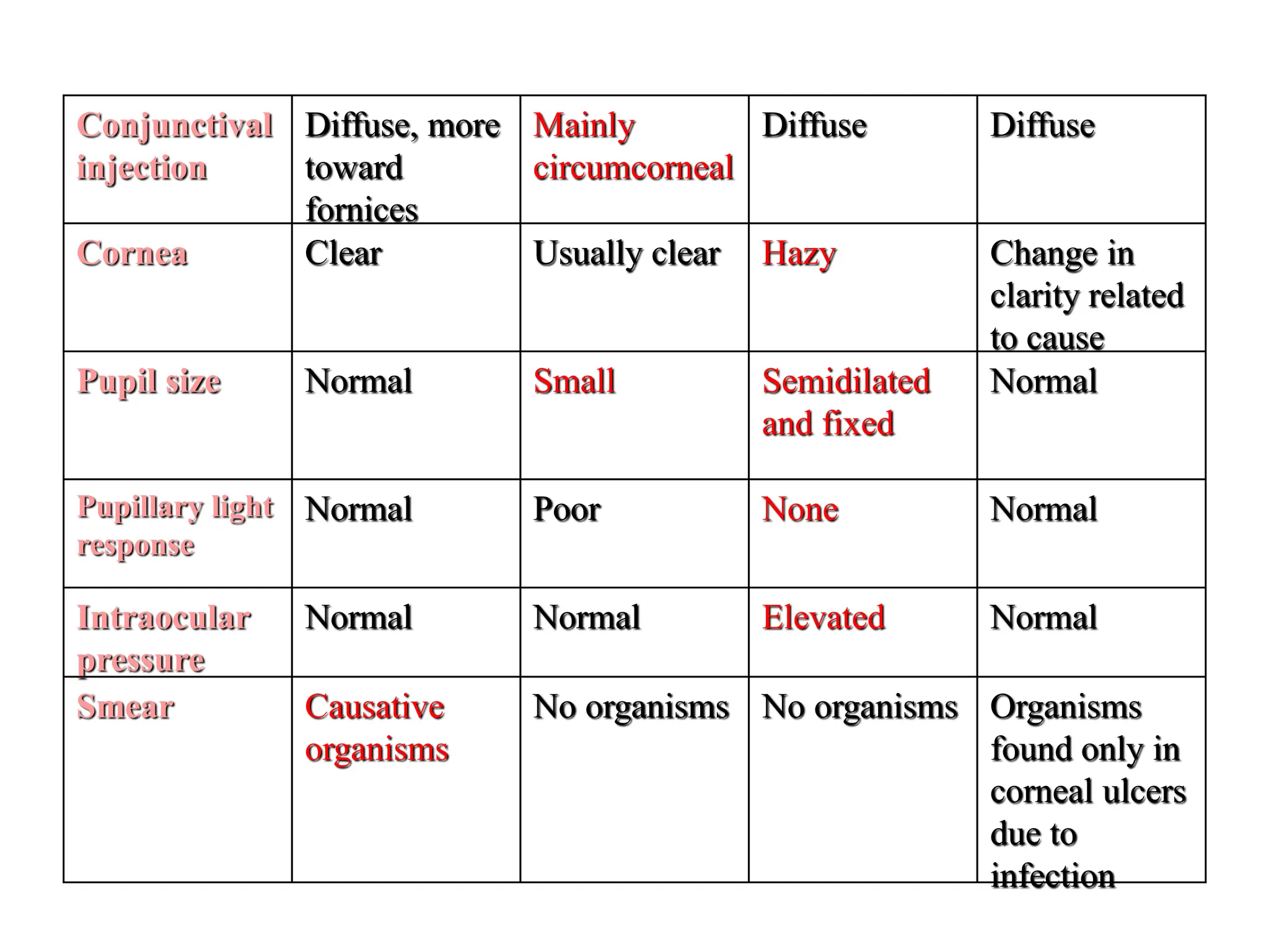
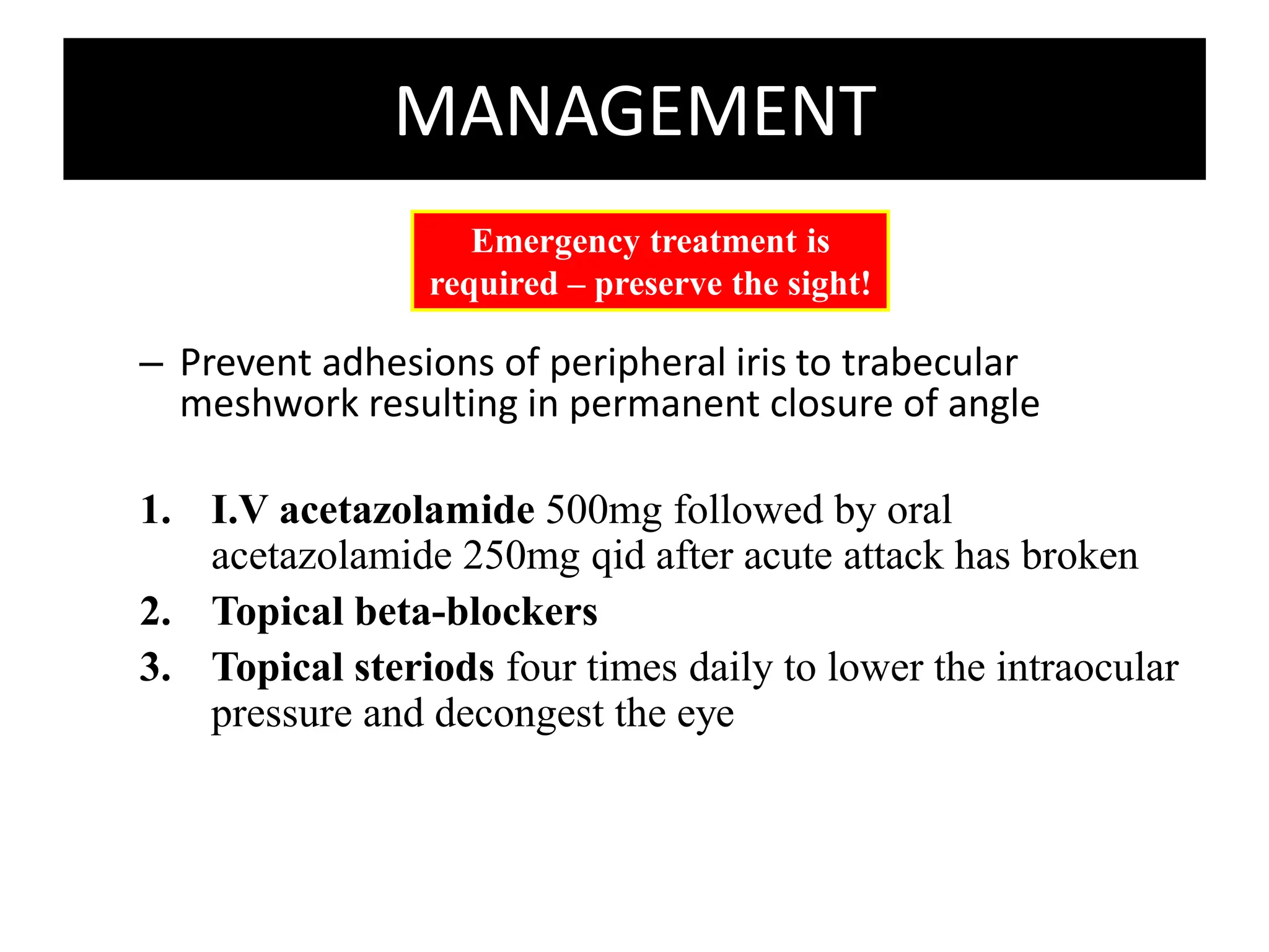
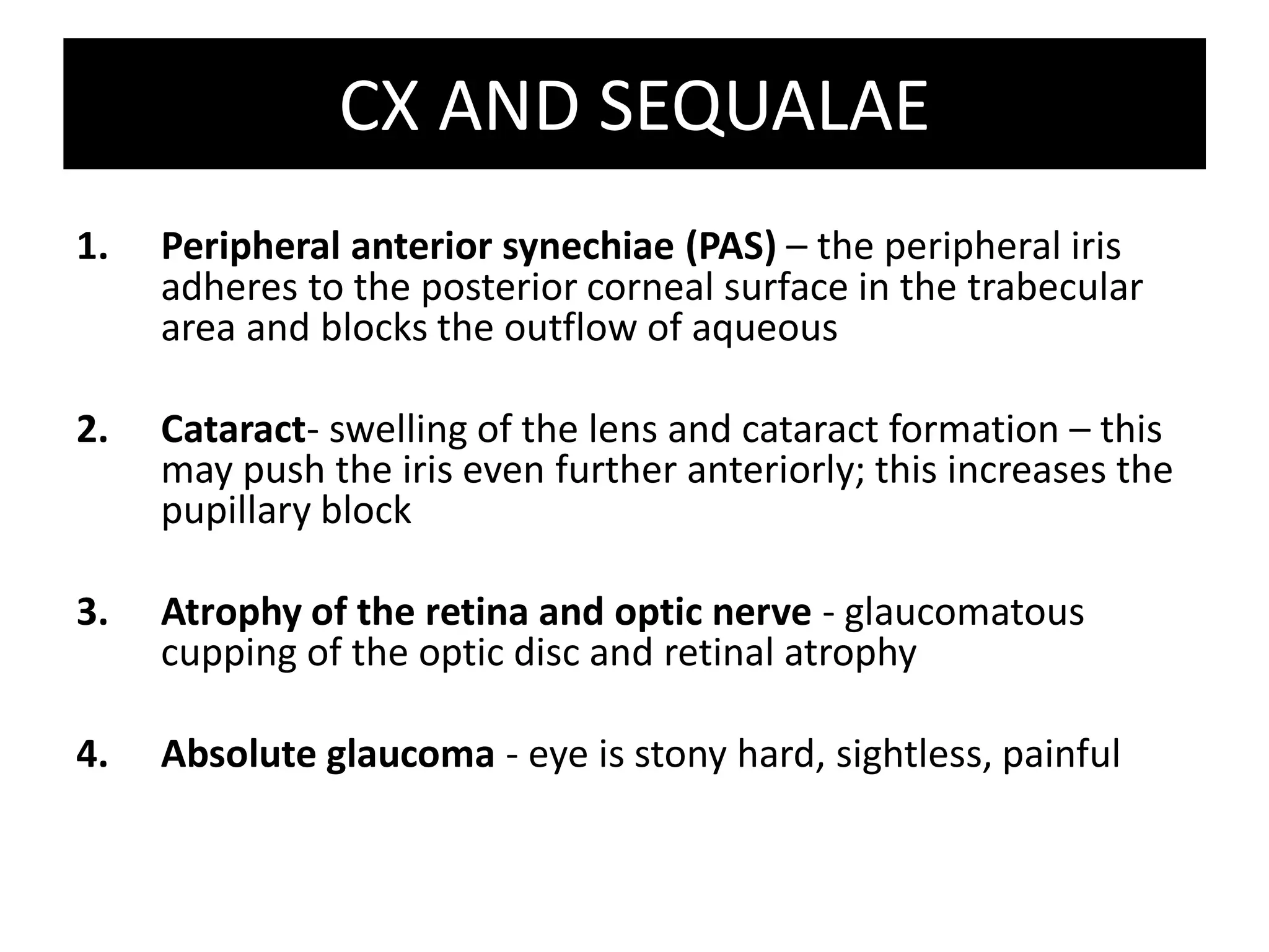
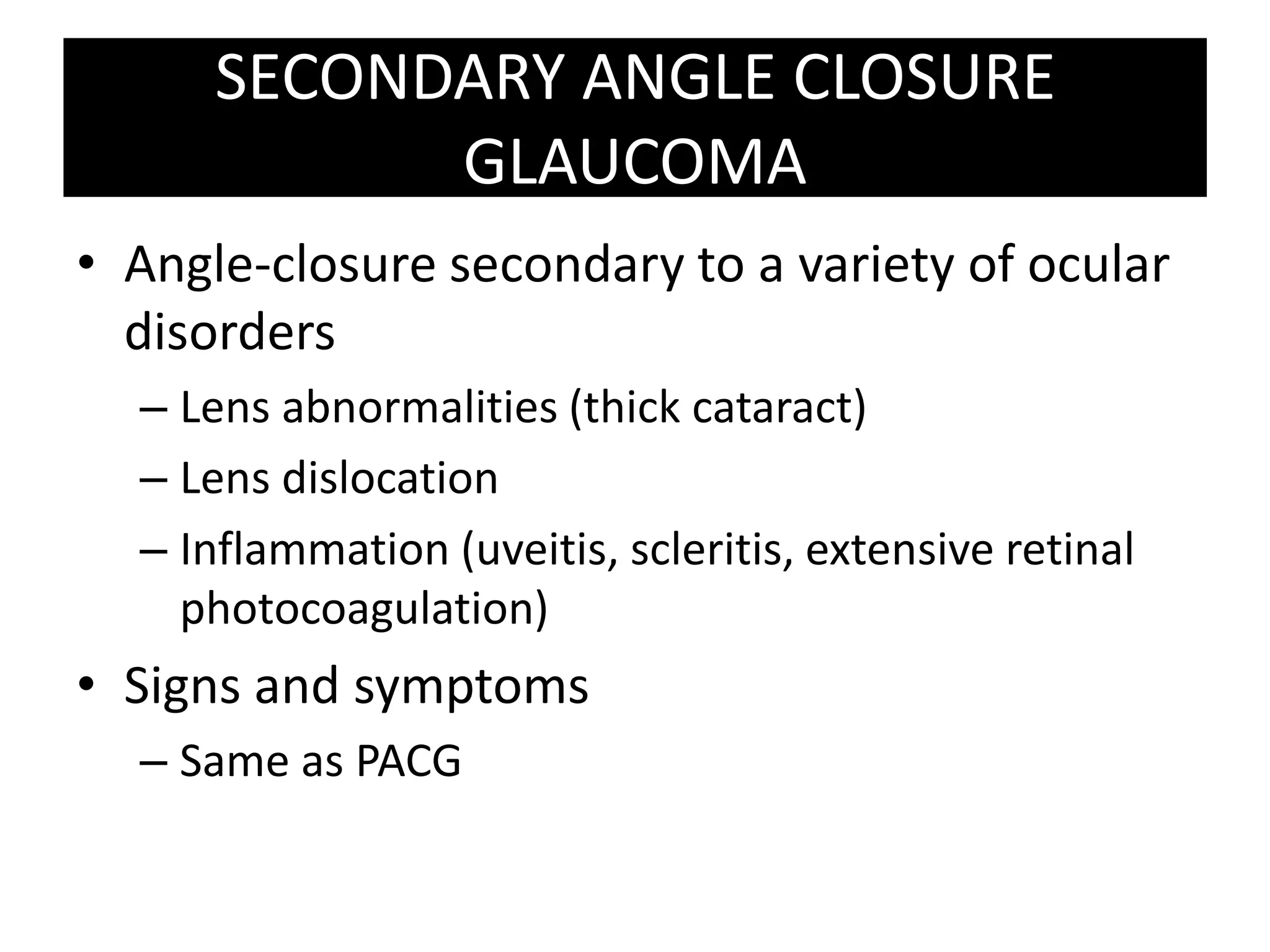
Angle Closure
Glaucoma
CLASSIFICATION
Angle-Closure Glaucoma
Primary
Acute (AACG) Chronic (CACG)
Secondary
Neovascular/
Inflammatory/
Iridocorneal
endothelial (ICE)
syndrome
DEFINITION
• Closed-angle glaucomas are characterized by a
shallow anterior chamber that forces the root of
the mid-dilated iris forward against the trabecular
network, obstructing the drainage of aqueous
humor and thereby increasing the intraocular
pressure.
Groups at Risks
1. Age >60 years
2. Gender: females > males (4:1)
3. Race: Asians
4. Family history: increased risk with 1st degree relatives
PREDISPOSING FACTORS
• Relative anterior position of iris-lens
diaphragm
• Shallow anterior chamber
• Narrow entrance to angle
Anatomical
• Physiological pupillary block
Physiological
PHYSIOLOGICAL PUPILLARY BLOCK
1. Iris has large arc of
contact with anterior
surface of lens
2. Resistance to
aqueous flow from
posterior to anterior
chamber (relative
pupil block)
3. Pupil dilates,
peripheral iris
becomes more
flaccid and pushed
anteriorly
4. Iris lies against
trabecular meshwork
 impede aqueous
humor drainage  ↑
IOP
SYMPTOMS
1. Rapidly progressive impairment of
vision
2. Painful eye
3. Red eye
4. Nausea, vomiting
5. Photophobia
6. Haloes, transient blurring – indicate
previous intermittent attacks
7. Hx of similar attacks in the past, aborted
by sleep
** CACG: usually asymptomatic due to slow onset
of disease
SIGNS
1. Reduced visual acuity
2. Cornea cloudy and oedematous
3. Pupil oval, fixed and moderately dilated
4. Ciliary injection
5. Eye feels hard on palpation
6. Elevated IOP (50-100 mmHg)
7. Narrow chamber angle with peripheral
iridocorneal contact
8. Aqueous flare and cells
9. Gonioscopy – complete peripheral
iridocorneal contact
10. Ophthalmoscopy – optic disc odema and
hyperaemia
ACUTE CONGESTIVE ANGLE CLOSURE
GLAUCOMA
• Due to rapid ↑ in IOP
• Defined as:
At least 2 of the
following SYMPTOMS:
• Ocular pain
• Nausea/ vomiting
• Hx of intermittent
BOV with halos
Plus 3 of the following
SIGNS
• IOP > 21mmHg
• Conjunctival injection
• Corneal epithelial
edema
• Mid-dilated non
reactive pupil
• Shallower chamber in
presence of occlusion
Severe
edematous
cornea, Dilated,
unreactive,
vertically oval
pupil
Ciliary injection,
Shallow
anterior
chamber
Complete angle
closure
DIFFERENTIAL DIAGNOSIS
Usually
blurred
Markedly
blurred
Slightly
blurred
No effect on
vision
Vision
Moderate to
severe
Severe
Moderate
variable
Pain
Watery or
purulent
None
None
Moderate to
copious
(mucopurulent
)
Discharge
Common
Uncommon
Common
Extremely
common
Incidence
Corneal
trauma or
infection
Acute
congestive
glaucoma
Acute
iridocyclitis
Acute
conjunctivitis
Organisms
found only in
corneal ulcers
due to
infection
No organisms
No organisms
Causative
organisms
Smear
Normal
Elevated
Normal
Normal
Intraocular
pressure
Normal
None
Poor
Normal
Pupillary light
response
Normal
Semidilated
and fixed
Small
Normal
Pupil size
Change in
clarity related
to cause
Hazy
Usually clear
Clear
Cornea
Diffuse
Diffuse
Mainly
circumcorneal
Diffuse, more
toward
fornices
Conjunctival
injection
MANAGEMENT
– Prevent adhesions of peripheral iris to trabecular
meshwork resulting in permanent closure of angle
1. I.V acetazolamide 500mg followed by oral
acetazolamide 250mg qid after acute attack has broken
2. Topical beta-blockers
3. Topical steriods four times daily to lower the intraocular
pressure and decongest the eye
Emergency treatment is
required – preserve the sight!
Reassessment
• Evaluate IOP
• Evaluate adjunct drops
• May need osmotic agents? Immediate
iridotomy?
Approx 1 hr
after initial RX
• Start with Pilocarpine (myotic drug)
every 15mins x 2 doses
SURGICAL MANAGEMENT
1. Peripheral laser iridotomy (LPI)
(YAG Laser)
– To establish the communication between the posterior and anterior
chambers by making an opening in the peripheral iris
– This will be successful only if less than 50% of the angle is closed by
permanent peripheral anterior synechiae
1. Peripheral Iridectomy
CX AND SEQUALAE
1. Peripheral anterior synechiae (PAS) – the peripheral iris
adheres to the posterior corneal surface in the trabecular
area and blocks the outflow of aqueous
2. Cataract- swelling of the lens and cataract formation – this
may push the iris even further anteriorly; this increases the
pupillary block
3. Atrophy of the retina and optic nerve - glaucomatous
cupping of the optic disc and retinal atrophy
4. Absolute glaucoma - eye is stony hard, sightless, painful
SECONDARY ANGLE CLOSURE
GLAUCOMA
• Angle-closure secondary to a variety of ocular
disorders
– Lens abnormalities (thick cataract)
– Lens dislocation
– Inflammation (uveitis, scleritis, extensive retinal
photocoagulation)
• Signs and symptoms
– Same as PACG
Angle Closure Glaucoma primary_061832.pdf

Angle Closure Glaucoma primary_061832.pdf

  • 1.
  • 2.
    CLASSIFICATION Angle-Closure Glaucoma Primary Acute (AACG)Chronic (CACG) Secondary Neovascular/ Inflammatory/ Iridocorneal endothelial (ICE) syndrome
  • 3.
    DEFINITION • Closed-angle glaucomasare characterized by a shallow anterior chamber that forces the root of the mid-dilated iris forward against the trabecular network, obstructing the drainage of aqueous humor and thereby increasing the intraocular pressure. Groups at Risks 1. Age >60 years 2. Gender: females > males (4:1) 3. Race: Asians 4. Family history: increased risk with 1st degree relatives
  • 7.
    PREDISPOSING FACTORS • Relativeanterior position of iris-lens diaphragm • Shallow anterior chamber • Narrow entrance to angle Anatomical • Physiological pupillary block Physiological
  • 8.
    PHYSIOLOGICAL PUPILLARY BLOCK 1.Iris has large arc of contact with anterior surface of lens 2. Resistance to aqueous flow from posterior to anterior chamber (relative pupil block) 3. Pupil dilates, peripheral iris becomes more flaccid and pushed anteriorly 4. Iris lies against trabecular meshwork  impede aqueous humor drainage  ↑ IOP
  • 9.
    SYMPTOMS 1. Rapidly progressiveimpairment of vision 2. Painful eye 3. Red eye 4. Nausea, vomiting 5. Photophobia 6. Haloes, transient blurring – indicate previous intermittent attacks 7. Hx of similar attacks in the past, aborted by sleep ** CACG: usually asymptomatic due to slow onset of disease
  • 10.
    SIGNS 1. Reduced visualacuity 2. Cornea cloudy and oedematous 3. Pupil oval, fixed and moderately dilated 4. Ciliary injection 5. Eye feels hard on palpation 6. Elevated IOP (50-100 mmHg) 7. Narrow chamber angle with peripheral iridocorneal contact 8. Aqueous flare and cells 9. Gonioscopy – complete peripheral iridocorneal contact 10. Ophthalmoscopy – optic disc odema and hyperaemia
  • 12.
    ACUTE CONGESTIVE ANGLECLOSURE GLAUCOMA • Due to rapid ↑ in IOP • Defined as: At least 2 of the following SYMPTOMS: • Ocular pain • Nausea/ vomiting • Hx of intermittent BOV with halos Plus 3 of the following SIGNS • IOP > 21mmHg • Conjunctival injection • Corneal epithelial edema • Mid-dilated non reactive pupil • Shallower chamber in presence of occlusion
  • 13.
    Severe edematous cornea, Dilated, unreactive, vertically oval pupil Ciliaryinjection, Shallow anterior chamber Complete angle closure
  • 14.
    DIFFERENTIAL DIAGNOSIS Usually blurred Markedly blurred Slightly blurred No effecton vision Vision Moderate to severe Severe Moderate variable Pain Watery or purulent None None Moderate to copious (mucopurulent ) Discharge Common Uncommon Common Extremely common Incidence Corneal trauma or infection Acute congestive glaucoma Acute iridocyclitis Acute conjunctivitis
  • 15.
    Organisms found only in cornealulcers due to infection No organisms No organisms Causative organisms Smear Normal Elevated Normal Normal Intraocular pressure Normal None Poor Normal Pupillary light response Normal Semidilated and fixed Small Normal Pupil size Change in clarity related to cause Hazy Usually clear Clear Cornea Diffuse Diffuse Mainly circumcorneal Diffuse, more toward fornices Conjunctival injection
  • 16.
    MANAGEMENT – Prevent adhesionsof peripheral iris to trabecular meshwork resulting in permanent closure of angle 1. I.V acetazolamide 500mg followed by oral acetazolamide 250mg qid after acute attack has broken 2. Topical beta-blockers 3. Topical steriods four times daily to lower the intraocular pressure and decongest the eye Emergency treatment is required – preserve the sight!
  • 17.
    Reassessment • Evaluate IOP •Evaluate adjunct drops • May need osmotic agents? Immediate iridotomy? Approx 1 hr after initial RX • Start with Pilocarpine (myotic drug) every 15mins x 2 doses
  • 18.
    SURGICAL MANAGEMENT 1. Peripherallaser iridotomy (LPI) (YAG Laser) – To establish the communication between the posterior and anterior chambers by making an opening in the peripheral iris – This will be successful only if less than 50% of the angle is closed by permanent peripheral anterior synechiae 1. Peripheral Iridectomy
  • 19.
    CX AND SEQUALAE 1.Peripheral anterior synechiae (PAS) – the peripheral iris adheres to the posterior corneal surface in the trabecular area and blocks the outflow of aqueous 2. Cataract- swelling of the lens and cataract formation – this may push the iris even further anteriorly; this increases the pupillary block 3. Atrophy of the retina and optic nerve - glaucomatous cupping of the optic disc and retinal atrophy 4. Absolute glaucoma - eye is stony hard, sightless, painful
  • 20.
    SECONDARY ANGLE CLOSURE GLAUCOMA •Angle-closure secondary to a variety of ocular disorders – Lens abnormalities (thick cataract) – Lens dislocation – Inflammation (uveitis, scleritis, extensive retinal photocoagulation) • Signs and symptoms – Same as PACG