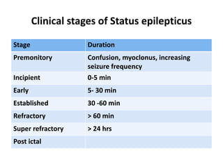
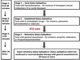
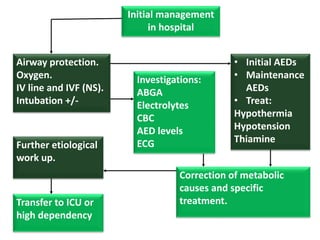
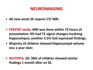
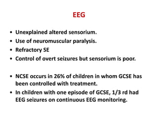
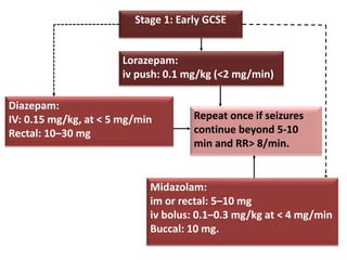
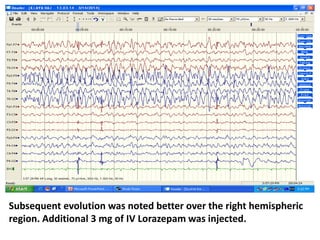
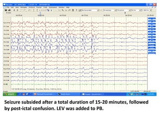
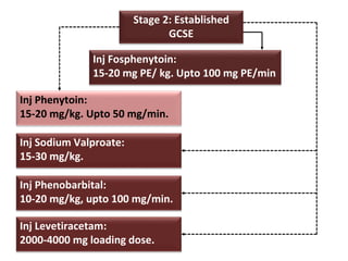
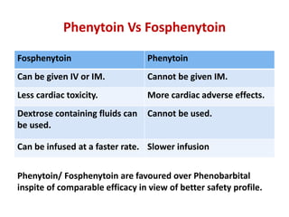
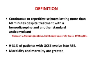
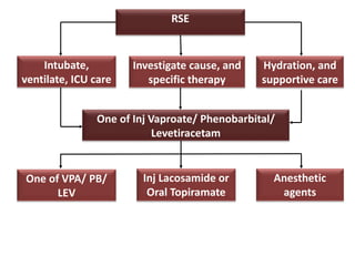
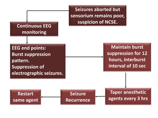
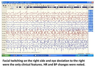
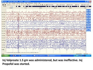
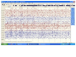
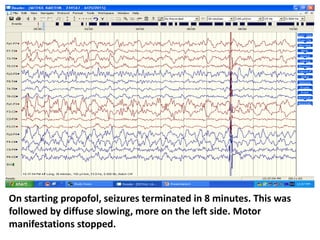
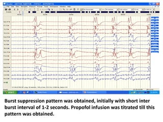
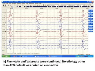
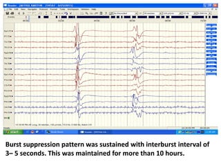
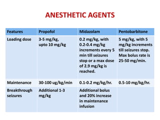
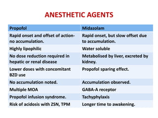
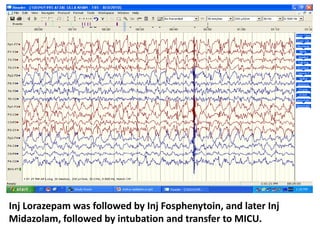
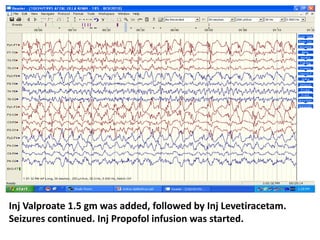
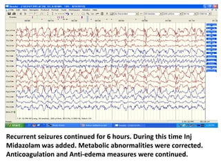
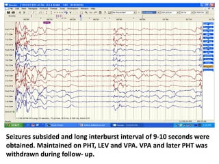
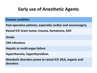
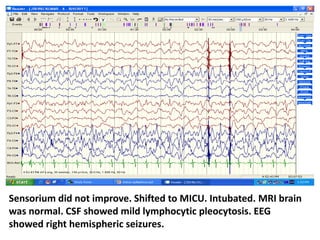
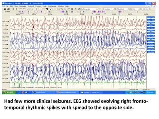
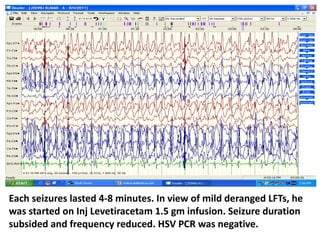
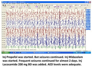
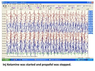
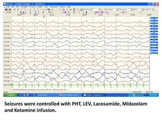
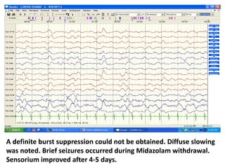
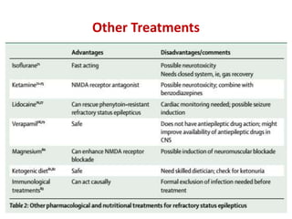
PEDIATRIC STATUS EPILEPTICUS
Management
Dr Pramod Krishnan
Consultant Neurologist and Epileptologist.
Manipal Hospital, Bangalore.
DEFINITION
• “more than 30 minutes of a continuous seizure or
two or more sequential seizures without full
recovery of consciousness’
Working Group on Status Epilepticus of the Epilepsy Foundation of America.
• In clinical practice, a single seizure of >5 min (in
adults and children > 5 years of age) should be
considered as status epilepticus.
Lowenstein DH et al. NEJM 1998; 338: 970-6.
RATIONALE
• Increased risk of neuronal injury after 30 min when
homeostatic mechanism fail.
• A typical CPS/ GTCS in children lasts 62 sec (16- 108
sec) followed by upto 4 minutes of post ictal phase.
Theodore WH, et al. Neurology 1994; 44: 1403-07.
• If a seizure lasts >5 min, it tends to be prolonged.
• Response to treatment is better when commenced
early.
Mayer SA, et al. Arch Neurol 2002; 59: 205- 210.
EPIDEMIOLOGY
• North London Status Epilepticus Childhood
Surveillance Study (NLSTEPSS).
• Community based prospective study.
• Incidence: 17- 23/100,000.
Chin RF, et al. Lancet 2006; 368: 222- 229.
• In children, SE occurs within 2 years of the onset of
epilepsy.
• Recurrent SE is usually associated with underlying
neurologic disorder.
Sillanpaa M, et al. Ann Neurol 2002; 52: 303- 310.
EPIDEMIOLOGY
• Incidence in children < 5 years of age was
7.5/100,000.
• SE is most common in those < 2 years of age.
Shinnar S, et al. Epilepsia 1997; 38: 907-14.
• 25% of patients with SE have pre-existing epilepsy.
• 4-16% of patients with epilepsy have atleast 1
episode of SE.
• SE is the presenting symptoms in upto 1/3 rd of
such patients.
Hauser WA, et al. Neurology 1990; 40: 9-13.
ETIOLOGY
ETIOLOGY
• SE is not a disease in itself but rather a
manifestation of a primary CNS insult or a systemic
disorder with secondary CNS effects.
• Cause may be acute or remote or part of a genetic
epileptic encephalopathy.
• It is important to identify and specifically treat the
precipitating cause, thus preventing ongoing
neurologic injury and seizure recurrence.
North London Status Epilepticus in Childhood Surveillance Study
Group Incidence
Acute symptomatic SE < 1 year: 16 %
1- 4 years: 2.5 %
5-15 years: 0.1 %
Acute on remote < 1 year: 6 %
1- 4 years: 5.3 %
5- 15 years: 0.7 %
Prolonged febrile seizure 4.1/ 100,000
Acute symptomatic SE 2.2/ 100,000
Remote symptomatic 2.3/ 100,000
Acute on remote 2.1/ 100,000
Idiopathic 1.4/ 100,000
Cryptogenic 0.2/ 100,000
Unclassified 1/ 100,000
Etiology in Children and Adults (Richmond Study)
Etiology % of children
(<16 years)
% of adults
(> 16 years)
Cerebrovascular 3.3 25.2
Medication change 19.8 18.9
Anoxia 5.3 10.7
Substance abuse 2.4 12.2
Metabolic 8.2 8.8
Unknown 9.3 8.1
Fever/ infection 35.7 4.6
Trauma 3.5 4.6
Tumor 0.7 4.3
CNS infection 4.8 1.8
Congenital 7.0 0.8
PATHOPHYSIOLOGY
MECHANISMS
• SE occurs when factors that normally terminate
seizures fail.
• There is decreased cerebral inhibition, excessive
excitation or both.
• Altered receptor trafficking causing reduced
expression of GABA-a receptor and increased
expression of amino acid receptors play a role.
• Excessive excitation can cause neuronal injury.
blood flow
glucose conc in brain
glucose metabolism
oxygen supply to
brain
cardiac output.
catecholamine output
Stage of decompensation
(seizures > 30 min)
Failure of cerebral
autoregulation
Hypoxia, hypoglycemia
Acidosis,
dyselectrolytemia
DIC, Leucocytosis
Falling BP, Falling CO
Stage of compensation
(seizures < 30 min)
CLASSIFICATION
Classification of SE
Generalised
Partial
Aura status
Autonomic
status
Dyscognitive
status
Motor status
Special
status
Simple
motor
Complex
motor
Generalised
NCSE
(13%)
Convulsive
status
Focal Motor
status (10%)
Generalised
convulsive
status (77%)
Clinical stages of Status epilepticus
Stage Duration
Premonitory Confusion, myoclonus, increasing
seizure frequency
Incipient 0-5 min
Early 5- 30 min
Established 30 -60 min
Refractory > 60 min
Super refractory > 24 hrs
Post ictal
EEG progression of status epilepticus
Discrete seizures with
inter-ictal slowing
Waxing and waning of
inter- ictal discharges
Continuous ictal
discharges
Continuous ictal discharges
punctuated by flat periods
PED on a flat background
Lateralised Bilateral
MANAGEMENT
ICU care
PRE-HOSPITAL
TREATMENT
RAMPART trial (Rapid Anticonvulsant
Medication Prior to Arrival Trial)
IM Midazolam 10
mg + IV placebo
IV Lorazepam 4
mg + IM placebo
Termination of clinical seizures prior to arrival
to ER. Need for a second rescue medication.
73.4% 63.4%
Not statistically significant. No difference in
seizure recurrence or adverse effects
PREHOSPITAL TREATMENT
• Rectal Diazepam
• Sublingual Lorazepam
• Intranasal/ buccal Midazolam.
Midazolam vs Diazepam
Study Type N Conclusion
McIntyre
et al 2005
Buccal Midazolam
(0.5 mg/kg) vs
Rectal Diazepam
(0.5 mg/kg)
177 56% of Midazolam group
had seizure control
compared to 27% for
diazepam.
Mpimbaza
et al 2008
Buccal Midazolam
Vs rectal
Diazepam.
330 Midazolam was
as well tolerated and more
effective than diazepam.
STAGE 1
Early GCSE
Initial management
in hospital
• Initial AEDs
• Maintenance
AEDs
• Treat:
Hypothermia
Hypotension
Thiamine
Investigations:
ABGA
Electrolytes
CBC
AED levels
ECG
Airway protection.
Oxygen.
IV line and IVF (NS).
Intubation +/-
Further etiological
work up.
Correction of metabolic
causes and specific
treatment.Transfer to ICU or
high dependency
NEUROIMAGING
• All new onset SE require CT/ MRI.
• FEBSTAT study: MRI was done within 72 hours of
presentation: 9% had T2 signal changes involving
hippocampus; another 2.5% had equivocal findings.
• Majority of children showed Hippocampal volume
loss a year later.
• NLSTEPSS: 20- 30% of children showed similar
findings 1 month after an SE.
EEG
• Unexplained altered sensorium.
• Use of neuromuscular paralysis.
• Refractory SE
• Control of overt seizures but sensorium is poor.
• NCSE occurs in 26% of children in whom GCSE has
been controlled with treatment.
• In children with one episode of GCSE, 1/3 rd had
EEG seizures on continuous EEG monitoring.
Stage 1: Early GCSE
Midazolam:
im or rectal: 5–10 mg
iv bolus: 0.1–0.3 mg/kg at < 4 mg/min
Buccal: 10 mg.
Lorazepam:
iv push: 0.1 mg/kg (<2 mg/min)
Diazepam:
IV: 0.15 mg/kg, at < 5 mg/min
Rectal: 10–30 mg
Repeat once if seizures
continue beyond 5-10
min and RR> 8/min.
Veterans Affairs Cooperative Trial
N= 384 GCSE
Inj Phenobarbital
15 mg/kg
Inj Phenytoin
18 mg/kg
Inj Diazepam
0.15 mg/kg +
Inj Phenytoin 18 mg/kg
Inj Lorazepam
0.1 mg/kg
64.9% 43.6% 58.2% 55.8%
Clinical and EEG termination of seizures.
No difference in recurrence rates between the 4 groups.
Treiman DM et al. NEJM 1998; 339: 792-8
Lorazepam Vs Diazepam
Lorazepam Diazepam
Longer duration of action
(>6 hrs).
Shorter duration (20 min) due
to rapid redistribution.
Onset of action upto 5 min. 1 min.
Risk of respiratory depression
is less.
Risk is more.
Light proof storage. Not required.
Refrigeration is recommended. Not required.
IYJ, 54 yrs/ M, with epilepsy, on Phenobarbitone, had a seizure
during EEG with loss of consciousness. CT brain showed multiple
NCC.
Frequent rhythmic bifrontal (Lt> Rt) spikes and sharp waves noted.
Fast activity seen building up over both hemispheric regions,
during which patient became unresponsiveness with stare.
Inj Lorazepam 2 mg IV was administered.
Seizures continued.
Initial evolution was better over the left hemispheric region,
mainly frontal. Eye deviation to the right was noted.
Subsequent evolution was noted better over the right hemispheric
region. Additional 3 mg of IV Lorazepam was injected.
Seizure subsided after a total duration of 15-20 minutes, followed
by post-ictal confusion. LEV was added to PB.
STAGE 2
ESTABLISHED GCSE
Stage 2: Established
GCSE
Inj Fosphenytoin:
15-20 mg PE/ kg. Upto 100 mg PE/min
Inj Phenytoin:
15-20 mg/kg. Upto 50 mg/min.
Inj Sodium Valproate:
15-30 mg/kg.
Inj Phenobarbital:
10-20 mg/kg, upto 100 mg/min.
Inj Levetiracetam:
2000-4000 mg loading dose.
Phenytoin Vs Fosphenytoin
Fosphenytoin Phenytoin
Can be given IV or IM. Cannot be given IM.
Less cardiac toxicity. More cardiac adverse effects.
Dextrose containing fluids can
be used.
Cannot be used.
Can be infused at a faster rate. Slower infusion
Phenytoin/ Fosphenytoin are favoured over Phenobarbital
inspite of comparable efficacy in view of better safety profile.
GCSE
n= 68
Inj Valproate
30 mg/kg
Inj Phenytoin
18 mg/kg
Inj Valproate
30 mg/kg
Inj Phenytoin
18 mg/kg
66% 42%25% 79%
Not
controlled
Seizures controlled
Misra UK et al. Neurology 2006; 67: 340-2.
STAGE 3
REFRACTORY STATUS EPILEPTICUS (RSE)
DEFINITION
• Continuous or repetitive seizures lasting more than
60 minutes despite treatment with a
benzodiazepine and another standard
anticonvulsant
Shorvon S. Status Epilepticus. Cambridge University Press, 1994: p201.
• 9-31% of patients with GCSE evolve into RSE.
• Morbidity and mortality are greater.
NORSE SYNDROME
New onset refractory status epilepticus (NORSE)
• Patients who develop refractory status epilepticus
with no prior history of epilepsy, or identifiable
causative factors.
RSE
Intubate,
ventilate, ICU care
Investigate cause, and
specific therapy
Hydration, and
supportive care
One of Inj Vaproate/ Phenobarbital/
Levetiracetam
Inj Lacosamide or
Oral Topiramate
Anesthetic
agents
One of VPA/ PB/
LEV
Continuous EEG
monitoring
Seizures aborted but
sensorium remains poor,
suspicion of NCSE.
EEG end points:
Burst suppression
pattern.
Suppression of
electrographic seizures.
Taper anesthetic
agents every 3 hrs
Maintain burst
suppression for 12
hours, interburst
interval of 10 sec
Seizure
Recurrence
Restart
same agent
23 yrs/ M, epilepsy patient with normal MRI brain on phenytoin
presented with status epilepticus following drug default. Clinical
seizures subsided after Lorazepam and Phenytoin injection.
Sensorium did not improve. EEG showed electrographic seizures of
left fronto-temporal origin in the form of evolving rhythmic spike
and wave discharge with rapid generalisation.
Each seizure lasted 2-4 minutes followed by diffuse slowing. 15- 20
seizures were recorded over the next one hour.
Facial twitching on the right side and eye deviation to the right
were the only clinical features. HR and BP changes were noted.
Inj Valproate 1.5 gm was administered, but was ineffective. Inj
Propofol was started.
On starting propofol, seizures terminated in 8 minutes. This was
followed by diffuse slowing, more on the left side. Motor
manifestations stopped.
Burst suppression pattern was obtained, initially with short inter
burst interval of 1-2 seconds. Propofol infusion was titrated till this
pattern was obtained.
Inj Phenytoin and Valproate were continued. No etiology other
than AED default was noted on evaluation.
Burst suppression pattern was sustained with interburst interval of
3– 5 seconds. This was maintained for more than 10 hours.
Long inter burst interval of 7-8 seconds was obtained. Inj
Levetiracetam was started prior to propofol withdrawal.
ANESTHETIC AGENTS
Features Propofol Midazolam Pentobarbitone
Loading dose 3-5 mg/kg,
upto 10 mg/kg
0.2 mg/kg, with
0.2-0.4 mg/kg
increments every 5
min till seizures
stop or a max dose
of 2.9 mg/kg is
reached.
5 mg/kg, with 5
mg/kg increments
till seizures stop.
Max bolus rate is
25-50 mg/min.
Maintenance 30-100 ug/kg/min 0.1-0.2 mg/kg/hr. 0.5-10 mg/kg/hr.
Breakthrough
seizures
Additional 1-3
mg/kg
Additional bolus
and 20% increase
in maintenance
infusion
ANESTHETIC AGENTS
Propofol Midazolam
Rapid onset and offset of action-
no accumulation.
Rapid onset, but slow offset due
to accumulation.
Highly lipophilic Water soluble
No dose reduction required in
hepatic or renal disease
Metabolised by liver, excreted by
kidney.
Lower doses with concomitant
BZD use
Propofol sparing effect.
No accumulation noted. Accumulation observed.
Multiple MOA GABA-A receptor
Propofol infusion syndrome. Tachyphylaxis
Risk of acidosis with ZSN, TPM Longer time to awakening.
AZU, 55 yrs/ M, with left transverse sinus thrombosis with left
fronto- parietal hemorrhagic infarct and status epilepticus.
EEG shows seizure evolution over the left hemispheric region.
Right facial jerking and right sided stiffening and head deviation
was noted.
Inj Lorazepam was followed by Inj Fosphenytoin, and later Inj
Midazolam, followed by intubation and transfer to MICU.
Inj Valproate 1.5 gm was added, followed by Inj Levetiracetam.
Seizures continued. Inj Propofol infusion was started.
Recurrent seizures continued for 6 hours. During this time Inj
Midazolam was added. Metabolic abnormalities were corrected.
Anticoagulation and Anti-edema measures were continued.
Seizures subsided and long interburst interval of 9-10 seconds were
obtained. Maintained on PHT, LEV and VPA. VPA and later PHT was
withdrawn during follow- up.
Early use of Anesthetic Agents
Disease condition
Post-operative patients, especially cardiac and neurosurgery.
Raised ICP, brain tumor, trauma, hematoma, SAH
Stroke
CNS infections
Hepatic or multi-organ failure
Hyperthermia, hyperthyroidism.
Metabolic disorders prone to raised ICP, DKA, organic acid
disorders.
SUPER REFRACTORY
STATUS EPILEPTICUS
DEFINITION
• Status epilepticus that continues or recurs 24 h or
more after the onset of anaesthetic therapy, or
recurs on the reduction or withdrawal of
anaesthesia.
• 15% of all the cases with status epilepticus
admitted to hospital will become super-refractory.
JK, 31 yrs/ M, with short history of fever, followed by altered
sensorium and 2-3 episodes of GTCS. Had seizures at arrival and
was given Inj Lorazepam followed by Inj Phenytoin.
Sensorium did not improve. Shifted to MICU. Intubated. MRI brain
was normal. CSF showed mild lymphocytic pleocytosis. EEG
showed right hemispheric seizures.
Had few more clinical seizures. EEG showed evolving right fronto-
temporal rhythmic spikes with spread to the opposite side.
Each seizures lasted 4-8 minutes. In view of mild deranged LFTs, he
was started on Inj Levetiracetam 1.5 gm infusion. Seizure duration
subsided and frequency reduced. HSV PCR was negative.
Inj Propofol was started. But seizures continued. Inj Midazolam
was started. Frequent seizures continued for almost 2 days. Inj
Lacosamide 200 mg BD was added. AED levels were adequate.
Inj Ketamine was started and propofol was stopped.
Seizures were controlled with PHT, LEV, Lacosamide, Midazolam
and Ketamine infusion.
A definite burst suppression could not be obtained. Diffuse slowing
was noted. Brief seizures occurred during Midazolam withdrawal.
Sensorium improved after 4-5 days.
Other Treatments
Non pharmacological treatments in RSE
Common reasons for treatment failure
• Inadequate drug treatment
• Failure to initiate or continue maintenance
antiepileptic drug therapy.
• Medical factors can exacerbate seizures.
• Failure to treat (or identify) the underlying cause
• Misdiagnosis- pseudostatus epilepticus.
GCSE in special situations
Patient group Treatment
Wilson Disease,
INH toxicity
Pyridoxine supplementation at 25
mg/day (Penicillamine dependent
pyridoxine deficiency).
Porphyrias Use non enzyme inducing AEDs like
LEV. Mg, Lorazepam can be used.
Coexisting liver disease Avoid VPA. LEV is best.
Hepatic encephalopathy AEDs are largely ineffective.
PROGNOSIS
• Mortality rate: 3- 11 %
• Related to age, etiology, acute/ progressive
symptomatic causes.
• Richmond Study: highest mortality was noted in the
first 6 months of life, followed by the next 6
months.
DeLorenzo RJ, et al. Neurology 1996; 46: 1029-1035.
• 34% of children with SE duration of > 30 min
developed neuro developmental deterioration.
Barnard C, et al. J Child Neurol 1999; 14: 787- 794.
Netherlands Study.
N= 346
Favourable
outcome.
263/346
Sequelae.
45/346
Death
38/346
Sequelae
due to SE
itself
Death due
to SE itself
Inadequate treatment:
Wrong AED dose or route
Unnecessary delay
Mech Vent not used on time
NM paralysis without EEG
monitoring
10.3% 22.2% 50%
55.3%
62%
THANK YOU

Pediatric status epilepticus

  • 1.
    PEDIATRIC STATUS EPILEPTICUS Management DrPramod Krishnan Consultant Neurologist and Epileptologist. Manipal Hospital, Bangalore.
  • 2.
    DEFINITION • “more than30 minutes of a continuous seizure or two or more sequential seizures without full recovery of consciousness’ Working Group on Status Epilepticus of the Epilepsy Foundation of America. • In clinical practice, a single seizure of >5 min (in adults and children > 5 years of age) should be considered as status epilepticus. Lowenstein DH et al. NEJM 1998; 338: 970-6.
  • 3.
    RATIONALE • Increased riskof neuronal injury after 30 min when homeostatic mechanism fail. • A typical CPS/ GTCS in children lasts 62 sec (16- 108 sec) followed by upto 4 minutes of post ictal phase. Theodore WH, et al. Neurology 1994; 44: 1403-07. • If a seizure lasts >5 min, it tends to be prolonged. • Response to treatment is better when commenced early. Mayer SA, et al. Arch Neurol 2002; 59: 205- 210.
  • 4.
    EPIDEMIOLOGY • North LondonStatus Epilepticus Childhood Surveillance Study (NLSTEPSS). • Community based prospective study. • Incidence: 17- 23/100,000. Chin RF, et al. Lancet 2006; 368: 222- 229. • In children, SE occurs within 2 years of the onset of epilepsy. • Recurrent SE is usually associated with underlying neurologic disorder. Sillanpaa M, et al. Ann Neurol 2002; 52: 303- 310.
  • 5.
    EPIDEMIOLOGY • Incidence inchildren < 5 years of age was 7.5/100,000. • SE is most common in those < 2 years of age. Shinnar S, et al. Epilepsia 1997; 38: 907-14. • 25% of patients with SE have pre-existing epilepsy. • 4-16% of patients with epilepsy have atleast 1 episode of SE. • SE is the presenting symptoms in upto 1/3 rd of such patients. Hauser WA, et al. Neurology 1990; 40: 9-13.
  • 6.
  • 7.
    ETIOLOGY • SE isnot a disease in itself but rather a manifestation of a primary CNS insult or a systemic disorder with secondary CNS effects. • Cause may be acute or remote or part of a genetic epileptic encephalopathy. • It is important to identify and specifically treat the precipitating cause, thus preventing ongoing neurologic injury and seizure recurrence.
  • 8.
    North London StatusEpilepticus in Childhood Surveillance Study Group Incidence Acute symptomatic SE < 1 year: 16 % 1- 4 years: 2.5 % 5-15 years: 0.1 % Acute on remote < 1 year: 6 % 1- 4 years: 5.3 % 5- 15 years: 0.7 % Prolonged febrile seizure 4.1/ 100,000 Acute symptomatic SE 2.2/ 100,000 Remote symptomatic 2.3/ 100,000 Acute on remote 2.1/ 100,000 Idiopathic 1.4/ 100,000 Cryptogenic 0.2/ 100,000 Unclassified 1/ 100,000
  • 9.
    Etiology in Childrenand Adults (Richmond Study) Etiology % of children (<16 years) % of adults (> 16 years) Cerebrovascular 3.3 25.2 Medication change 19.8 18.9 Anoxia 5.3 10.7 Substance abuse 2.4 12.2 Metabolic 8.2 8.8 Unknown 9.3 8.1 Fever/ infection 35.7 4.6 Trauma 3.5 4.6 Tumor 0.7 4.3 CNS infection 4.8 1.8 Congenital 7.0 0.8
  • 10.
  • 11.
    MECHANISMS • SE occurswhen factors that normally terminate seizures fail. • There is decreased cerebral inhibition, excessive excitation or both. • Altered receptor trafficking causing reduced expression of GABA-a receptor and increased expression of amino acid receptors play a role. • Excessive excitation can cause neuronal injury.
  • 12.
    blood flow glucose concin brain glucose metabolism oxygen supply to brain cardiac output. catecholamine output Stage of decompensation (seizures > 30 min) Failure of cerebral autoregulation Hypoxia, hypoglycemia Acidosis, dyselectrolytemia DIC, Leucocytosis Falling BP, Falling CO Stage of compensation (seizures < 30 min)
  • 13.
  • 14.
    Classification of SE Generalised Partial Aurastatus Autonomic status Dyscognitive status Motor status Special status Simple motor Complex motor Generalised NCSE (13%) Convulsive status Focal Motor status (10%) Generalised convulsive status (77%)
  • 15.
    Clinical stages ofStatus epilepticus Stage Duration Premonitory Confusion, myoclonus, increasing seizure frequency Incipient 0-5 min Early 5- 30 min Established 30 -60 min Refractory > 60 min Super refractory > 24 hrs Post ictal
  • 16.
    EEG progression ofstatus epilepticus Discrete seizures with inter-ictal slowing Waxing and waning of inter- ictal discharges Continuous ictal discharges Continuous ictal discharges punctuated by flat periods PED on a flat background Lateralised Bilateral
  • 17.
  • 18.
  • 19.
  • 20.
    RAMPART trial (RapidAnticonvulsant Medication Prior to Arrival Trial) IM Midazolam 10 mg + IV placebo IV Lorazepam 4 mg + IM placebo Termination of clinical seizures prior to arrival to ER. Need for a second rescue medication. 73.4% 63.4% Not statistically significant. No difference in seizure recurrence or adverse effects
  • 21.
    PREHOSPITAL TREATMENT • RectalDiazepam • Sublingual Lorazepam • Intranasal/ buccal Midazolam.
  • 22.
    Midazolam vs Diazepam StudyType N Conclusion McIntyre et al 2005 Buccal Midazolam (0.5 mg/kg) vs Rectal Diazepam (0.5 mg/kg) 177 56% of Midazolam group had seizure control compared to 27% for diazepam. Mpimbaza et al 2008 Buccal Midazolam Vs rectal Diazepam. 330 Midazolam was as well tolerated and more effective than diazepam.
  • 23.
  • 24.
    Initial management in hospital •Initial AEDs • Maintenance AEDs • Treat: Hypothermia Hypotension Thiamine Investigations: ABGA Electrolytes CBC AED levels ECG Airway protection. Oxygen. IV line and IVF (NS). Intubation +/- Further etiological work up. Correction of metabolic causes and specific treatment.Transfer to ICU or high dependency
  • 25.
    NEUROIMAGING • All newonset SE require CT/ MRI. • FEBSTAT study: MRI was done within 72 hours of presentation: 9% had T2 signal changes involving hippocampus; another 2.5% had equivocal findings. • Majority of children showed Hippocampal volume loss a year later. • NLSTEPSS: 20- 30% of children showed similar findings 1 month after an SE.
  • 26.
    EEG • Unexplained alteredsensorium. • Use of neuromuscular paralysis. • Refractory SE • Control of overt seizures but sensorium is poor. • NCSE occurs in 26% of children in whom GCSE has been controlled with treatment. • In children with one episode of GCSE, 1/3 rd had EEG seizures on continuous EEG monitoring.
  • 27.
    Stage 1: EarlyGCSE Midazolam: im or rectal: 5–10 mg iv bolus: 0.1–0.3 mg/kg at < 4 mg/min Buccal: 10 mg. Lorazepam: iv push: 0.1 mg/kg (<2 mg/min) Diazepam: IV: 0.15 mg/kg, at < 5 mg/min Rectal: 10–30 mg Repeat once if seizures continue beyond 5-10 min and RR> 8/min.
  • 28.
    Veterans Affairs CooperativeTrial N= 384 GCSE Inj Phenobarbital 15 mg/kg Inj Phenytoin 18 mg/kg Inj Diazepam 0.15 mg/kg + Inj Phenytoin 18 mg/kg Inj Lorazepam 0.1 mg/kg 64.9% 43.6% 58.2% 55.8% Clinical and EEG termination of seizures. No difference in recurrence rates between the 4 groups. Treiman DM et al. NEJM 1998; 339: 792-8
  • 29.
    Lorazepam Vs Diazepam LorazepamDiazepam Longer duration of action (>6 hrs). Shorter duration (20 min) due to rapid redistribution. Onset of action upto 5 min. 1 min. Risk of respiratory depression is less. Risk is more. Light proof storage. Not required. Refrigeration is recommended. Not required.
  • 30.
    IYJ, 54 yrs/M, with epilepsy, on Phenobarbitone, had a seizure during EEG with loss of consciousness. CT brain showed multiple NCC.
  • 31.
    Frequent rhythmic bifrontal(Lt> Rt) spikes and sharp waves noted.
  • 32.
    Fast activity seenbuilding up over both hemispheric regions, during which patient became unresponsiveness with stare.
  • 33.
    Inj Lorazepam 2mg IV was administered.
  • 34.
  • 35.
    Initial evolution wasbetter over the left hemispheric region, mainly frontal. Eye deviation to the right was noted.
  • 36.
    Subsequent evolution wasnoted better over the right hemispheric region. Additional 3 mg of IV Lorazepam was injected.
  • 37.
    Seizure subsided aftera total duration of 15-20 minutes, followed by post-ictal confusion. LEV was added to PB.
  • 38.
  • 39.
    Stage 2: Established GCSE InjFosphenytoin: 15-20 mg PE/ kg. Upto 100 mg PE/min Inj Phenytoin: 15-20 mg/kg. Upto 50 mg/min. Inj Sodium Valproate: 15-30 mg/kg. Inj Phenobarbital: 10-20 mg/kg, upto 100 mg/min. Inj Levetiracetam: 2000-4000 mg loading dose.
  • 40.
    Phenytoin Vs Fosphenytoin FosphenytoinPhenytoin Can be given IV or IM. Cannot be given IM. Less cardiac toxicity. More cardiac adverse effects. Dextrose containing fluids can be used. Cannot be used. Can be infused at a faster rate. Slower infusion Phenytoin/ Fosphenytoin are favoured over Phenobarbital inspite of comparable efficacy in view of better safety profile.
  • 41.
    GCSE n= 68 Inj Valproate 30mg/kg Inj Phenytoin 18 mg/kg Inj Valproate 30 mg/kg Inj Phenytoin 18 mg/kg 66% 42%25% 79% Not controlled Seizures controlled Misra UK et al. Neurology 2006; 67: 340-2.
  • 42.
    STAGE 3 REFRACTORY STATUSEPILEPTICUS (RSE)
  • 43.
    DEFINITION • Continuous orrepetitive seizures lasting more than 60 minutes despite treatment with a benzodiazepine and another standard anticonvulsant Shorvon S. Status Epilepticus. Cambridge University Press, 1994: p201. • 9-31% of patients with GCSE evolve into RSE. • Morbidity and mortality are greater.
  • 44.
    NORSE SYNDROME New onsetrefractory status epilepticus (NORSE) • Patients who develop refractory status epilepticus with no prior history of epilepsy, or identifiable causative factors.
  • 45.
    RSE Intubate, ventilate, ICU care Investigatecause, and specific therapy Hydration, and supportive care One of Inj Vaproate/ Phenobarbital/ Levetiracetam Inj Lacosamide or Oral Topiramate Anesthetic agents One of VPA/ PB/ LEV
  • 46.
    Continuous EEG monitoring Seizures abortedbut sensorium remains poor, suspicion of NCSE. EEG end points: Burst suppression pattern. Suppression of electrographic seizures. Taper anesthetic agents every 3 hrs Maintain burst suppression for 12 hours, interburst interval of 10 sec Seizure Recurrence Restart same agent
  • 47.
    23 yrs/ M,epilepsy patient with normal MRI brain on phenytoin presented with status epilepticus following drug default. Clinical seizures subsided after Lorazepam and Phenytoin injection.
  • 48.
    Sensorium did notimprove. EEG showed electrographic seizures of left fronto-temporal origin in the form of evolving rhythmic spike and wave discharge with rapid generalisation.
  • 49.
    Each seizure lasted2-4 minutes followed by diffuse slowing. 15- 20 seizures were recorded over the next one hour.
  • 50.
    Facial twitching onthe right side and eye deviation to the right were the only clinical features. HR and BP changes were noted.
  • 51.
    Inj Valproate 1.5gm was administered, but was ineffective. Inj Propofol was started.
  • 53.
    On starting propofol,seizures terminated in 8 minutes. This was followed by diffuse slowing, more on the left side. Motor manifestations stopped.
  • 54.
    Burst suppression patternwas obtained, initially with short inter burst interval of 1-2 seconds. Propofol infusion was titrated till this pattern was obtained.
  • 55.
    Inj Phenytoin andValproate were continued. No etiology other than AED default was noted on evaluation.
  • 56.
    Burst suppression patternwas sustained with interburst interval of 3– 5 seconds. This was maintained for more than 10 hours.
  • 58.
    Long inter burstinterval of 7-8 seconds was obtained. Inj Levetiracetam was started prior to propofol withdrawal.
  • 59.
    ANESTHETIC AGENTS Features PropofolMidazolam Pentobarbitone Loading dose 3-5 mg/kg, upto 10 mg/kg 0.2 mg/kg, with 0.2-0.4 mg/kg increments every 5 min till seizures stop or a max dose of 2.9 mg/kg is reached. 5 mg/kg, with 5 mg/kg increments till seizures stop. Max bolus rate is 25-50 mg/min. Maintenance 30-100 ug/kg/min 0.1-0.2 mg/kg/hr. 0.5-10 mg/kg/hr. Breakthrough seizures Additional 1-3 mg/kg Additional bolus and 20% increase in maintenance infusion
  • 60.
    ANESTHETIC AGENTS Propofol Midazolam Rapidonset and offset of action- no accumulation. Rapid onset, but slow offset due to accumulation. Highly lipophilic Water soluble No dose reduction required in hepatic or renal disease Metabolised by liver, excreted by kidney. Lower doses with concomitant BZD use Propofol sparing effect. No accumulation noted. Accumulation observed. Multiple MOA GABA-A receptor Propofol infusion syndrome. Tachyphylaxis Risk of acidosis with ZSN, TPM Longer time to awakening.
  • 61.
    AZU, 55 yrs/M, with left transverse sinus thrombosis with left fronto- parietal hemorrhagic infarct and status epilepticus.
  • 62.
    EEG shows seizureevolution over the left hemispheric region. Right facial jerking and right sided stiffening and head deviation was noted.
  • 63.
    Inj Lorazepam wasfollowed by Inj Fosphenytoin, and later Inj Midazolam, followed by intubation and transfer to MICU.
  • 64.
    Inj Valproate 1.5gm was added, followed by Inj Levetiracetam. Seizures continued. Inj Propofol infusion was started.
  • 65.
    Recurrent seizures continuedfor 6 hours. During this time Inj Midazolam was added. Metabolic abnormalities were corrected. Anticoagulation and Anti-edema measures were continued.
  • 66.
    Seizures subsided andlong interburst interval of 9-10 seconds were obtained. Maintained on PHT, LEV and VPA. VPA and later PHT was withdrawn during follow- up.
  • 67.
    Early use ofAnesthetic Agents Disease condition Post-operative patients, especially cardiac and neurosurgery. Raised ICP, brain tumor, trauma, hematoma, SAH Stroke CNS infections Hepatic or multi-organ failure Hyperthermia, hyperthyroidism. Metabolic disorders prone to raised ICP, DKA, organic acid disorders.
  • 68.
  • 69.
    DEFINITION • Status epilepticusthat continues or recurs 24 h or more after the onset of anaesthetic therapy, or recurs on the reduction or withdrawal of anaesthesia. • 15% of all the cases with status epilepticus admitted to hospital will become super-refractory.
  • 70.
    JK, 31 yrs/M, with short history of fever, followed by altered sensorium and 2-3 episodes of GTCS. Had seizures at arrival and was given Inj Lorazepam followed by Inj Phenytoin.
  • 71.
    Sensorium did notimprove. Shifted to MICU. Intubated. MRI brain was normal. CSF showed mild lymphocytic pleocytosis. EEG showed right hemispheric seizures.
  • 72.
    Had few moreclinical seizures. EEG showed evolving right fronto- temporal rhythmic spikes with spread to the opposite side.
  • 73.
    Each seizures lasted4-8 minutes. In view of mild deranged LFTs, he was started on Inj Levetiracetam 1.5 gm infusion. Seizure duration subsided and frequency reduced. HSV PCR was negative.
  • 74.
    Inj Propofol wasstarted. But seizures continued. Inj Midazolam was started. Frequent seizures continued for almost 2 days. Inj Lacosamide 200 mg BD was added. AED levels were adequate.
  • 75.
    Inj Ketamine wasstarted and propofol was stopped.
  • 76.
    Seizures were controlledwith PHT, LEV, Lacosamide, Midazolam and Ketamine infusion.
  • 77.
    A definite burstsuppression could not be obtained. Diffuse slowing was noted. Brief seizures occurred during Midazolam withdrawal. Sensorium improved after 4-5 days.
  • 78.
  • 79.
  • 80.
    Common reasons fortreatment failure • Inadequate drug treatment • Failure to initiate or continue maintenance antiepileptic drug therapy. • Medical factors can exacerbate seizures. • Failure to treat (or identify) the underlying cause • Misdiagnosis- pseudostatus epilepticus.
  • 81.
    GCSE in specialsituations Patient group Treatment Wilson Disease, INH toxicity Pyridoxine supplementation at 25 mg/day (Penicillamine dependent pyridoxine deficiency). Porphyrias Use non enzyme inducing AEDs like LEV. Mg, Lorazepam can be used. Coexisting liver disease Avoid VPA. LEV is best. Hepatic encephalopathy AEDs are largely ineffective.
  • 82.
    PROGNOSIS • Mortality rate:3- 11 % • Related to age, etiology, acute/ progressive symptomatic causes. • Richmond Study: highest mortality was noted in the first 6 months of life, followed by the next 6 months. DeLorenzo RJ, et al. Neurology 1996; 46: 1029-1035. • 34% of children with SE duration of > 30 min developed neuro developmental deterioration. Barnard C, et al. J Child Neurol 1999; 14: 787- 794.
  • 83.
    Netherlands Study. N= 346 Favourable outcome. 263/346 Sequelae. 45/346 Death 38/346 Sequelae dueto SE itself Death due to SE itself Inadequate treatment: Wrong AED dose or route Unnecessary delay Mech Vent not used on time NM paralysis without EEG monitoring 10.3% 22.2% 50% 55.3% 62%
  • 84.

Editor's Notes

  • #12 ANIMAL studies support this.
  • #15 Netherlands study. In NCSe patients 40 had complex partial SE and 25 had absence SE.
  • #17 Response to treatment depends on the EEG stage. Discrete seizures were all controlled with diazepam. In the PED stage, only 20 % or less patients were controlled with treatment.
  • #51 23
  • #83 NLSTEPSS= 3% Richmond study: 6.2%