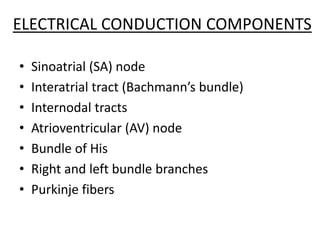
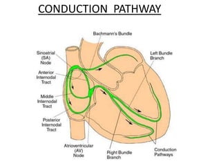
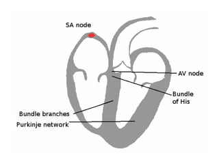
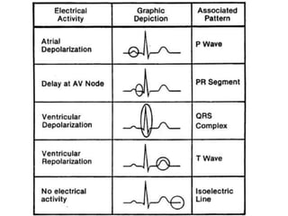
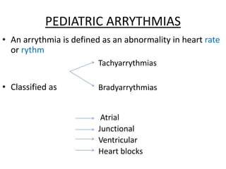
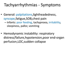
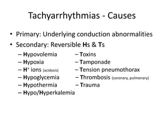
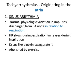
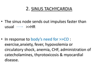
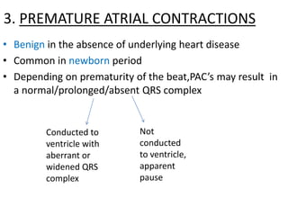
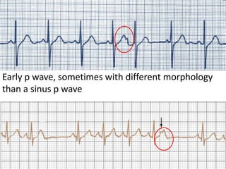
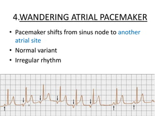
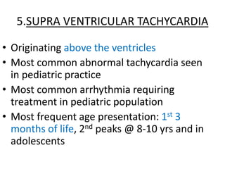
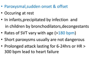
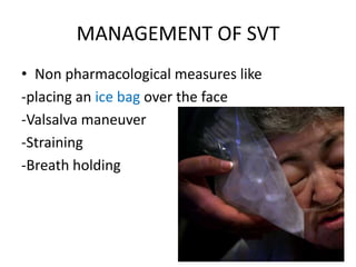
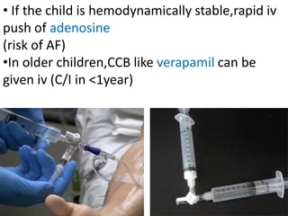
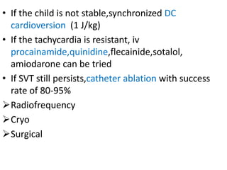
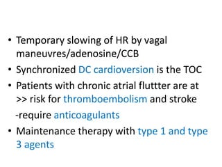
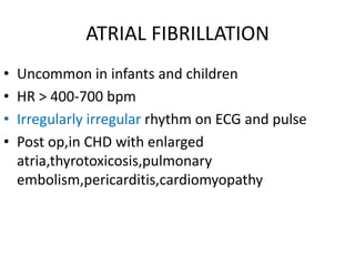
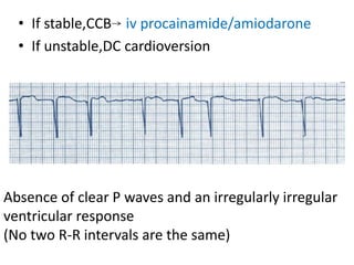
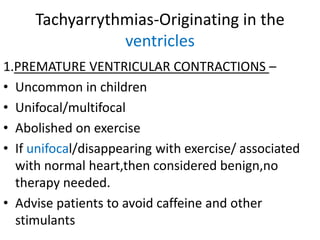
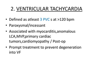
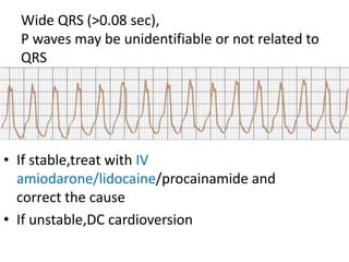
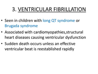
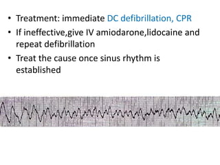
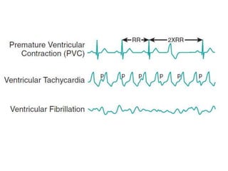
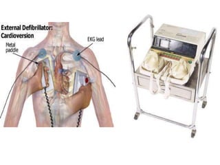
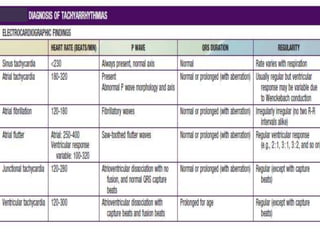
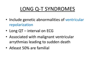
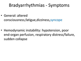
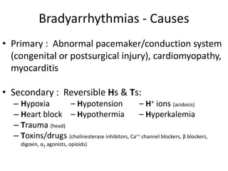
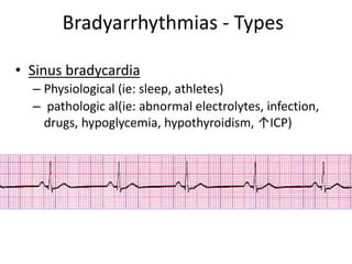
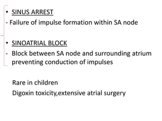
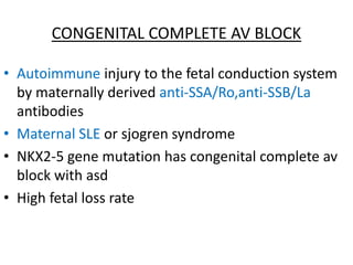
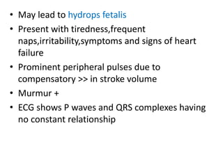
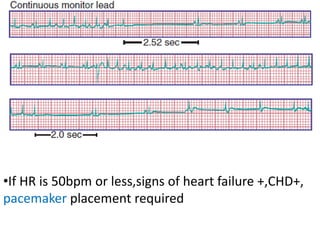
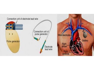
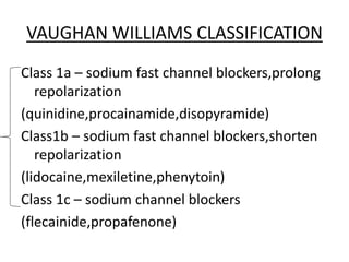
ARRYTHMIAS IN
CHILDREN
-Dr.Apoorva.E
PG,DCMS
ELECTRICAL ANATOMY OF
THE HEART
• The heart is a functional syncytium
• Network of myocytes connected to each other
by intercalated discs which have gap junctions
• Through which electrical impulses propagate
allowing rapid,synchronous depolarization of the
myocardium
• Sinoatrial (SA) node
• Interatrial tract (Bachmann’s bundle)
• Internodal tracts
• Atrioventricular (AV) node
• Bundle of His
• Right and left bundle branches
• Purkinje fibers
ELECTRICAL CONDUCTION COMPONENTS
CONDUCTION PATHWAY
PEDIATRIC ARRYTHMIAS
• An arrythmia is defined as an abnormality in heart rate
or rythm
• Classified as
Tachyarrythmias
Bradyarrythmias
Atrial
Junctional
Ventricular
Heart blocks
Tachyarrhythmias - Symptoms
• General: palpitations,lightheadedness,
syncope,fatigue,SOB,chest pain
– Infants: poor feeding, tachypnoea, irritability,
sleepiness, pallor, vomiting
• Hemodynamic instability: respiratory
distress/failure,hypotension,poor end-organ
perfusion,LOC,sudden collapse
Tachyarrhythmias - Causes
• Primary: Underlying conduction abnormalities
• Secondary: Reversible Hs & Ts
– Hypovolemia – Toxins
– Hypoxia – Tamponade
– H+ ions (acidosis) – Tension pneumothorax
– Hypoglycemia – Thrombosis (coronary, pulmonary)
– Hypothermia – Trauma
– Hypo/Hyperkalemia
Tachyarrhythmias - Originating in the
atria
1. SINUS ARRYTHMIA
• Normal physiologic variation in impulses
discharged from SA node in relation to
respiration
• HR slows during expiration,increases during
inspiration
• Drugs like digoxin exaggerate it
• Abolished by exercise
Varied PP interval.No significant change in
P wave morphology/PR interval
2. SINUS TACHYCARDIA
• The sinus node sends out impulses faster than
usual >>HR
• In response to body’s need for >>CO :
exercise,anxiety, fever, hypovolemia or
circulatory shock, anemia, CHF, administration of
catecholamines, thyrotoxicosis & myocardial
disease.
P waves are present and normal , narrow
QRS, beat to beat variability
3. PREMATURE ATRIAL CONTRACTIONS
• Benign in the absence of underlying heart disease
• Common in newborn period
• Depending on prematurity of the beat,PAC’s may result in
a normal/prolonged/absent QRS complex
Conducted to
ventricle with
aberrant or
widened QRS
complex
Not
conducted
to ventricle,
apparent
pause
Early p wave, sometimes with different morphology
than a sinus p wave
• Pacemaker shifts from sinus node to another
atrial site
• Normal variant
• Irregular rhythm
4.WANDERING ATRIAL PACEMAKER
5.SUPRA VENTRICULAR TACHYCARDIA
• Originating above the ventricles
• Most common abnormal tachycardia seen
in pediatric practice
• Most common arrhythmia requiring
treatment in pediatric population
• Most frequent age presentation: 1st 3
months of life, 2nd peaks @ 8-10 yrs and in
adolescents
• Paroxysmal,sudden onset & offset
• Occuring at rest
• In infants,precipitated by infection and
in children by bronchodilators,decongestants
• Rates of SVT vary with age (>180 bpm)
• Short paroxysms usually are not dangerous
• Prolonged attack lasting for 6-24hrs or HR >
300 bpm lead to heart failure
• Older children present with palpitations
• Younger children – Basal HR higher for that age,HR
>> greatly with crying
• P waves difficult to define, but 1:1 with normal QRS
• ECG similar to sinus tachycardia
• Differentiating features :
HR>230bpm,unvarying HR,abnormal P wave
axis if seen
• 3 major types
- re-entrant tachycardia with an accessory pathway
- Re-entrant tachycardia without an accessory pathway
- Ectopic/automatic tachycardias
AVRT
AVNRT
ATRIAL ECTOPICS
JUNCTIONAL ECTOPICS
ATRIAL FLUTTER
ATRIAL FIBRILLATION
ATRIOVENTRICULAR RECIPROCATING
TACHYCARDIA (AVRT)
• Most common mechanism of SVT in infants
• Re-entrant tachycardia with an accessory
pathway
• Flow of impulses may be bidirectional or
retrograde only
• Wolff-Parkinson-White syndrome :
- Characterized by the presence of a muscular bridge
connecting atria and ventricles on either the right
or left side of AV ring
- Flow of impulses is bidirectional
- Flow of impulses is antegrade through the
AV node and retrograde through the
accessory pathway towards the atrium
SVT in a child with WPW showing normal QRS
complexes with P waves seen on upstroke of T waves
• Typical features of WPW are apparent when
tachycardia subsides
• Wide QRS complexes,delta waves,short PR interval
• Risk of sudden death
MANAGEMENT OF SVT
• Non pharmacological measures like
-placing an ice bag over the face
-Valsalva maneuver
-Straining
-Breath holding
• If the child is hemodynamically stable,rapid iv
push of adenosine
(risk of AF)
•In older children,CCB like verapamil can be
given iv (C/I in <1year)
• If the child is not stable,synchronized DC
cardioversion (1 J/kg)
• If the tachycardia is resistant, iv
procainamide,quinidine,flecainide,sotalol,
amiodarone can be tried
• If SVT still persists,catheter ablation with success
rate of 80-95%
Radiofrequency
Cryo
Surgical
• Maintenance therapy
- When sinus rhythm is restored,
for long term maintenance,
DOC is beta blockers in both WPW and
Non WPW syndromes
- Digoxin can be given in infants with no
accessory pathway
AV NODAL RE-ENTRANT TACHYCARDIA
• Common form of SVT in adolescents
• Involves the use of 2 pathways within the AV
node
• Precipitated by exercise
• Present with syncopal attacks
• Good control on antiarrythmic therapy
• Beta blockers remain the drug of choice for
maintenance
ATRIAL ECTOPIC TACHYCARDIA
• Uncommon in children
• Variable HR,usually >200bpm
• Due to a single focus of automaticity
• On starting pharmacologic therapy,the
tachycardia gradually slows down only to
speed up again
• ECG shows ectopic p waves with an abnormal
axis
MULTIFOCAL ATRIAL TACHYCARDIA
• More common in infants than in older
children
• Characterized by 3 or more ectopic P
waves and varying PR intervals
• Spontaneous resolution occurs usually by
3 years of age
Chaotic ECG pattern with multiple ectopic P
waves with abnormal axes
JUNCTIONAL ECTOPIC TACHYCARDIA
• Due to an abnormal focus of automaticity
• The focus being a conducting tissue very close
to the AV node (junctional)
• Discharge of impulses from junctional tissue
exceeds SA nodal discharge leading to
AV dissociation
• Occurs in early post op period or may be
congenital
• IV amiodarone is the DOC for post-op JET
• Congenital JET requires catheter ablation
• Maintenance therapy with
amiodarone/sotalol
ATRIAL FLUTTER
• Also called intra-atrial re-entrant tachycardia
• HR > 400-600 bpm in neonates
>250-300 bpm in children
• Due to re-entrant pathway located in the right
atrium circling the tricuspid valve annulus
• AV dissociation occurs and ventricles respond
to 2nd - 4th atrial beat
• Occurs in neonates with normal hearts and in
children with CHD (with large stretched atria)
and post-op
Rapid and regular saw-toothed flutter
waves
• Temporary slowing of HR by vagal
maneuvres/adenosine/CCB
• Synchronized DC cardioversion is the TOC
• Patients with chronic atrial fluttter are at
>> risk for thromboembolism and stroke
-require anticoagulants
• Maintenance therapy with type 1 and type
3 agents
ATRIAL FIBRILLATION
• Uncommon in infants and children
• HR > 400-700 bpm
• Irregularly irregular rhythm on ECG and pulse
• Post op,in CHD with enlarged
atria,thyrotoxicosis,pulmonary
embolism,pericarditis,cardiomyopathy
• If stable,CCB iv procainamide/amiodarone
• If unstable,DC cardioversion
Absence of clear P waves and an irregularly irregular
ventricular response
(No two R-R intervals are the same)
Tachyarrythmias-Originating in the
ventricles
1.PREMATURE VENTRICULAR CONTRACTIONS –
• Uncommon in children
• Unifocal/multifocal
• Abolished on exercise
• If unifocal/disappearing with exercise/ associated
with normal heart,then considered benign,no
therapy needed.
• Advise patients to avoid caffeine and other
stimulants
• Early, wide QRS complexes
• T waves in opposite direction of QRS
• Bigeminy, sinus beat followed by PVC,this
repeating as a pattern also frequently seen
2. VENTRICULAR TACHYCARDIA
• Defined as atleast 3 PVC s at >120 bpm
• Paroxysmal/incessant
• Associated with myocarditis,anomalous
LCA,MVP,primary cardiac
tumors,cardiomyopathy / Post-op
• Prompt treatment to prevent degeneration
into VF
• If stable,treat with IV
amiodarone/lidocaine/procainamide and
correct the cause
• If unstable,DC cardioversion
Wide QRS (>0.08 sec),
P waves may be unidentifiable or not related to
QRS
3. VENTRICULAR FIBRILLATION
• Seen in children with long QT syndrome or
Brugada syndrome
• Associated with cardiomyopathies,structural
heart diseases causing ventricular dysfunction
• Sudden death occurs unless an effective
ventricular beat is reestablished rapidly
• Treatment: immediate DC defibrillation, CPR
• If ineffective,give IV amiodarone,lidocaine and
repeat defibrillation
• Treat the cause once sinus rhythm is
established
LONG Q-T SYNDROMES
• Include genetic abnormalities of ventricular
repolarization
• Long QT – interval on ECG
• Associated with malignant ventricular
arrythmias leading to sudden death
• Atleast 50% are familial
• Precipitated by exercise
• LQT1 events are stress induced
• LQT3 occur during sleep
• LQT2 have an intermediate pattern
• LQT3 has highest probability of sudden death
• Diagnostic criteria
Present with syncope,seizures,palpitations
Corrected QT interval >0.47sec or a QT
interval >0.44sec
Notched T waves
Low HR for age
Familial history of LQTS/sudden death
• Treatment - Beta blockers which blunt the
HR s response to exercise
• If drug induced profound bradycardia –
pacemaker
• If drug resistant,LQT3- implantable cardiac
defibrillator
Bradyarrhythmias - Symptoms
• General: altered
consciousness,fatigue,dizziness,syncope
• Hemodynamic instability: hypotension, poor
end-organ perfusion, respiratory distress/failure,
sudden collapse
Bradyarrhythmias - Causes
• Primary : Abnormal pacemaker/conduction system
(congenital or postsurgical injury), cardiomyopathy,
myocarditis
• Secondary : Reversible Hs & Ts:
– Hypoxia – Hypotension – H+ ions (acidosis)
– Heart block – Hypothermia – Hyperkalemia
– Trauma (head)
– Toxins/drugs (cholinesterase inhibitors, Ca++ channel blockers, β blockers,
digoxin, α2 agonists, opioids)
Bradyarrhythmias - Types
• Sinus bradycardia
– Physiological (ie: sleep, athletes)
– pathologic al(ie: abnormal electrolytes, infection,
drugs, hypoglycemia, hypothyroidism, ↑ICP)
• SINUS ARREST
- Failure of impulse formation within SA node
• SINOATRIAL BLOCK
- Block between SA node and surrounding atrium
preventing conduction of impulses
Rare in children
Digoxin toxicity,extensive atrial surgery
• SICK SINUS SYNDROME
- Due to abnormalities in either the SA node /
atrial conduction pathways / both
- Post surgery for CHD (fontan,mustard,senning
procedures) or even in patients with normal
heart
- Usually asymptomatic and don’t require
treatment
- Periods of marked sinus slowing present with
dizziness and syncope pacemaker if symptoms
recur
• AV BLOCKS
Type EKG Findings Causes & Clinical Significance
1st
degree
Prolonged PR interval Causes include AV nodal disease,myocarditis,↑K+, drugs (ie:
Ca++ channel blockers, β-blockers, digoxin), acute rheumatic
fever. Usually asymptomatic.
2nd
degree
Mobitz type I
Wenchebach
Progressive prolongation
of PR interval until a P
wave is not
conducted.After this
dropped beat cycle starts
again with a short PR
interval
Usually due to block within AV node. Caused by drugs (ie: Ca++
channel blockers, β-blockers, digoxin).
Can cause dizziness. Typically transient and benign; Rarely
progresses to 3rd degree heart block.
2nd
Degree
Mobitz type II
Prolonged constant PR
interval, inhibition of a set
proportion of atrial
impulses
Usually caused by defect in conduction pathway or acute
coronary syndrome, leading to block below AV node & His
bundle. Symptoms include palpitations, presyncope, syncope.
Can progress to 3rd degree heart block; often requires
pacemaker.
3rd
Degree
complete
AV dissociation. No atrial
impulses are conducted to
the ventricle
Congenital or caused by conduction system disease(myocardial
tumors/abscess/myocarditis) or injury (surgery for vsd). Most
symptomatic form of heart block: fatigue, presyncope, syncope.
Usually requires pacemaker
CONGENITAL COMPLETE AV BLOCK
• Autoimmune injury to the fetal conduction system
by maternally derived anti-SSA/Ro,anti-SSB/La
antibodies
• Maternal SLE or sjogren syndrome
• NKX2-5 gene mutation has congenital complete av
block with asd
• High fetal loss rate
• May lead to hydrops fetalis
• Present with tiredness,frequent
naps,irritability,symptoms and signs of heart
failure
• Prominent peripheral pulses due to
compensatory >> in stroke volume
• Murmur +
• ECG shows P waves and QRS complexes having
no constant relationship
•If HR is 50bpm or less,signs of heart failure +,CHD+,
pacemaker placement required
VAUGHAN WILLIAMS CLASSIFICATION
Class 1a – sodium fast channel blockers,prolong
repolarization
(quinidine,procainamide,disopyramide)
Class1b – sodium fast channel blockers,shorten
repolarization
(lidocaine,mexiletine,phenytoin)
Class 1c – sodium channel blockers
(flecainide,propafenone)
Class 2 – beta blockers
(propranolol,atenolol)
Class 3 – potassium channel openers,prolong
repolarization
(amiodarone)
Class 4 – miscellaneous
(verapamil,adenosine,digoxin)
THAT’S ALL
FOLKS !

Arrhythmias in children

  • 1.
  • 2.
  • 3.
    • The heartis a functional syncytium • Network of myocytes connected to each other by intercalated discs which have gap junctions • Through which electrical impulses propagate allowing rapid,synchronous depolarization of the myocardium
  • 4.
    • Sinoatrial (SA)node • Interatrial tract (Bachmann’s bundle) • Internodal tracts • Atrioventricular (AV) node • Bundle of His • Right and left bundle branches • Purkinje fibers ELECTRICAL CONDUCTION COMPONENTS
  • 5.
  • 8.
    PEDIATRIC ARRYTHMIAS • Anarrythmia is defined as an abnormality in heart rate or rythm • Classified as Tachyarrythmias Bradyarrythmias Atrial Junctional Ventricular Heart blocks
  • 9.
    Tachyarrhythmias - Symptoms •General: palpitations,lightheadedness, syncope,fatigue,SOB,chest pain – Infants: poor feeding, tachypnoea, irritability, sleepiness, pallor, vomiting • Hemodynamic instability: respiratory distress/failure,hypotension,poor end-organ perfusion,LOC,sudden collapse
  • 10.
    Tachyarrhythmias - Causes •Primary: Underlying conduction abnormalities • Secondary: Reversible Hs & Ts – Hypovolemia – Toxins – Hypoxia – Tamponade – H+ ions (acidosis) – Tension pneumothorax – Hypoglycemia – Thrombosis (coronary, pulmonary) – Hypothermia – Trauma – Hypo/Hyperkalemia
  • 11.
    Tachyarrhythmias - Originatingin the atria 1. SINUS ARRYTHMIA • Normal physiologic variation in impulses discharged from SA node in relation to respiration • HR slows during expiration,increases during inspiration • Drugs like digoxin exaggerate it • Abolished by exercise
  • 12.
    Varied PP interval.Nosignificant change in P wave morphology/PR interval
  • 13.
    2. SINUS TACHYCARDIA •The sinus node sends out impulses faster than usual >>HR • In response to body’s need for >>CO : exercise,anxiety, fever, hypovolemia or circulatory shock, anemia, CHF, administration of catecholamines, thyrotoxicosis & myocardial disease.
  • 14.
    P waves arepresent and normal , narrow QRS, beat to beat variability
  • 15.
    3. PREMATURE ATRIALCONTRACTIONS • Benign in the absence of underlying heart disease • Common in newborn period • Depending on prematurity of the beat,PAC’s may result in a normal/prolonged/absent QRS complex Conducted to ventricle with aberrant or widened QRS complex Not conducted to ventricle, apparent pause
  • 16.
    Early p wave,sometimes with different morphology than a sinus p wave
  • 17.
    • Pacemaker shiftsfrom sinus node to another atrial site • Normal variant • Irregular rhythm 4.WANDERING ATRIAL PACEMAKER
  • 18.
    5.SUPRA VENTRICULAR TACHYCARDIA •Originating above the ventricles • Most common abnormal tachycardia seen in pediatric practice • Most common arrhythmia requiring treatment in pediatric population • Most frequent age presentation: 1st 3 months of life, 2nd peaks @ 8-10 yrs and in adolescents
  • 19.
    • Paroxysmal,sudden onset& offset • Occuring at rest • In infants,precipitated by infection and in children by bronchodilators,decongestants • Rates of SVT vary with age (>180 bpm) • Short paroxysms usually are not dangerous • Prolonged attack lasting for 6-24hrs or HR > 300 bpm lead to heart failure
  • 20.
    • Older childrenpresent with palpitations • Younger children – Basal HR higher for that age,HR >> greatly with crying • P waves difficult to define, but 1:1 with normal QRS
  • 21.
    • ECG similarto sinus tachycardia • Differentiating features : HR>230bpm,unvarying HR,abnormal P wave axis if seen
  • 22.
    • 3 majortypes - re-entrant tachycardia with an accessory pathway - Re-entrant tachycardia without an accessory pathway - Ectopic/automatic tachycardias AVRT AVNRT ATRIAL ECTOPICS JUNCTIONAL ECTOPICS ATRIAL FLUTTER ATRIAL FIBRILLATION
  • 23.
    ATRIOVENTRICULAR RECIPROCATING TACHYCARDIA (AVRT) •Most common mechanism of SVT in infants • Re-entrant tachycardia with an accessory pathway • Flow of impulses may be bidirectional or retrograde only
  • 24.
    • Wolff-Parkinson-White syndrome: - Characterized by the presence of a muscular bridge connecting atria and ventricles on either the right or left side of AV ring - Flow of impulses is bidirectional
  • 26.
    - Flow ofimpulses is antegrade through the AV node and retrograde through the accessory pathway towards the atrium SVT in a child with WPW showing normal QRS complexes with P waves seen on upstroke of T waves
  • 27.
    • Typical featuresof WPW are apparent when tachycardia subsides • Wide QRS complexes,delta waves,short PR interval • Risk of sudden death
  • 28.
    MANAGEMENT OF SVT •Non pharmacological measures like -placing an ice bag over the face -Valsalva maneuver -Straining -Breath holding
  • 29.
    • If thechild is hemodynamically stable,rapid iv push of adenosine (risk of AF) •In older children,CCB like verapamil can be given iv (C/I in <1year)
  • 30.
    • If thechild is not stable,synchronized DC cardioversion (1 J/kg) • If the tachycardia is resistant, iv procainamide,quinidine,flecainide,sotalol, amiodarone can be tried • If SVT still persists,catheter ablation with success rate of 80-95% Radiofrequency Cryo Surgical
  • 31.
    • Maintenance therapy -When sinus rhythm is restored, for long term maintenance, DOC is beta blockers in both WPW and Non WPW syndromes - Digoxin can be given in infants with no accessory pathway
  • 32.
    AV NODAL RE-ENTRANTTACHYCARDIA • Common form of SVT in adolescents • Involves the use of 2 pathways within the AV node • Precipitated by exercise • Present with syncopal attacks • Good control on antiarrythmic therapy • Beta blockers remain the drug of choice for maintenance
  • 33.
    ATRIAL ECTOPIC TACHYCARDIA •Uncommon in children • Variable HR,usually >200bpm • Due to a single focus of automaticity • On starting pharmacologic therapy,the tachycardia gradually slows down only to speed up again • ECG shows ectopic p waves with an abnormal axis
  • 34.
    MULTIFOCAL ATRIAL TACHYCARDIA •More common in infants than in older children • Characterized by 3 or more ectopic P waves and varying PR intervals • Spontaneous resolution occurs usually by 3 years of age
  • 35.
    Chaotic ECG patternwith multiple ectopic P waves with abnormal axes
  • 36.
    JUNCTIONAL ECTOPIC TACHYCARDIA •Due to an abnormal focus of automaticity • The focus being a conducting tissue very close to the AV node (junctional) • Discharge of impulses from junctional tissue exceeds SA nodal discharge leading to AV dissociation
  • 37.
    • Occurs inearly post op period or may be congenital • IV amiodarone is the DOC for post-op JET • Congenital JET requires catheter ablation • Maintenance therapy with amiodarone/sotalol
  • 38.
    ATRIAL FLUTTER • Alsocalled intra-atrial re-entrant tachycardia • HR > 400-600 bpm in neonates >250-300 bpm in children • Due to re-entrant pathway located in the right atrium circling the tricuspid valve annulus • AV dissociation occurs and ventricles respond to 2nd - 4th atrial beat
  • 39.
    • Occurs inneonates with normal hearts and in children with CHD (with large stretched atria) and post-op Rapid and regular saw-toothed flutter waves
  • 40.
    • Temporary slowingof HR by vagal maneuvres/adenosine/CCB • Synchronized DC cardioversion is the TOC • Patients with chronic atrial fluttter are at >> risk for thromboembolism and stroke -require anticoagulants • Maintenance therapy with type 1 and type 3 agents
  • 41.
    ATRIAL FIBRILLATION • Uncommonin infants and children • HR > 400-700 bpm • Irregularly irregular rhythm on ECG and pulse • Post op,in CHD with enlarged atria,thyrotoxicosis,pulmonary embolism,pericarditis,cardiomyopathy
  • 42.
    • If stable,CCBiv procainamide/amiodarone • If unstable,DC cardioversion Absence of clear P waves and an irregularly irregular ventricular response (No two R-R intervals are the same)
  • 43.
    Tachyarrythmias-Originating in the ventricles 1.PREMATUREVENTRICULAR CONTRACTIONS – • Uncommon in children • Unifocal/multifocal • Abolished on exercise • If unifocal/disappearing with exercise/ associated with normal heart,then considered benign,no therapy needed. • Advise patients to avoid caffeine and other stimulants
  • 44.
    • Early, wideQRS complexes • T waves in opposite direction of QRS • Bigeminy, sinus beat followed by PVC,this repeating as a pattern also frequently seen
  • 45.
    2. VENTRICULAR TACHYCARDIA •Defined as atleast 3 PVC s at >120 bpm • Paroxysmal/incessant • Associated with myocarditis,anomalous LCA,MVP,primary cardiac tumors,cardiomyopathy / Post-op • Prompt treatment to prevent degeneration into VF
  • 46.
    • If stable,treatwith IV amiodarone/lidocaine/procainamide and correct the cause • If unstable,DC cardioversion Wide QRS (>0.08 sec), P waves may be unidentifiable or not related to QRS
  • 47.
    3. VENTRICULAR FIBRILLATION •Seen in children with long QT syndrome or Brugada syndrome • Associated with cardiomyopathies,structural heart diseases causing ventricular dysfunction • Sudden death occurs unless an effective ventricular beat is reestablished rapidly
  • 48.
    • Treatment: immediateDC defibrillation, CPR • If ineffective,give IV amiodarone,lidocaine and repeat defibrillation • Treat the cause once sinus rhythm is established
  • 52.
    LONG Q-T SYNDROMES •Include genetic abnormalities of ventricular repolarization • Long QT – interval on ECG • Associated with malignant ventricular arrythmias leading to sudden death • Atleast 50% are familial
  • 54.
    • Precipitated byexercise • LQT1 events are stress induced • LQT3 occur during sleep • LQT2 have an intermediate pattern • LQT3 has highest probability of sudden death
  • 55.
    • Diagnostic criteria Presentwith syncope,seizures,palpitations Corrected QT interval >0.47sec or a QT interval >0.44sec Notched T waves Low HR for age Familial history of LQTS/sudden death
  • 56.
    • Treatment -Beta blockers which blunt the HR s response to exercise • If drug induced profound bradycardia – pacemaker • If drug resistant,LQT3- implantable cardiac defibrillator
  • 57.
    Bradyarrhythmias - Symptoms •General: altered consciousness,fatigue,dizziness,syncope • Hemodynamic instability: hypotension, poor end-organ perfusion, respiratory distress/failure, sudden collapse
  • 58.
    Bradyarrhythmias - Causes •Primary : Abnormal pacemaker/conduction system (congenital or postsurgical injury), cardiomyopathy, myocarditis • Secondary : Reversible Hs & Ts: – Hypoxia – Hypotension – H+ ions (acidosis) – Heart block – Hypothermia – Hyperkalemia – Trauma (head) – Toxins/drugs (cholinesterase inhibitors, Ca++ channel blockers, β blockers, digoxin, α2 agonists, opioids)
  • 59.
    Bradyarrhythmias - Types •Sinus bradycardia – Physiological (ie: sleep, athletes) – pathologic al(ie: abnormal electrolytes, infection, drugs, hypoglycemia, hypothyroidism, ↑ICP)
  • 60.
    • SINUS ARREST -Failure of impulse formation within SA node • SINOATRIAL BLOCK - Block between SA node and surrounding atrium preventing conduction of impulses Rare in children Digoxin toxicity,extensive atrial surgery
  • 61.
    • SICK SINUSSYNDROME - Due to abnormalities in either the SA node / atrial conduction pathways / both - Post surgery for CHD (fontan,mustard,senning procedures) or even in patients with normal heart - Usually asymptomatic and don’t require treatment - Periods of marked sinus slowing present with dizziness and syncope pacemaker if symptoms recur
  • 62.
    • AV BLOCKS TypeEKG Findings Causes & Clinical Significance 1st degree Prolonged PR interval Causes include AV nodal disease,myocarditis,↑K+, drugs (ie: Ca++ channel blockers, β-blockers, digoxin), acute rheumatic fever. Usually asymptomatic. 2nd degree Mobitz type I Wenchebach Progressive prolongation of PR interval until a P wave is not conducted.After this dropped beat cycle starts again with a short PR interval Usually due to block within AV node. Caused by drugs (ie: Ca++ channel blockers, β-blockers, digoxin). Can cause dizziness. Typically transient and benign; Rarely progresses to 3rd degree heart block. 2nd Degree Mobitz type II Prolonged constant PR interval, inhibition of a set proportion of atrial impulses Usually caused by defect in conduction pathway or acute coronary syndrome, leading to block below AV node & His bundle. Symptoms include palpitations, presyncope, syncope. Can progress to 3rd degree heart block; often requires pacemaker. 3rd Degree complete AV dissociation. No atrial impulses are conducted to the ventricle Congenital or caused by conduction system disease(myocardial tumors/abscess/myocarditis) or injury (surgery for vsd). Most symptomatic form of heart block: fatigue, presyncope, syncope. Usually requires pacemaker
  • 65.
    CONGENITAL COMPLETE AVBLOCK • Autoimmune injury to the fetal conduction system by maternally derived anti-SSA/Ro,anti-SSB/La antibodies • Maternal SLE or sjogren syndrome • NKX2-5 gene mutation has congenital complete av block with asd • High fetal loss rate
  • 66.
    • May leadto hydrops fetalis • Present with tiredness,frequent naps,irritability,symptoms and signs of heart failure • Prominent peripheral pulses due to compensatory >> in stroke volume • Murmur + • ECG shows P waves and QRS complexes having no constant relationship
  • 67.
    •If HR is50bpm or less,signs of heart failure +,CHD+, pacemaker placement required
  • 69.
    VAUGHAN WILLIAMS CLASSIFICATION Class1a – sodium fast channel blockers,prolong repolarization (quinidine,procainamide,disopyramide) Class1b – sodium fast channel blockers,shorten repolarization (lidocaine,mexiletine,phenytoin) Class 1c – sodium channel blockers (flecainide,propafenone)
  • 70.
    Class 2 –beta blockers (propranolol,atenolol) Class 3 – potassium channel openers,prolong repolarization (amiodarone) Class 4 – miscellaneous (verapamil,adenosine,digoxin)
  • 71.

Editor's Notes

  • #4 obeying the “all or none” law.enabling it to function as a single unit
  • #9 Site of origin
  • #10 Pathophysiology of tachyarrhythmias Insufficient time for diastolic filling leads to decreased stroke volume and thus decreased cardiac output Impaired coronary artery perfusion (which occurs during diastole) Increased myocardial oxygen demand as HR increases
  • #12 Indicates healthy interaction between autonomic respi and cardiac control in cns..drugs >>vagal tone
  • #13 6 boxes 4 boxes
  • #14 <220 bpm in infants, <180 bpm in children
  • #16 Refractoriness of av node……………………………….Resets sa node pacemaker
  • #20 Range of 240-300bpm…..tachypnoea ,ashenchild,hepatomegaly,irritable
  • #21 Diagnosis is diff in younger children
  • #22 Normal p wave is positive in lead 1 and avf
  • #24 Concealed accessory pathway……….bundle of kent
  • #27 Orthodromic..antidromic
  • #29 Vagal maneuvres…blowing against obstructed distal end of straw..standing on their head..ocular massage..carotid sinus massage
  • #30 0.1 mg/kg; max 1st dose 6 mg; additional 0.2 mg/kg if needed (max 2nd dose 12 mg)
  • #31 Definitive rx
  • #32 Not effective in adults…in wpw it increases risk of af therefore c/i
  • #34 Unlike reentrant tachycardias which break suddenly therefore more resistant to therapy
  • #37 Therefore complications will be more
  • #39 Because the AV NODE CANNOT TRANSMIT IMPULSES AT THE SAME RATE
  • #40 Like in tricuspid and mitral insufficiency,ebstein s anamoly,rheumatic mitral stenosis
  • #42 Very rarely familial
  • #44 Usually unifocal….Medications usually not needed
  • #46 After intraventricular surgery like for TOF
  • #48 Thump on chest
  • #49 Brugada syndrome – inherited arrhythmia, autosomal dominant person goes into v-fib, faints, dies suddenly
  • #53 Torsades de pointes and vf..gene+ qt may not be prolonged…viceversa…helps to indicate asymptomatic relatives with prolonged qt
  • #55 Romano ward syndrome- heterozygous Jervell lange nielsen syndrome - homozygous
  • #56 Ecg criteria + clinical criteria…..genotyping
  • #60 Sinus bradycardia Can be a physiologic consequence of decreased metabolic demand (ie: while sleeping) or increased stroke volume (ie: athletes) Pathologic etiologies include electrolyte disorders, infection, drug effects, hypoglycemia, hypothyroidism, increased ICP Sinus node arrest - absent pacemaker activity in the sinus node with subsidiary pacemaker in the atrium, AV junction, or ventricles initiating depolarization (leading to atrial escape, junctional escape, and idioventricular escape rhythms, respectively) Atrial escape: late P wave, different P wave morphology Junctional escape: originates in AV node, narrow-complex, +/- retrograde P waves Idioventricular escape: wide-complex, typical rate 30-40 beats/min
  • #62 Tachy brady syndrome- sa node dysfunction with episodes of svt - require drug therapy for tachy and pacemaker for sudden brady
  • #63 AV block – disturbance of electrical conduction through AV node
  • #68 REGULAR VENTRICULAR RATE.VARIABLE ATRIAL RATE COMPLETELY DISSOCIATED FROM VENTRICLE.NORMAL QRS MORPHOLOGY