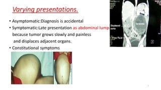
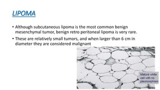
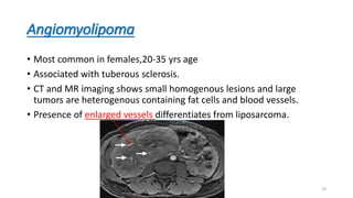
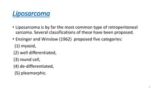
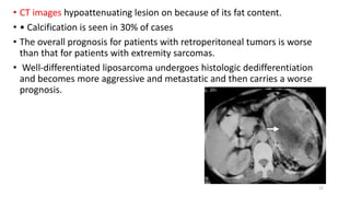
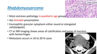
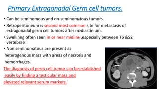
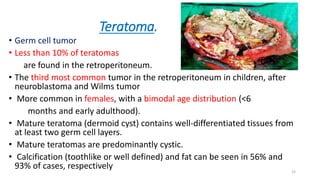
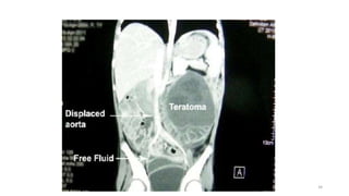
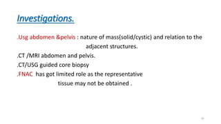
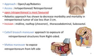
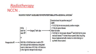
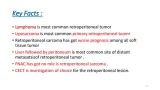
RETROPERITONEAL
TUMORS
By:Dr.B.Vinod
Dept. of General surgery,
Gandhi medical college and hospital,
Hyderabad, Telangana.
1
• Introduction
• Risk factors
• Clinical features
• Classification
• Investigation
• Management
2
Introduction
• Primary retroperitoneal tumors (RPTs) refer to a group of rare
neoplasms arising in the retroperitoneum and pelvis
• Tendency for extensive growth before becoming clinically evident.
• Most cases tumors do not originate in a specific organ but rather
grow from connective tissues normally present in the
retroperitoneum and pelvis.
• RPTs represent a heterogeneous group of neoplasms comprising a
majority of malignant mesenchymal tumors and a minority of benign
lesions.
3
• Incidence is approximately 2.7 cases /10 persons/ pear.
• Most of then are malignant & accounts for 0.1 to 0.2 % of all
malignancies in body.
• Sarcoms are second most common tumor of retroperitoneum & first
most primary retroperitoneal malignancy.
4
• As a group RPTs represent a combination of sarcomas and other benign and
malignant lesions
• Liposarcoma
Leiomyosarcoma account for 80% of all retroperitoneal
Malignant fibrous histiocytoma sarcomas
• Benign mesenchymal tumors almost never transform into malignant
counterparts.
5
Risk Factors
• Radiation exposure
• Genetic causes
• Carcinogen
• Immunodeficiency
• Viral infections
6
Varying presentations.
• Asymptomatic:Diagnosis is accidental
• Symptomatic:Late presentation as abdominal lump
because tumor grows slowly and painless
and displaces adjacent organs.
• Constitutional symptoms
7
•Due to compression on adjacent organs:
i) Back Pain- Severe back pain often following pressure by
• tumor mass over muscles, facet joint and vertebral column.
• Radicular Pain- Radiating type of pain along the nerve root due to its
compression.
ii) Obstruction of Viscera and Tubular Organs – usually of duodenum , colon ,
ureter , pancreas, kidney resulting in
• Nausea and Vomiting-
• Colicky Pain
• Constipation/ obstipation
• Urinary Retention
8
iii) Compression of Aorta
• Hypertension-
• Renal Insufficiency-
• Mesenteric Ischemia-
• Intermittent Claudication
iv) Compression of Vena Cava
• Edema of Feet
• Low Blood Pressure
v)Nerve Lesions
• Tingling and Numbness in Lower limbs
• Weakness of the Lower
9
On examination.
• No clinical findings unless the swelling is very large
• Consistency:Firm to hard mass
• Surface :Smooth , but in lymphoma it is nodular
• Borders: can not be traced properly because of deep position of the
swellings
• Not moving with respiration.
• Non mobile.
• Non tender
• Does not fall forward
10
Classification of Primary Mesenchymal Tumors
11
12
Secondaries(Metastatic).
• Prostate
• Lung(SCC)
• SCC of cervix, vagina
• Pancreas.
13
Benign tumors
• Benign RPTs are much less common than retroperitoneal sarcomas.
The more frequent of these are
Lipoma,
Myelolipoma,
Schwannoma,
Extra-adrenal pheochromocytoma,
Paraganglioma,and cystadenoma.
14
LIPOMA
• Although subcutaneous lipoma is the most common benign
mesenchymal tumor, benign retro peritoneal lipoma is very rare.
• These are relatively small tumors, and when larger than 6 cm in
diameter they are considered malignant
15
MRI imaging.
1
2
16
Schwannoma.
• Benign tumor that arises from the perineural sheath
of Schwann cell.
• 6% of retroperitoneal neoplasms
• Common in females (2:1)
• Usually asymptomatic.
• CT, small schwannomas are round, well defined,
and homogeneous,
but large schwannomas may be
heterogeneous in appearance.
• Calcification(23%)
• Cystic degenerations (66%).
• Risk of malignancy 5% 17
18
Angiomyolipoma
• Most common in females,20-35 yrs age
• Associated with tuberous sclerosis.
• CT and MR imaging shows small homogenous lesions and large
tumors are heterogenous containing fat cells and blood vessels.
• Presence of enlarged vessels differentiates from liposarcoma.
19
MALIGNANT TUMOR.
Other retroperitoneal masses need to be differentiated from
retroperitoneal sarcomas which includes:
Lymphomas
Retroperitoneal fibrosis
Germ cell tumors
20
Liposarcoma
• Liposarcoma is by far the most common type of retroperitoneal
sarcoma. Several classifications of these have been proposed.
• Enzinger and Winslow (1962) proposed five categories:
(1) myxoid,
(2) well differentiated,
(3) round cell,
(4) de-differentiated,
(5) pleomorphic.
21
• CT images hypoattenuating lesion on because of its fat content.
• • Calcification is seen in 30% of cases
• The overall prognosis for patients with retroperitoneal tumors is worse
than that for patients with extremity sarcomas.
• Well-differentiated liposarcoma undergoes histologic dedifferentiation
and becomes more aggressive and metastatic and then carries a worse
prognosis.
22
Myxoid
• Composed of primitive lipoblasts that do not have the typical fat-
laden cytoplasm but rather resemble primitive mesenchymal cells.
Abundant capillary network and myxoid matrix are other typical
components.
• Balanced translocation of chromosomes 12 and 16 t(12:16).
23
Well-differentiated liposarcoma
• The histologic appearance closely resembles that of a benign lipoma,
and the distinction between the two by imaging and even under the
microscope is a challenge.
• In fact, many well-differentiated liposarcomas are misdiagnosed as
deeply seated lipomas.
• Ring chromosome 12 is typical of well-differentiated liposarcomas
24
Round cell liposarcoma
• composed of small round cells uniform in size and closely packed
together. There is no specific pattern of cellular arrangement and
intracellular lipid content is scarce.
25
De-differentiated liposarcoma
• characterized by the coexistence of well-differentiated and poorly
differentiated areas within the same tumor.
26
Pleomorphic liposarcoma.
• Features include a disorderly growth pattern with cellular
pleomorphism, giant cells,and anaplastic pyknotic nuclei.
• Because this anaplastic tumor resembles other undifferentiated
sarcomas, some lipoblastic presence must be documented to confirm
this diagnosis.
27
Leiomyosarcomas.
• Second most common (28%).
• Can grow to a large size >10 cm before compromising adjacent organs and
precipitating clinical symptoms such as venous thrombosis.
• M/C in females in 5 to 7 decades.
• Histology cells arranged in wavy sheets with cigar shaped nuclei
• It can be extravascular(62%),intravascular(5%),or
• combination(33%). of extra and intrvascular.
• At CT ,small tumors may be homogeneously solid,
but large tumors have
extensive areas of necrosis and occasional hemorrhage
28
Axial MR imaging HPE
29
Malignant fibrous histiocytoma
• Third most common(19%).
• Males(3:1)
• 5th to 6th decade
• CT and MR imaging appearances are nonspecific ,and present as
heterogenous mass wit areas of necrosis and hemorrhage
• Calcification seen in 10% of cases
30
Rhabdomyosarcoma
• Most common pathology in paediatric age group.
• Has bimodal presentation.
• Eosinophilic granular cytoplasm either round or elongated
cells(tadpole).
• CT or MR imaging shows areas of calcification and areas of necrosis
with hemorrhages
• Metastasis occurs in 10 to 20 % cases
31
Primary Extragonadal Germ cell tumors.
• Can be seminomous and on-seminomatous tumors.
• Retroperitoneum is second most common site for metastasis of
extragonadal germ cell tumors after mediastinium.
• Swellinng often seen in or near midline ,especially between T6 &S2
vertebrae
• Non seminomatous are present as
heterogenous mass with areas of necrosis and
hemorrhages.
The diagnosis of germ cell tumor can be established
easily by finding a testicular mass and
elevated relevant serum markers. 32
Teratoma.
• Germ cell tumor
• Less than 10% of teratomas
are found in the retroperitoneum.
• The third most common tumor in the retroperitoneum in children, after
neuroblastoma and Wilms tumor
• More common in females, with a bimodal age distribution (<6
months and early adulthood).
• Mature teratoma (dermoid cyst) contains well-differentiated tissues from
at least two germ cell layers.
• Mature teratomas are predominantly cystic.
• Calcification (toothlike or well defined) and fat can be seen in 56% and
93% of cases, respectively 33
34
Lymphoma
• Most common retroperitoneal malignancy, accounting for 33%.
• Seen in the 40–70-year age group
• Frequently manifests with extranodal disease in the liver,spleen, or
bowel, often at an advanced stage.
• History of fever , myalgia , night sweats , weight loss
• Paraaortic lymph nodes are involved in 25% of the patients with
Hodgkin lymphoma and 55% of the patients with non-Hodgkin
lymphoma.
35
• At CT, lymphoma is seen as a well-defined homogeneous mass.
• The aorta and IVC can be anteriorly displaced, producing the
“floating aorta” sign.
36
Retroperitoneal and pelvic sarcomas
are classified as deep tumors
• Superficial tumor is located
exclusively above the superficial
fascia without invasion of the
fascia;
• Deep tumor is located exclusively
beneath the superficial fascia,
superficial to the fascia with
invasion of or through the fascia,
or both superficial yet beneath the
fascia
37
STAGING OF RETROPERITONEAL SARCOMAS(TNM)
• T0 - No demonstrable tumor
• T1- Tumor measuring <5 cm in maximal diameter
T1a- superficial tumor
T1b- Deep tumor
• T2 - Tumor measuring =>5 cm in maximal diameter
T2a- superficial tumor
T2b- Deep tumor
• T3 - Evidence of macroscopic invasion of nearby structures by the
tumor
• N0 - No histologic evidence of regional lymph node involvement
• N1 - Histologically proved regional lymph node involvement
• M0 - No distant metastasis
• M1- Distant metastasis present
38
Grading
• Gx: Cannot be assessed
• G1:Grade 1
• G2:Grade 2
• G3:Grade 3
• AJCC incorporates a three tired grading
system determined by
Mitotic activity(1-3)
Differentiation(1-3)
Necrosis(0-2)
Above 3 parameters are summed to grade
• Grade 1 2 or 3
• Grade 2 4 or 5
• Grade 3 6 or 6
39
40
Based on the extent of surgical resection of the
primary tumor(R)
• R0 - Tumor was entirely resected with no residual tumor and
negative surgical margins
• R1 - Microscopic residual tumor positive surgical margins
• R2 - Macroscopic residual tumor
• R3- Tumor spillage and dissemination during resection
41
Investigations.
.Usg abdomen &pelvis : nature of mass(solid/cystic) and relation to the
adjacent structures.
.CT /MRI abdomen and pelvis.
.CT/USG guided core biopsy
.FNAC has got limited role as the representative
tissue may not be obtained .
42
Routine blood investigations:
.Hemoglobin: anemia
.Blood and serum creatinine- raised on compression of kidney and
ureter
.Liver function test
. Effect of paraneoplastic syndrome
Hypoglycemia:- due to increased insulin like hormone
Catecholamines:- paraganglioma
43
.Plane X ray abdomen:- signs of intestinal obstruction, obliterated
psoas shadow, calcification of tumor mass
.CT scan of chest.
lung metastasis
.Chest X ray PA view
.IVU :- Can show obstruction and displacement of
kidney and ureter, distortion of renal pelvis and bladder
compression.
44
Indications of preoperative biopsy.
• Lymphomas
• Metastasis from a preexisting cancer is suspected.
• Patients with a suspected sarcoma in whom metastatic disease is
noted on imaging and a biopsy may guide subsequent systemic
therapy.
• A surgically resectable retroperitoneal/intra-abdominal sarcoma for
diagnosis and grading
45
Management of primary localized RPS
• Surgery
• Radiation
• Chemotherapy
• Combined chemo-radiotherapy.
46
Surgery
• En bloc resection with complete clearance of margin is standard
treatment for sarcomas .
• 40 to 60% are amenable to complete surgical resection.
• Nephrectomy (42%) followed by colectomy (30%) resection of intestine
are most common adjunctive surgery .
• Positive surgical margins are associated with high local
recurrence.(50% in 5 YRS ).
47
• Approach : Open/Lap/Robotics
• Access : Inrtaperitoneal/ Retroperitoneal
• open intraperitoneal is most favoured
• Robotics approach has shown to decrease morbidity and mortality in
retroperitoneal tumor of size less than 3 cm.
• Incision : midline, rooftop (cheveron) , thoracoabdominal, Subcoastal.
• Cattell braasch maneuver approach to exposure of
retroperitoneal structures from Right-sided.
• Mattox maneuver to expose
retroperitoneum from left side
48
Contraindications of surgery :
• Tumor invading major vascular structure
• Multiple Distant metastasis
• Gross peritoneal invasion / peritoneal disease
• Patient not fit for major surgery.
49
Radiotherapy
NCCN .
50
51
Chemotherapy
• Indications:-
Neoadjuvant(stage 3)
Unresectable tumor ( Palliative )
Distant metastasis
• Treatment regimens :
AIM :-Adriyamycin(20-25mg/m2 IV push on days 1-3) ,
Ifosfamide(2000-3000mg/m2 IV push bolus for 3 hrs on days 1-3 ,
Mesna(225mg/m2 IV over 1hr before Ifosfamide and 4 to 8 hrs
after ifosfamide)
Repeat every 3 – 4 weeks.
MAID:-Mesna , Iphosphamide, Adriyamycin, Dacarbazine(300mg/m2/day IV
infusion on days 1-3)
52
Combined chemo-radiation.
• Doxorubicin dose: 20mg/m2/week for 4-5 weeks infusion.
• Radiation:- 18 to 50.4 Gy total radiation followed by 15 Gy IORT at
bed of resected tumour.
53
Overveiw of management of
Retoperitoneal sarcoma.
• Stage I –surgery
• Stage II -pre-op radiation + surgery + post op radiation
• Stage III -Neoadjuvant chemo-radiotherapy +Surgery
• Stage IV –Palliative CTRT.
54
Key Facts :
• Lymphoma is most common retroperitoneal tumor
• Liposarcoma is most common primary retroperitoneal tuomr
• Retroperitoneal sarcoma has got worse prognosis among all soft
tissue tumor
• Liver followed by peritoneum is most common site of distant
metasatsisof retroperitoneal tumor .
• FNAC has got no role is retroperitoneal sarcoma .
• CECT is investigation of choice for the retroperitoneal lesion.
55
56

Retroperitoneal tumors

  • 1.
    RETROPERITONEAL TUMORS By:Dr.B.Vinod Dept. of Generalsurgery, Gandhi medical college and hospital, Hyderabad, Telangana. 1
  • 2.
    • Introduction • Riskfactors • Clinical features • Classification • Investigation • Management 2
  • 3.
    Introduction • Primary retroperitonealtumors (RPTs) refer to a group of rare neoplasms arising in the retroperitoneum and pelvis • Tendency for extensive growth before becoming clinically evident. • Most cases tumors do not originate in a specific organ but rather grow from connective tissues normally present in the retroperitoneum and pelvis. • RPTs represent a heterogeneous group of neoplasms comprising a majority of malignant mesenchymal tumors and a minority of benign lesions. 3
  • 4.
    • Incidence isapproximately 2.7 cases /10 persons/ pear. • Most of then are malignant & accounts for 0.1 to 0.2 % of all malignancies in body. • Sarcoms are second most common tumor of retroperitoneum & first most primary retroperitoneal malignancy. 4
  • 5.
    • As agroup RPTs represent a combination of sarcomas and other benign and malignant lesions • Liposarcoma Leiomyosarcoma account for 80% of all retroperitoneal Malignant fibrous histiocytoma sarcomas • Benign mesenchymal tumors almost never transform into malignant counterparts. 5
  • 6.
    Risk Factors • Radiationexposure • Genetic causes • Carcinogen • Immunodeficiency • Viral infections 6
  • 7.
    Varying presentations. • Asymptomatic:Diagnosisis accidental • Symptomatic:Late presentation as abdominal lump because tumor grows slowly and painless and displaces adjacent organs. • Constitutional symptoms 7
  • 8.
    •Due to compressionon adjacent organs: i) Back Pain- Severe back pain often following pressure by • tumor mass over muscles, facet joint and vertebral column. • Radicular Pain- Radiating type of pain along the nerve root due to its compression. ii) Obstruction of Viscera and Tubular Organs – usually of duodenum , colon , ureter , pancreas, kidney resulting in • Nausea and Vomiting- • Colicky Pain • Constipation/ obstipation • Urinary Retention 8
  • 9.
    iii) Compression ofAorta • Hypertension- • Renal Insufficiency- • Mesenteric Ischemia- • Intermittent Claudication iv) Compression of Vena Cava • Edema of Feet • Low Blood Pressure v)Nerve Lesions • Tingling and Numbness in Lower limbs • Weakness of the Lower 9
  • 10.
    On examination. • Noclinical findings unless the swelling is very large • Consistency:Firm to hard mass • Surface :Smooth , but in lymphoma it is nodular • Borders: can not be traced properly because of deep position of the swellings • Not moving with respiration. • Non mobile. • Non tender • Does not fall forward 10
  • 11.
    Classification of PrimaryMesenchymal Tumors 11
  • 12.
  • 13.
    Secondaries(Metastatic). • Prostate • Lung(SCC) •SCC of cervix, vagina • Pancreas. 13
  • 14.
    Benign tumors • BenignRPTs are much less common than retroperitoneal sarcomas. The more frequent of these are Lipoma, Myelolipoma, Schwannoma, Extra-adrenal pheochromocytoma, Paraganglioma,and cystadenoma. 14
  • 15.
    LIPOMA • Although subcutaneouslipoma is the most common benign mesenchymal tumor, benign retro peritoneal lipoma is very rare. • These are relatively small tumors, and when larger than 6 cm in diameter they are considered malignant 15
  • 16.
  • 17.
    Schwannoma. • Benign tumorthat arises from the perineural sheath of Schwann cell. • 6% of retroperitoneal neoplasms • Common in females (2:1) • Usually asymptomatic. • CT, small schwannomas are round, well defined, and homogeneous, but large schwannomas may be heterogeneous in appearance. • Calcification(23%) • Cystic degenerations (66%). • Risk of malignancy 5% 17
  • 18.
  • 19.
    Angiomyolipoma • Most commonin females,20-35 yrs age • Associated with tuberous sclerosis. • CT and MR imaging shows small homogenous lesions and large tumors are heterogenous containing fat cells and blood vessels. • Presence of enlarged vessels differentiates from liposarcoma. 19
  • 20.
    MALIGNANT TUMOR. Other retroperitonealmasses need to be differentiated from retroperitoneal sarcomas which includes: Lymphomas Retroperitoneal fibrosis Germ cell tumors 20
  • 21.
    Liposarcoma • Liposarcoma isby far the most common type of retroperitoneal sarcoma. Several classifications of these have been proposed. • Enzinger and Winslow (1962) proposed five categories: (1) myxoid, (2) well differentiated, (3) round cell, (4) de-differentiated, (5) pleomorphic. 21
  • 22.
    • CT imageshypoattenuating lesion on because of its fat content. • • Calcification is seen in 30% of cases • The overall prognosis for patients with retroperitoneal tumors is worse than that for patients with extremity sarcomas. • Well-differentiated liposarcoma undergoes histologic dedifferentiation and becomes more aggressive and metastatic and then carries a worse prognosis. 22
  • 23.
    Myxoid • Composed ofprimitive lipoblasts that do not have the typical fat- laden cytoplasm but rather resemble primitive mesenchymal cells. Abundant capillary network and myxoid matrix are other typical components. • Balanced translocation of chromosomes 12 and 16 t(12:16). 23
  • 24.
    Well-differentiated liposarcoma • Thehistologic appearance closely resembles that of a benign lipoma, and the distinction between the two by imaging and even under the microscope is a challenge. • In fact, many well-differentiated liposarcomas are misdiagnosed as deeply seated lipomas. • Ring chromosome 12 is typical of well-differentiated liposarcomas 24
  • 25.
    Round cell liposarcoma •composed of small round cells uniform in size and closely packed together. There is no specific pattern of cellular arrangement and intracellular lipid content is scarce. 25
  • 26.
    De-differentiated liposarcoma • characterizedby the coexistence of well-differentiated and poorly differentiated areas within the same tumor. 26
  • 27.
    Pleomorphic liposarcoma. • Featuresinclude a disorderly growth pattern with cellular pleomorphism, giant cells,and anaplastic pyknotic nuclei. • Because this anaplastic tumor resembles other undifferentiated sarcomas, some lipoblastic presence must be documented to confirm this diagnosis. 27
  • 28.
    Leiomyosarcomas. • Second mostcommon (28%). • Can grow to a large size >10 cm before compromising adjacent organs and precipitating clinical symptoms such as venous thrombosis. • M/C in females in 5 to 7 decades. • Histology cells arranged in wavy sheets with cigar shaped nuclei • It can be extravascular(62%),intravascular(5%),or • combination(33%). of extra and intrvascular. • At CT ,small tumors may be homogeneously solid, but large tumors have extensive areas of necrosis and occasional hemorrhage 28
  • 29.
  • 30.
    Malignant fibrous histiocytoma •Third most common(19%). • Males(3:1) • 5th to 6th decade • CT and MR imaging appearances are nonspecific ,and present as heterogenous mass wit areas of necrosis and hemorrhage • Calcification seen in 10% of cases 30
  • 31.
    Rhabdomyosarcoma • Most commonpathology in paediatric age group. • Has bimodal presentation. • Eosinophilic granular cytoplasm either round or elongated cells(tadpole). • CT or MR imaging shows areas of calcification and areas of necrosis with hemorrhages • Metastasis occurs in 10 to 20 % cases 31
  • 32.
    Primary Extragonadal Germcell tumors. • Can be seminomous and on-seminomatous tumors. • Retroperitoneum is second most common site for metastasis of extragonadal germ cell tumors after mediastinium. • Swellinng often seen in or near midline ,especially between T6 &S2 vertebrae • Non seminomatous are present as heterogenous mass with areas of necrosis and hemorrhages. The diagnosis of germ cell tumor can be established easily by finding a testicular mass and elevated relevant serum markers. 32
  • 33.
    Teratoma. • Germ celltumor • Less than 10% of teratomas are found in the retroperitoneum. • The third most common tumor in the retroperitoneum in children, after neuroblastoma and Wilms tumor • More common in females, with a bimodal age distribution (<6 months and early adulthood). • Mature teratoma (dermoid cyst) contains well-differentiated tissues from at least two germ cell layers. • Mature teratomas are predominantly cystic. • Calcification (toothlike or well defined) and fat can be seen in 56% and 93% of cases, respectively 33
  • 34.
  • 35.
    Lymphoma • Most commonretroperitoneal malignancy, accounting for 33%. • Seen in the 40–70-year age group • Frequently manifests with extranodal disease in the liver,spleen, or bowel, often at an advanced stage. • History of fever , myalgia , night sweats , weight loss • Paraaortic lymph nodes are involved in 25% of the patients with Hodgkin lymphoma and 55% of the patients with non-Hodgkin lymphoma. 35
  • 36.
    • At CT,lymphoma is seen as a well-defined homogeneous mass. • The aorta and IVC can be anteriorly displaced, producing the “floating aorta” sign. 36
  • 37.
    Retroperitoneal and pelvicsarcomas are classified as deep tumors • Superficial tumor is located exclusively above the superficial fascia without invasion of the fascia; • Deep tumor is located exclusively beneath the superficial fascia, superficial to the fascia with invasion of or through the fascia, or both superficial yet beneath the fascia 37
  • 38.
    STAGING OF RETROPERITONEALSARCOMAS(TNM) • T0 - No demonstrable tumor • T1- Tumor measuring <5 cm in maximal diameter T1a- superficial tumor T1b- Deep tumor • T2 - Tumor measuring =>5 cm in maximal diameter T2a- superficial tumor T2b- Deep tumor • T3 - Evidence of macroscopic invasion of nearby structures by the tumor • N0 - No histologic evidence of regional lymph node involvement • N1 - Histologically proved regional lymph node involvement • M0 - No distant metastasis • M1- Distant metastasis present 38
  • 39.
    Grading • Gx: Cannotbe assessed • G1:Grade 1 • G2:Grade 2 • G3:Grade 3 • AJCC incorporates a three tired grading system determined by Mitotic activity(1-3) Differentiation(1-3) Necrosis(0-2) Above 3 parameters are summed to grade • Grade 1 2 or 3 • Grade 2 4 or 5 • Grade 3 6 or 6 39
  • 40.
  • 41.
    Based on theextent of surgical resection of the primary tumor(R) • R0 - Tumor was entirely resected with no residual tumor and negative surgical margins • R1 - Microscopic residual tumor positive surgical margins • R2 - Macroscopic residual tumor • R3- Tumor spillage and dissemination during resection 41
  • 42.
    Investigations. .Usg abdomen &pelvis: nature of mass(solid/cystic) and relation to the adjacent structures. .CT /MRI abdomen and pelvis. .CT/USG guided core biopsy .FNAC has got limited role as the representative tissue may not be obtained . 42
  • 43.
    Routine blood investigations: .Hemoglobin:anemia .Blood and serum creatinine- raised on compression of kidney and ureter .Liver function test . Effect of paraneoplastic syndrome Hypoglycemia:- due to increased insulin like hormone Catecholamines:- paraganglioma 43
  • 44.
    .Plane X rayabdomen:- signs of intestinal obstruction, obliterated psoas shadow, calcification of tumor mass .CT scan of chest. lung metastasis .Chest X ray PA view .IVU :- Can show obstruction and displacement of kidney and ureter, distortion of renal pelvis and bladder compression. 44
  • 45.
    Indications of preoperativebiopsy. • Lymphomas • Metastasis from a preexisting cancer is suspected. • Patients with a suspected sarcoma in whom metastatic disease is noted on imaging and a biopsy may guide subsequent systemic therapy. • A surgically resectable retroperitoneal/intra-abdominal sarcoma for diagnosis and grading 45
  • 46.
    Management of primarylocalized RPS • Surgery • Radiation • Chemotherapy • Combined chemo-radiotherapy. 46
  • 47.
    Surgery • En blocresection with complete clearance of margin is standard treatment for sarcomas . • 40 to 60% are amenable to complete surgical resection. • Nephrectomy (42%) followed by colectomy (30%) resection of intestine are most common adjunctive surgery . • Positive surgical margins are associated with high local recurrence.(50% in 5 YRS ). 47
  • 48.
    • Approach :Open/Lap/Robotics • Access : Inrtaperitoneal/ Retroperitoneal • open intraperitoneal is most favoured • Robotics approach has shown to decrease morbidity and mortality in retroperitoneal tumor of size less than 3 cm. • Incision : midline, rooftop (cheveron) , thoracoabdominal, Subcoastal. • Cattell braasch maneuver approach to exposure of retroperitoneal structures from Right-sided. • Mattox maneuver to expose retroperitoneum from left side 48
  • 49.
    Contraindications of surgery: • Tumor invading major vascular structure • Multiple Distant metastasis • Gross peritoneal invasion / peritoneal disease • Patient not fit for major surgery. 49
  • 50.
  • 51.
  • 52.
    Chemotherapy • Indications:- Neoadjuvant(stage 3) Unresectabletumor ( Palliative ) Distant metastasis • Treatment regimens : AIM :-Adriyamycin(20-25mg/m2 IV push on days 1-3) , Ifosfamide(2000-3000mg/m2 IV push bolus for 3 hrs on days 1-3 , Mesna(225mg/m2 IV over 1hr before Ifosfamide and 4 to 8 hrs after ifosfamide) Repeat every 3 – 4 weeks. MAID:-Mesna , Iphosphamide, Adriyamycin, Dacarbazine(300mg/m2/day IV infusion on days 1-3) 52
  • 53.
    Combined chemo-radiation. • Doxorubicindose: 20mg/m2/week for 4-5 weeks infusion. • Radiation:- 18 to 50.4 Gy total radiation followed by 15 Gy IORT at bed of resected tumour. 53
  • 54.
    Overveiw of managementof Retoperitoneal sarcoma. • Stage I –surgery • Stage II -pre-op radiation + surgery + post op radiation • Stage III -Neoadjuvant chemo-radiotherapy +Surgery • Stage IV –Palliative CTRT. 54
  • 55.
    Key Facts : •Lymphoma is most common retroperitoneal tumor • Liposarcoma is most common primary retroperitoneal tuomr • Retroperitoneal sarcoma has got worse prognosis among all soft tissue tumor • Liver followed by peritoneum is most common site of distant metasatsisof retroperitoneal tumor . • FNAC has got no role is retroperitoneal sarcoma . • CECT is investigation of choice for the retroperitoneal lesion. 55
  • 56.

Editor's Notes

  • #4 Two thirds of the patients are diagnosed with high-grade disease and 10% with metas tasis, mainly to the lungs and liver
  • #6 However, sarcomas are the most prevalent entity in this group.
  • #7 Gardner syndrome Familial retinoblastoma- associated with osteogenic sarcoma Deletion of retinoblastic gene-ass with leiomyosarcoma TS,VHL,PJ,Li-fraumeni
  • #17 MRI coronal section showing large Homogenous hyperintense lesion on rt side displacing kidney superomedially ,and extending upto pelvis inferiorly.
  • #18 Axial section image cect abdomen shows large well defined heterogeneously enhancing lesion with central non enhancing necrotic area noted.
  • #19 S-100 most widely used marker for schwannoma. Also seen in leiomyosarcoma,synovial sarcoma.
  • #20 MRI: T1 fat suppressed post constrast axial section image of lower abdomen level showing large hypointense lesion with intrlesion flow voids.
  • #23 Cect axial image of abdomen showing large heterogeneously enhancing lesion with intralesional fat attenuation (hypodense) areas in left rp
  • #27 Irregular cells with hyperchromatic nuclei
  • #30 HPE:- Cells are arranged in wavy sheet with cigar shaped nuclei.
  • #31 HPE:-Charecteristic storiform arrangement of fibroblast cell, large histiocytes, abnormal atypical nuclei.
  • #35 Reformed coronal section image of CECT abdomen showing large well defined hypodense fat attenuating lesion in left paraaortic region
  • #37 Axial image cect abdomen at renal levels showing multiple hypodense enlarged lymph nodes in B/L para aortic region enhancing B/L renal veins and lifting aorta anteriorly