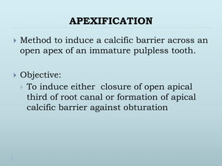
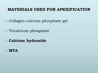
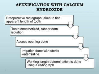
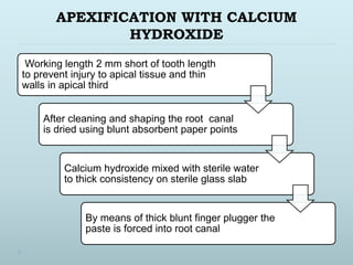
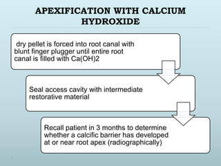
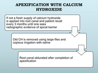
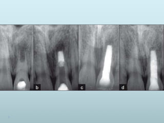
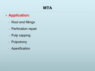
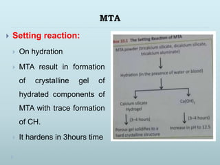
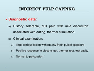
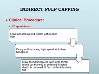
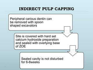
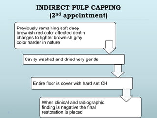
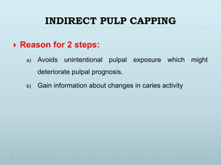
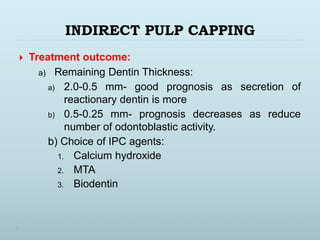
APEXIFICATION
 Method to induce a calcific barrier across an
open apex of an immature pulpless tooth.
 Objective:
 To induce either closure of open apical
third of root canal or formation of apical
calcific barrier against obturation
MATERIALS USED FOR APEXIFICATION
 Collagen calcium phosphate gel
 Tricalcium phosphate
 Calcium hydroxide
 MTA
APEXIFICATION WITH CALCIUM
HYDROXIDE
Preoperative radiograph taken to find
apparent length of tooth
Tooth anesthetized, rubber dam
isolation
Access opening done
Irrigation done with sterile
water/saline
Working length determination is done
using a radiograph
APEXIFICATION WITH CALCIUM
HYDROXIDE
Working length 2 mm short of tooth length
to prevent injury to apical tissue and thin
walls in apical third
After cleaning and shaping the root canal
is dried using blunt absorbent paper points
Calcium hydroxide mixed with sterile water
to thick consistency on sterile glass slab
By means of thick blunt finger plugger the
paste is forced into root canal
APEXIFICATION WITH CALCIUM
HYDROXIDE
dry pellet is forced into root canal with
blunt finger plugger until entire root
canal is filled with Ca(OH)2
Seal access cavity with intermediate
restorative material
Recall patient in 3 months to determine
whether a calcific barrier has developed
at or near root apex (radiographically)
APEXIFICATION WITH CALCIUM
HYDROXIDE
If not a fresh supply of calcium hydroxide
is applied into root canal and patient recall
every 3 months until one sees
radiographic evidence of apical barrier
Old CH is removed using large files and
copious irrigation with saline
Root canal obturated after completion of
apexification
APEXIFICATION WITH MTA
After proper isolation and anesthesia
access cavity is prepared to allow
debridement of necrotic pulp tissue
Working length kept 2 mm short of working
length and gentle circular filing is done
Irrigation with sodium hypochlorite
Paper points to dry the canals
APEXIFICATION WITH MTA
MTA is mixed according to manufactures
instruction and introduced into canal using
MTA carrier
MTA is condensed into apical 3-4mm
Moist cotton pellet is placed on top of MTA
to ensure setting and temporary
restoration is placed
Patient recalled after 48hours for
obturation and permanent restoration.
MTA
MINERAL TRIOXDE AGGREGATE
MTA
 Mohammad Torabinejad (1993)
 Composition:
• Tricalcium silicate
• Dicalcium silicate
• Tricalcium aluminate
• Tetracalcium aluminoferrite
• Bismuth oxides
• Traces of free crystalline silica
• Traces of calcium oxide magnesium oxide potassium
and sodium sulfate compounds
MTA
 Available in two forms
 Gray and white MTA
 Difference between two is lack of iron in
tetracalcium aluminoferrite in white MTA.
MTA
 Advantage:
 Biocompatible
 Acceptable biological performance
 Superior structural integrity of dentinal bridging
 Resist future bacterial penetration
 Antimicrobial property
 Hydrophillic
 Alkaline ph – induce dentinogenesis
 Less microleakage
MTA
 Application:
 Root end fillings
 Perforation repair
 Pulp capping
 Pulpotomy
 Apexification
MTA
 Setting reaction:
 On hydration
 MTA result in formation
of crystalline gel of
hydrated components of
MTA with trace formation
of CH.
 It hardens in 3hours time
IPC
INDIRECT PULP CAPPING
 Procedure wherein the deepest layer of
remaining affected carious dentin is covered with
layer of biocompatible material in order to
prevent pulpal exposure and further trauma to
pulp
INDIRECT PULP CAPPING
 Objective:
 To preserve vitality of pulp by completing removing
carious infected dentin
INDIRECT PULP CAPPING
 Diagnostic data:
a) History: tolerable, dull pain with mild discomfort
associated with eating, thermal stimulation.
b) Clinical examination:
a) large carious lesion without any frank pulpal exposure
b) Positive response to electric test, thermal test, test cavity
c) Normal to percussion
INDIRECT PULP CAPPING
 Diagnostic data:
c) Radiographic examination:
 large carious lesion
 Involves 3/4th thickness of dentin
 Lamina dura is intact
INDIRECT PULP CAPPING
 Clinical Procedure:
 1st appointment:
Local anesthesia and isolate with rubber
dam
Cavity outlined using high speed air turbine
handpiece
Slow speed handpiece with large #6/#8
round bur majority of softened infected
dentin is removed till firm resilient dentin is
felt
INDIRECT PULP CAPPING
Peripheral carious dentin can
be removed with spoon
shaped excavators
Site is covered with hard set
calcium hydroxide preparation
and sealed with overlying base
of ZOE
Sealed cavity is not disturbed
for 6-8weeks
INDIRECT PULP CAPPING
(2nd appointment)
Between 2 appointment history
must be negative and IRM should
be intact
Bitewing radiograph of treated
tooth is obtained
LA and rubber dam isolation
Careful removal of temporary
restoration and CH
INDIRECT PULP CAPPING
(2nd appointment)
Previously remaining soft deep
brownish red color affected dentin
changes to lighter brownish gray
color harder in nature
Cavity washed and dried very gentle
Entire floor is cover with hard set CH
When clinical and radiographic
finding is negative the final
restoration is placed
INDIRECT PULP CAPPING
 Reason for 2 steps:
a) Avoids unintentional pulpal exposure which might
deteriorate pulpal prognosis.
b) Gain information about changes in caries activity
INDIRECT PULP CAPPING
 Treatment outcome:
a) Remaining Dentin Thickness:
a) 2.0-0.5 mm- good prognosis as secretion of
reactionary dentin is more
b) 0.5-0.25 mm- prognosis decreases as reduce
number of odontoblastic activity.
b) Choice of IPC agents:
1. Calcium hydroxide
2. MTA
3. Biodentin

Apexification

  • 1.
    APEXIFICATION  Method toinduce a calcific barrier across an open apex of an immature pulpless tooth.  Objective:  To induce either closure of open apical third of root canal or formation of apical calcific barrier against obturation
  • 2.
    MATERIALS USED FORAPEXIFICATION  Collagen calcium phosphate gel  Tricalcium phosphate  Calcium hydroxide  MTA
  • 3.
    APEXIFICATION WITH CALCIUM HYDROXIDE Preoperativeradiograph taken to find apparent length of tooth Tooth anesthetized, rubber dam isolation Access opening done Irrigation done with sterile water/saline Working length determination is done using a radiograph
  • 4.
    APEXIFICATION WITH CALCIUM HYDROXIDE Workinglength 2 mm short of tooth length to prevent injury to apical tissue and thin walls in apical third After cleaning and shaping the root canal is dried using blunt absorbent paper points Calcium hydroxide mixed with sterile water to thick consistency on sterile glass slab By means of thick blunt finger plugger the paste is forced into root canal
  • 5.
    APEXIFICATION WITH CALCIUM HYDROXIDE drypellet is forced into root canal with blunt finger plugger until entire root canal is filled with Ca(OH)2 Seal access cavity with intermediate restorative material Recall patient in 3 months to determine whether a calcific barrier has developed at or near root apex (radiographically)
  • 6.
    APEXIFICATION WITH CALCIUM HYDROXIDE Ifnot a fresh supply of calcium hydroxide is applied into root canal and patient recall every 3 months until one sees radiographic evidence of apical barrier Old CH is removed using large files and copious irrigation with saline Root canal obturated after completion of apexification
  • 7.
    APEXIFICATION WITH MTA Afterproper isolation and anesthesia access cavity is prepared to allow debridement of necrotic pulp tissue Working length kept 2 mm short of working length and gentle circular filing is done Irrigation with sodium hypochlorite Paper points to dry the canals
  • 8.
    APEXIFICATION WITH MTA MTAis mixed according to manufactures instruction and introduced into canal using MTA carrier MTA is condensed into apical 3-4mm Moist cotton pellet is placed on top of MTA to ensure setting and temporary restoration is placed Patient recalled after 48hours for obturation and permanent restoration.
  • 10.
  • 11.
    MTA  Mohammad Torabinejad(1993)  Composition: • Tricalcium silicate • Dicalcium silicate • Tricalcium aluminate • Tetracalcium aluminoferrite • Bismuth oxides • Traces of free crystalline silica • Traces of calcium oxide magnesium oxide potassium and sodium sulfate compounds
  • 12.
    MTA  Available intwo forms  Gray and white MTA  Difference between two is lack of iron in tetracalcium aluminoferrite in white MTA.
  • 13.
    MTA  Advantage:  Biocompatible Acceptable biological performance  Superior structural integrity of dentinal bridging  Resist future bacterial penetration  Antimicrobial property  Hydrophillic  Alkaline ph – induce dentinogenesis  Less microleakage
  • 14.
    MTA  Application:  Rootend fillings  Perforation repair  Pulp capping  Pulpotomy  Apexification
  • 15.
    MTA  Setting reaction: On hydration  MTA result in formation of crystalline gel of hydrated components of MTA with trace formation of CH.  It hardens in 3hours time
  • 16.
  • 17.
    INDIRECT PULP CAPPING Procedure wherein the deepest layer of remaining affected carious dentin is covered with layer of biocompatible material in order to prevent pulpal exposure and further trauma to pulp
  • 18.
    INDIRECT PULP CAPPING Objective:  To preserve vitality of pulp by completing removing carious infected dentin
  • 19.
    INDIRECT PULP CAPPING Diagnostic data: a) History: tolerable, dull pain with mild discomfort associated with eating, thermal stimulation. b) Clinical examination: a) large carious lesion without any frank pulpal exposure b) Positive response to electric test, thermal test, test cavity c) Normal to percussion
  • 20.
    INDIRECT PULP CAPPING Diagnostic data: c) Radiographic examination:  large carious lesion  Involves 3/4th thickness of dentin  Lamina dura is intact
  • 21.
    INDIRECT PULP CAPPING Clinical Procedure:  1st appointment: Local anesthesia and isolate with rubber dam Cavity outlined using high speed air turbine handpiece Slow speed handpiece with large #6/#8 round bur majority of softened infected dentin is removed till firm resilient dentin is felt
  • 22.
    INDIRECT PULP CAPPING Peripheralcarious dentin can be removed with spoon shaped excavators Site is covered with hard set calcium hydroxide preparation and sealed with overlying base of ZOE Sealed cavity is not disturbed for 6-8weeks
  • 23.
    INDIRECT PULP CAPPING (2ndappointment) Between 2 appointment history must be negative and IRM should be intact Bitewing radiograph of treated tooth is obtained LA and rubber dam isolation Careful removal of temporary restoration and CH
  • 24.
    INDIRECT PULP CAPPING (2ndappointment) Previously remaining soft deep brownish red color affected dentin changes to lighter brownish gray color harder in nature Cavity washed and dried very gentle Entire floor is cover with hard set CH When clinical and radiographic finding is negative the final restoration is placed
  • 25.
    INDIRECT PULP CAPPING Reason for 2 steps: a) Avoids unintentional pulpal exposure which might deteriorate pulpal prognosis. b) Gain information about changes in caries activity
  • 26.
    INDIRECT PULP CAPPING Treatment outcome: a) Remaining Dentin Thickness: a) 2.0-0.5 mm- good prognosis as secretion of reactionary dentin is more b) 0.5-0.25 mm- prognosis decreases as reduce number of odontoblastic activity. b) Choice of IPC agents: 1. Calcium hydroxide 2. MTA 3. Biodentin