Downloaded 282 times
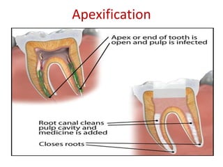
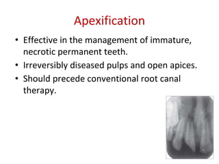
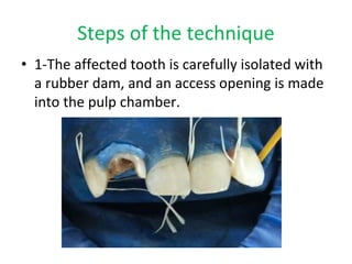
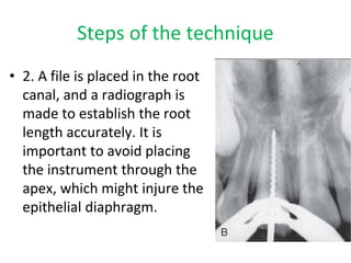
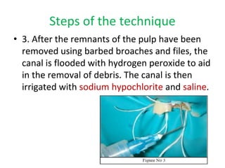
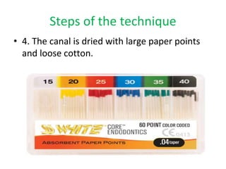


Apexification is a technique used to induce formation of a calcified barrier at the apex of a tooth with incomplete root development and non-vital pulp. It involves removal of pulp tissue, placement of calcium hydroxide or mineral trioxide aggregate (MTA) in the root canal to stimulate apical closure, and subsequent filling of the canal. The steps are accessing the canal, determining root length, cleaning and shaping, placing calcium hydroxide or MTA, and filling the canal once closure is achieved, usually within 6 months. Apexification aims to enable conventional root canal treatment in teeth that would otherwise be non-restorable due to open apices.


