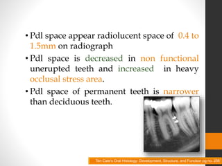
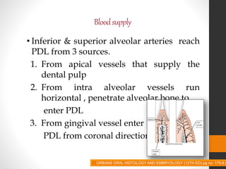
PERIODONTALLIGAMENT
CONTENTS
• Introduction
• Development
• Histology
• Pathological consideration
• Clinical consideration
• Conclusion
• References
INTRODUCTION
• The PDL is a layer of dense fibrous
connective tissue between the tooth root
(cementum) and the alveolar bone.
• Provide support , attachment, nutrition,
synthesis, protection & propioception.
ORAL ANATOMY, HISTOLOGY AND EMBRYOLOGY BY BERKOVITZ pg 180
• Pdl width ranges from 0.15 to 0.38mm
• Pdl is hour glass in shape
• Thinnest being at middle third of root
• Thickness of the Pdl reduces with age
Ten Cate's Oral Histology: Development, Structure, and Function pg no. 256
• Pdl space appear radiolucent space of 0.4 to
1.5mm on radiograph
• Pdl space is decreased in non functional
unerupted teeth and increased in heavy
occlusal stress area.
• Pdl space of permanent teeth is narrower
than deciduous teeth.
Ten Cate's Oral Histology: Development, Structure, and Function pg no. 256
DEVELOPMENT OF PDL
DEVELOPMENTOFPDL
• Begin with root formation ,prior to tooth
eruption
• Continues proliferation of the inner and
external enamel epithelium forms cervical
loop of tooth bud.
• This sheath grows apically, in the form of
HERS between dental papilla and dental
follicle.
ORBANS ORAL HISTOLOGY AND EMBRYOLOGY (12TH ED) pg no. 156
• HERS forms a circumferential structures
encompassing dental papilla, separating it
externally from dental follicle cells.
• Dental follicle cells located between
epithelial root sheath and alveolar bone are
composed of two subpopulation
• Mesenchymal cells of dental follicle
proper
• Perifollicular mesenchyme.
ORBANS ORAL HISTOLOGY AND EMBRYOLOGY (12TH ED) pg no. 156
• Mesenchymal cells are bounded by dental
follicle and developing alveolar bone are
stellate shaped , small and randomly
oriented .
• Perifollicular cells are more widely
separated , contain euchromatic nucleus,
very little cytoplasm.
• Long thin cytoplasmic process.
ORBANS ORAL HISTOLOGY AND EMBRYOLOGY (12TH ED) pg no. 156
• As the root formation progresses ,cells of
perifollicular mesenchyme gain polarity
and cellular volume & their synthetic
activity increases.
• As a result, there is active synthesis and
deposition of collagen fibrils and GAG in
developing PDL .
ORBANS ORAL HISTOLOGY AND EMBRYOLOGY (12TH ED) pg no. 156
• The developing PDL and mature PDL
contain undifferentiated stem cells which
has potential to differentiate into
osteoblasts, cementoblasts & fibroblasts.
ORBANS ORAL HISTOLOGY AND EMBRYOLOGY (12TH ED) pg no. 156
DEVELOPMENTOFPRINCIPALFIBERS
• Immediately before tooth eruption ,active
fibroblast adjacent to cementum of coronal
third of root, appears to align themselves
in oblique direction to long axis of tooth .
• These act as the precursors of the alveolar
crest fiber bundle group.
ORBANS ORAL HISTOLOGY AND EMBRYOLOGY (12TH ED) pg no. 156-7
• On examination of root surface reveals fine
brush like fibers extending from
cementum.
• Later , similar fibers are observed on
adjacent osseous surface of developing
alveolar process.
• Both set of fibers, continue to elongate
towards each other , ultimately to meet
each other as covalent bonding and cross
linking of individual collagen molecular
units occur. ORBANS ORAL HISTOLOGY AND EMBRYOLOGY (12TH ED) pg no. 156-7
• By the time of first occlusal contact of tooth
with its anatogonist.
• Horizontal group is almost completely
developed (coronal third)
• Oblique fibers are still being formed
(middle third)
• With the formation of apical fiber group
definitive PDL architecture is established.
ORBANS ORAL HISTOLOGY AND EMBRYOLOGY (12TH ED) pg no. 156-7
HISTOLOGY
PERIODONTAL
LIGAMENT
CELLS
FIBERS
GROUND
SUBSTANCE
CELLS
SYNTHETICCELLS
• Transcribe RNA
• Synthesize ribosomes in nucleolus
• Transport them to cytoplasm
• Increase its complement of RER
• Golgi membranes for translation and
transport of the protein
• Means to produce adequate supply of
energy
ORBANS ORAL HISTOLOGY AND EMBRYOLOGY (12TH ED) pg no. 160
FIBROBLASTS
• Principal cells of PDL
• Origin in part from ectomesenchyme of
investing layer of dental papilla & dental
follicle.
• Is different when compared to other cells
in connective tissue.
ORBANS ORAL HISTOLOGY AND EMBRYOLOGY (12TH ED) pg no. 161-3
• It is believed that PDL contain variety of
fibroblast cell population with different
functional characteristics.
• Responsible for formation & remodeling
of PDL Fibers
ORBANS ORAL HISTOLOGY AND EMBRYOLOGY (12TH ED) pg no. 161-3
OSTEOBLAST
• Bone forming cell lining the tooth socket.
• Basophilic appearance due to presence of
increased RER
• Active osteoblast contain extensive rough
endoplasmic reticulum
• Cells contact one another by
desmosomes .
ORBANS ORAL HISTOLOGY AND EMBRYOLOGY (12TH ED) pg no. 160
CEMENTOBLAST
• Line the surface of cementum
• Rich in cytoplasm & have large nuclei
• Cells depositing acellular cementum do
not prominent cytoplasmic process
• Cells depositing cellular cementum
exhibit
abundant basophilic cytoplasm and
cytoplasmic process.
ORBANS ORAL HISTOLOGY AND EMBRYOLOGY (12TH ED) pg no. 163
RESORPTIVE CELLS
Fibroblast
• These cells show rapid degradation of
collagen by fibroblast phagocytosis.
• The degradation of collagen may be
expected to occur as a result of
extracellular events and intracellular
events.
ORBANS ORAL HISTOLOGY AND EMBRYOLOGY (12TH ED) pg no. 163-4
OSTEOCLAST
• Cells that resorb bone and tend to be large
multinucleated.
• Howship’s lacunae are area of concavities
formed by resorption of bone.
• The part of plasma membrane lying
adjacent to bone that is being resorbed is
raised in characteristic folds
termed as
Ruffled or Striated borders
ORBANS ORAL HISTOLOGY AND EMBRYOLOGY (12TH ED) pg no. 163-4
CEMENTOCLAST
• Resemble osteoclasts
• Occassionally found in normal functioning
PDL
• Resorption of cementum occur under
certain circumstances, and in these
instances mononuclear cementoclasts /
multinucleated giant cells, often located in
Howship’s lacunae, are found on surface of
cementum.
ORBANS ORAL HISTOLOGY AND EMBRYOLOGY (12TH ED) pg no. 163-4
PROGINETOR CELL
• All connective tissue ,including PDL
contain progenitors for synthetic cells that
have capacity to undergo mitotic division.
• If these are not present there would be no
cells available to replace dying cells at end
of their life span.
ORBANS ORAL HISTOLOGY AND EMBRYOLOGY (12TH ED) pg no. 165
• Progenitor cells population appear to be in
highest concentration in location adjacent
to blood vessels.
• Exhibit some classical cytological features
of stem cells including small size,
responsiveness to stimulating factors and
slow cycle time.
• Investigators have found that ,there are
cells with characteristics of mesenchymal
stem cells capable of sustained renewal
and tissue regeneration.
ORBANS ORAL HISTOLOGY AND EMBRYOLOGY (12TH ED) pg no. 166-7
EPITHELIAL CELL REST OF MALASSEZ
• Described by Malassez in 1884
• Found close to cementum
• Are the remnants of HERS
• At the time of cementum formation , the
continuous layer of epithelium that covers
surface of newly formed dentin breaks
into lace like strands
ORBANS ORAL HISTOLOGY AND EMBRYOLOGY (12TH ED) pg no. 167
• Network, strands, islands, tube like
structure near or parallel to root surface
• These cells are abundant in furcation area.
• Involved in periodontal repair
• May proliferate to form cyst & tumor's
• Calcify to become cementicles
ORBANS ORAL HISTOLOGY AND EMBRYOLOGY (12TH ED) pg no. 167
DEFENCE CELLS
MAST CELLS
• Small , round having diameter about 12-
15 µm
• Often associated with blood vessel.
• Mast cell histamine plays a role in the
inflammatory reaction.
• Important role in regulating
endothelial & fibroblast cell
population.
ORBANS ORAL HISTOLOGY AND EMBRYOLOGY (12TH ED) pg no. 168-9
MACROPHAGES CELLS
• Predominantly located adjacent to blood
vessel.
• Wandering type of macrophages are are
derived from blood monocyte.
• It plays dual role in PDL
1. Phagocytosing dead cells.
2. Secreting growth factors.
ORBANS ORAL HISTOLOGY AND EMBRYOLOGY (12TH ED) pg no. 169-70
EOSINOPHILS
• Occasionally seen in PDL
• Capable of phagocytosis
ORBANS ORAL HISTOLOGY AND EMBRYOLOGY (12TH ED) pg no. 170
CELLBIOLOGY
GROWTH FACTOR
CYTOKINES
•IL-1
•FGF
•PDGF
•TGF
•INTERFERON
–Γ
•MMP’S
EXTRA CELLULAR SUBSTANCE
FIBERS
• Collagen
• Reticular
• Oxytalan
• Elastic
• Indifferent Fiber
Plexus
• Secondary
COLLAGEN FIBER
• Collagen is gathered to form bundles appx
5µm in dia and are termed as principal fiber
• Subunit of collagen c/s collagen fibrils
•Type I : uniformly distributed in ligament
•Type III: 20% collagen fiber
Found in peripheral
attachment to
alveolar bone.
ORBANS ORAL HISTOLOGY AND EMBRYOLOGY (12TH ED) pg no. 170-5
• Type IV &VII : associated with epithelial
cell rest & blood vessel.
• Type XII : is believed to occur only when
PDL is fully functional
ORBANS ORAL HISTOLOGY AND EMBRYOLOGY (12TH ED) pg no. 170-5
•The principal fiber group is the alvelodental
ligament
•Consist of 5 fiber groups
1. Alveolar crest fibers
2. Horizontal fibers
3. Oblique fibers
4. Apical fibers
5. Interradicular fibers
Alveolarcrestgroup
• Run in oblique direction from cementum
just beneath junctional epithelium to
alveolar crest.
• Resist tilting, intrusive, extrusive ,
rotational forces.
ORBANS ORAL HISTOLOGY AND EMBRYOLOGY (12TH ED) pg no. 170-5
Horizontalgroup
• Run at right angle to long axis of tooth from
cementum to alveolar bone.
• Roughly parallel to occlusal plane of the arch.
• Limited to coronal 1/4th of PDL space
• Resist horizontal & tipping force.
ORBANS ORAL HISTOLOGY AND EMBRYOLOGY (12TH ED) pg no. 170-5
Obliquegroup
• Most numerous
• Occupy nearly 2/3rd of ligament
• Insert into alveolar bone at position coronal
to their attachment to cementum, resulting
in oblique direction in PD space.
• Resist vertical & intrusive forces
ORBANS ORAL HISTOLOGY AND EMBRYOLOGY (12TH ED) pg no. 170-5
Apicalgroup
• Run from cementum at root tip.
• Fibers of apical group radiates through PD
space to become anchored into fundus of
bony socket.
• Resist forces of luxation , prevent root
tipping.
• Not present in incompletely
formed roots.
ORBANS ORAL HISTOLOGY AND EMBRYOLOGY (12TH ED) pg no. 170-5
Interradiculargroup
• Inserted into cementum from crest of
interradicular septum in multirooted tooth.
• Resist tipping, torquing , luxation
ORBANS ORAL HISTOLOGY AND EMBRYOLOGY (12TH ED) pg no. 170-5
SHARPEY’S FIBERS
• Collagen fiber are embedded into
cementum on one side & into alveolar bone
on other side of PD space .
• These embedded fibers are termed as
SHARPEY’S FIBER
• They are numerous but smaller at their
attachments into cementum
and alveolar bone.
ORBANS ORAL HISTOLOGY AND EMBRYOLOGY (12TH ED) pg no. 170-5
• The mineralized part of Sharpey’s fiber in
alveolar bone appears as projecting stubs
covered with mineral cluster.
• Mineralization occur at right angle to the
long axis of fibers, indicating that in
function , the fibers are subjected to
tensional forces.
• Few Sharpey’s fiber pass uninterruptedly
through bone known as Transalveolar
fibers.
ORBANS ORAL HISTOLOGY AND EMBRYOLOGY (12TH ED) pg no. 170-5
Intermediateplexus
• Earlier it was believed that , the periodontal
fiber follow a wavy course from cementum
to bone and are joined in mid region of
periodontal space giving rise to zone of
distinct appearance , called as intermediate
plexus.
• The recent concept is that, fibers cross the
entire width of PD space , but branch en
route and join neighboring fibers to form a
complex three dimensional network.
ELASTIC FIBER• MATURE ELASTIC FIBERS/ELASTIN:
Consist of microfibrillar component
surrounding the amorphous core of
elastin protein.
Observed only in walls of afferent blood
vessel.
• ELAUNIN :
Bundles of microfibrils
embedded in relatively
small amount of amorphous
elastin.
ORBANS ORAL HISTOLOGY AND EMBRYOLOGY (12TH ED) pg no. 170-5
Oxytalanfibers
• Type of immature elastic fibers
• Consist of microfibrillar component only.
 0.5-2.5µm diameter
 They run in axial direction
 One embedded in cementum and other
in alveolar bone
ORBANS ORAL HISTOLOGY AND EMBRYOLOGY (12TH ED) pg no. 170-5
• RETICULAR FIBER:
 Fine immature collagen fibers
 Related to basement membrane of
blood vessels & epithelial cell which
lie within PDL
• SECONDARY FIBERS:
 Represent newly formed collagenous
elements
 Relatively non directional and
randomly oriented
 These fiber appear to transverse the
PDL space corono-apically.
ORBANS ORAL HISTOLOGY AND EMBRYOLOGY (12TH ED) pg no. 170-5
• INDIFFERENT FIBER PLEXUS:
 Small collagen fiber associated with large
principal fibers
 Run in all direction forming plexus
ORBANS ORAL HISTOLOGY AND EMBRYOLOGY (12TH ED) pg no. 170-5
GROUND SUBSTANCE
GROUNDSUBSTANCE
• Contain 70 % water and thought to be
significantly effective on tooth’s ability to
withstand stress load.
• Gel like matrix in which cellular and
fibrous component are embedded
• It accounts for 65% of the volume of PDL
ORBANS ORAL HISTOLOGY AND EMBRYOLOGY (12TH ED) pg no. 170-5
• Consist of hyaluronate , GAG,
proteoglycan & glycoprotein.
• Proteoglycans are compound containing
anionic polysaccharides covalently attach
to protein core.
ORBANS ORAL HISTOLOGY AND EMBRYOLOGY (12TH ED) pg no. 170-5
• 2 types of PROTEOGLYCAN :
1. Proteodermatan sulphate
2. Proteoglycan containing chondroitin
sulphate/dermatan sulphate hybrids
that has been designated PG1.
• GAG:
 Linear polymer of disaccharides
 Contain hexosamine, heparin sulfate &
hexuronic acid.
ORBANS ORAL HISTOLOGY AND EMBRYOLOGY (12TH ED) pg no. 170-5
• Fibronectin:
 Promote attachment of cells to
substratum, especially to collagen
fibrils.
 Involved in cell migration & orientation.
• Tenascin:
 Commonly found in attachment zone
located near cementum & alveolar bone
 Act to transfer forces of mastication &
stresses of tooth support to specific
protein structures
ORBANS ORAL HISTOLOGY AND EMBRYOLOGY (12TH ED) pg no. 170-5
STRUCTURESPRESENTINCONNECTIVETISSUE
• Blood vessels
• Lymphatics
• Cementicles
• Nerves
Bloodsupply
• Inferior & superior alveolar arteries reach
PDL from 3 sources.
1. From apical vessels that supply the
dental pulp
2. From intra alveolar vessels run
horizontal , penetrate alveolar bone to
enter PDL
3. From gingival vessel enter
PDL from coronal direction.
ORBANS ORAL HISTOLOGY AND EMBRYOLOGY (12TH ED) pg no. 175-8
BLOOD VESSELS
• The PDL has some specialized feature in
vasculature namely, presence of large
number of fenestrations in capillaries and
a cervical plexus of capillary loops.
• The fenestration capillary beds have a
increased capacity for diffusion and
filteration.
ORBANS ORAL HISTOLOGY AND EMBRYOLOGY (12TH ED) pg no. 175-8
LYMPHATIC DRAINAGE
• Network of lymphatic vessel following
path of blood vessel provide lymph
drainage of PDL .
• The flow is from ligament towards and
into adjacent alveolar bone.
ORBANS ORAL HISTOLOGY AND EMBRYOLOGY (12TH ED) pg no. 175-8
NERVE SUPPLY
ORBANS ORAL HISTOLOGY AND EMBRYOLOGY (12TH ED) pg no. 175-8
NERVES
• 2 types of nerves
1. Sensory: touch, pressure, pain,
proprioceptive sensation
2. Autonomic : associated with PDL
vessels.
ORBANS ORAL HISTOLOGY AND EMBRYOLOGY (12TH ED) pg no. 175-8
CEMENTICLES
• These are the calcified bodies found in
PDL
• Remnants of HERS
• Are seen in older individuals
• They may remain free , fuse to large
calcified masses, or join to cementum
• When adherent to cementum
k/s excementoses
ORBANS ORAL HISTOLOGY AND EMBRYOLOGY (12TH ED) pg no. 175-8
FUNCTIONs
• Supportive
• Sensory
• Nutritive
• Eruptive
ORBANS ORAL HISTOLOGY AND EMBRYOLOGY (12TH ED) pg no. 178
PATHOLOGIC CONSIDERATION
TRAUMATIC INJURIES
TRAUMATICINJURIES
• Concussion
• Subluxation
• Extrusive luxation
• Intrusive luxation
• Lateral luxation
• Avulsion
CONCUSSION
• Injury to tooth supporting structure
• Without Abnormal
loosening/displacement
• Pdl changes after 1 hour of trauma
Hemorrhage
Stretched torn / compressed pdl fibers
Cell destruction & edema.
Textbook & color atlas of traumatic injuries to teeth 4th edition pg 372
CONCUSSION
• After 1 day cell free zone could be seen in
pdl , bordered by zone of inflammation.
• Marked reaction to percussion
Textbook & color atlas of traumatic injuries to teeth 4th edition pg 372
SUBLUXATION
• Loosening of tooth without clinical or
radiographic displacement of tooth
• Laceration of PDL fibers.
Textbook & color atlas of traumatic injuries to teeth 4th edition pg 372
EXTRUSIVELUXATION
• Peripheral displacement / partial avulsion
following axis of tooth out of socket but
without leaving socket.
• Complete rupture of pdl fibers.
Textbook & color atlas of traumatic injuries to teeth 4th edition pg 372
EXTRUSIVELUXATION
• After 3 days – split in pdl
• After 2 weeks – newly formed collagen
fibers are seen
• After 3 weeks – pdl appears normal.
Textbook & color atlas of traumatic injuries to teeth 4th edition pg 374
LATERALLUXATION
• Eccentric displacement of tooth.
• Increase in periodontal width space.
• Rupture / compression of PDL fibers.
Textbook & color atlas of traumatic injuries to teeth 4th edition pg 374
INTRUSIVE LUXATION
• Central dislocaton.
• Deeper into alveolar bone
• Fracture of alveolar bone
• Decrease in PDL space
• Crushing injury to PDL
Textbook & color atlas of traumatic injuries to teeth 4th edition pg 374
INTRUSIVE LUXATION
• After 3 mths - some show ankylosis
• Others may show surface resorption/
normal pdl
Textbook & color atlas of traumatic injuries to teeth 4th edition pg 374
AVULSION
• Total removal of tooth from socket
• Tearing of PDL
• Some of the viable cells leaves viable PDL
on root surface.
Textbook & color atlas of traumatic injuries to teeth 4th edition pg 374
• Cell and tissue culture solutions like
Hbss
 Saliva
Bovine milk and its variation
Green tea
Egg white
Coconut water
Braz Dent J. 2013 Sep-Oct;24(5):437-45]
ROOT RESORPTION
ROOTRESORPTION
• Late complication of luxation injuries
• It of 3 types
1. Surface (repair related resorption)
• Occurs as a result of localized
injury to the PDL
• Self limiting and spontaneous
repair
Textbook & color atlas of traumatic injuries to teeth 4th edition pg 386-390
1. Ankylosis (replacement resorption)
• Disappearance of PD space
• Union between bone and root substance
2. Inflammatory resorption
• Apparently related to presence of
infected necrotic pulp tissue in root canal.
• Bowel shaped resorption concavities in
cementum & dentin.
• Numerous lymphocytes ,plasma cells ,
PMN leukocytes.
Textbook & color atlas of traumatic injuries to teeth 4th edition pg 386-390
TRAUMAFROMOCCLUSION
• The effect of occlusal forces on the
periodontium is influenced by the
magnitude, direction, duration, and
frequency of the forces.
• If pressure is slightly excess results in
widening of PDL
• If pressure greatly excess results in
compression and hyalinization of PDL
fiber , PDL necrosis.
Newman M. trends in clinical periodontology and implant dentistryNewman M. trends in clinical periodontology and implant dentistry
TRAUMAFROMOCCLUSION
• If tension is slightly excess elongation of
periodontal fibers
• If tension is greatly excess results in
widening of PDL , tearing of PDL.
Newman M. trends in clinical periodontology and implant dentistry
CHRONIC PERIODONTITIS
CHRONICPERIODONTITIS
• The fibers of the PDL become
disorganized, and their attachments to
either the alveolar bone proper or
cementum through Sharpey fibers are lost
because of the resorption of these two
hard dental tissue.
Illustrated Dental Embryology, Histology, and Anatomy, Bath-Balogh and Fehrenbach, Elsevier,
2011, page 184.
PERIODONTITISOBSERVEDASAMANIFESTATIONOFSYSTEMIC
DISEASES:
1. Hematologic disorders
a. Acquired neutropenia
b. Leukemias
James E. Hinrichs and georgios kotsakis Carranza’s clinical periodontology, chapter 3, 45-67.
2. Genetic disorders
a. Familial and cyclic neutropenia
b. Down syndrome
c. Leukocyte adhesion deficiency syndromes
d. Papillon–Lefèvre syndrome
e. Chédiak–Higashi syndrome
f. Histiocytosis syndromes
James E. Hinrichs and georgios kotsakis Carranza’s clinical periodontology, chapter 3, 45-67.
g. Glycogen storage disease
h. Infantile genetic agranulocytosis
i. Cohen syndrome
j. Ehlers–Danlos syndrome (types IV and VIII,
autosomal dominant)
k. Hypophosphatasia
l. Cyclical neutropenia
m. Francoies syndrome: Absence of PDL
James E. Hinrichs and georgios kotsakis Carranza’s clinical periodontology, chapter 3, 45-67.
ORGANSYSTEMSANDCONDITIONSPOSSIBLYINFLUENCEDBY
PERIODONTALINFECTION
• Cardiovascular System
• Atherosclerosis
• Coronary heart disease
• Angina
• Myocardial infarction
James E. Hinrichs and georgios kotsakis Carranza’s clinical periodontology, chapter 3, 45-67.
• Endocrine System
• Diabetes mellitus
• Reproductive System
• Preterm low birth weight infants
• Respiratory System
• Chronic obstructive pulmonary disease
• Acute bacterial pneumonia
James E. Hinrichs and georgios kotsakis Carranza’s clinical periodontology, chapter 3, 45-67.
PERIODONTAL AND PERIAPICAL ABSCESS
Features Periapical abscess Periodontal abscess
Cause Pulpal problem Periodontal Pocket
Clinically Accumulation of pus
at root of infected
tooth
Edematous red and shiny
gingiva
Radiographically Slight thickening of
PDL space
Radiolucent area at
apex of root
Discrete radiolucency on
lateral aspect of root.
Bone loss is seen
Percussion +ve on VP +ve on LP
Pulp vitality Non vital vital
ENDO- PERIO LESION
ENDOPERIOLESION
• Persistent infection in the pulp tissue leads
to secondary infection and breakdown of
tissues in the periodontium.
• Conversely, severe periodontal disease
may initiate or exacerbate inflammatory
changes in the pulp tissue.
Kenneth C. Trabert and Mo K. KangCarranza’s Clinical Periodontology, Chapter 43, 470-479.
• This mutuality of infection between pulp
and periodontium is mediated through
physical routes, allowing for
communication between the two structures.
• The main and obvious route of
communication is the apical foramina.
Kenneth C. Trabert and Mo K. KangCarranza’s Clinical Periodontology, Chapter 43, 470-479.
• Retrograde periodontitis ,it represents the
periodontal tissue breakdown from an
apical to a cervical direction and is the
opposite of orthograde periodontitis that
results from a sulcular infection.
• Alternatively, lateral or accessory canals
may also be the route of periodontal and
pulpal communications.
• The third route of communication between
the periodontium and the pulp is through
the dentinal tubules.
• Also, bacterial invasion into dentinal
tubules from the periodontal pocket has
been demonstrated, suggesting that
dentinal tubules may allow pulpal
irritation from chronic periodontal
infections
Kenneth C. Trabert and Mo K. KangCarranza’s Clinical Periodontology, Chapter 43, 470-479.
CLASSIFICATION BASED ON ETIOLOGY
OF DISEASE
EFFECT OF PERIODONTAL DISEASE ON
PULP
• Atrophic changes(pulposis):
• Due to interference with blood supply
through lateral canals
• Which lead to death of pulp cells
• Resulting in fewer number of cells
• Increased collagen deposition
• Dystrophic mineralization
Seltzer & bender edi 3rdpg no. 309-314
• Inflammatory changes:
• Chronic inflammatory cells consist
primarily of lymphocytes were seen near
radicular portion of pulp at the level of
lateral canal
• Resorption:
• Frequently found sub-adjacent to
granulation of tissue overlying the roots
Seltzer & bender edi 3rdpg no. 309-314
CLINICAL CONSIDERATIONS
CLASSVRESTORATION
• Supragingival margin
• Least impact on the periodontium
• Equigingival margin
• More plaque accumulation
• A periodontal viewpoint, both supragingival
and equigingival margins are well
tolerated
Babitha Nugala etal J Conserv Dent 2012 Jan-Mar; 15(1): 12–17
•Subgingival margin
•Restorative margin placement within the
biologic width is unfavorable to periodontal
health and acts as a plaque retentive factor
•A constant inflammation is created and
made worse by the patient's inability to
clean this area.
Babitha Nugala etal J Conserv Dent 2012 Jan-Mar; 15(1): 12–17
CLASSIIRESTORATIONS
• Alteration of the interproximal contact
surface entails food retention, gingival
inflammation, pocket formation, bone loss
and finally dental mobility
Syed Sirajuddin etalOpen Dent J 2015; 9: 217–222
TOOTHSEPARATION
• Slow tooth separation there is less injury to
PDL maintaining the integrity of tooth.
• Rapid tooth separation there are chances of
rupturing pdl fibers accompanied by
soreness and pain.
Textbook of Operative Dentistry By Nisha Garg, Amiit Garg pg no.205-211
INTRALIGAMENTARYANESTHESIA
• Anesthetizing only the tooth being
worked on, eliminating the usual face and
tongue numbness
• Immediately effective
• Eliminate the need for uncomfortable
injections
• Uses only a fraction of the
anesthetic drug
• 2
Hristina L etal Journal of IMAB - Annual Proceeding (Scientific Papers) 2005, vol. 11, book
ENDODONTICCLINICALCONSIDERATION
• Effect of thermal changes during
endodontic treatment
• Root canal sealers
• Root canal irrigants
EFFECTOFRISEOFTEMPERATURE
• Endodontic treatment generates heat
within the dentin of the tooth that might
be transmitted to the attachment
apparatus.
• Thermoplasticized obturation techniques
cause rise in root surface temperature.
Chauhan A et al Indian Journal of Oral Health and Research / Vol. 1 / Issue 2 / Jul-Dec 2015
ROOTCANALSEALERS
• Cytotoxicity to PDL fibroblasts
• Reduction in PDL fibroblasts
Chang MC1 et alInt Endod J. 2010 Mar;43(3):251-7
• Inhibit mitochondrial dehydrogenase
activity of PDL fibroblast .
(Lin CP J Biomed Mater Res B. Appl Biomater.2004Nov;vol.71(2):429-40)
ROOTCANALIRRIGANTS
• Inhibited protein synthesis
• Cellular cytotoxicity
• Inhibitory effect on mitochondrial activity on
human PDL cell
Chang et al ORAL SURGERY ORAL MEDICINE ORAL PATHOLOGY 447 Volume 92( 4)
CONCLUSION
• PDL is the fibrous connective tissue with
reparative ,supportive , protective &
sensory characteristic.
REFERENCES
• Orbans oral histology and embryology
(12th ed)
• Oral histology,tencate (5 th edition)
• Oral anatomy, histology and embryology:
berkovitz
• Carranza’s clinical periodontology,10th
edition
• Seltzer & bender edi 3rd
• Bakland & andreasen book of
traumatology
• Hristina L etal journal of IMAB - annual
proceeding (scientific papers) 2005, vol. 11
• Chang M. et al int endod J. 2010
mar;43(3):251-7
• Chang et al oral surgery oral medicine
oral pathology 447 volume 92, number 4
• Christoph A. Ramseier1 periodontol 2000.
2012 june ; 59(1): 185–202.
THANK YOU

Periodontal ligament (PDL)

  • 1.
  • 2.
    CONTENTS • Introduction • Development •Histology • Pathological consideration • Clinical consideration • Conclusion • References
  • 3.
    INTRODUCTION • The PDLis a layer of dense fibrous connective tissue between the tooth root (cementum) and the alveolar bone. • Provide support , attachment, nutrition, synthesis, protection & propioception. ORAL ANATOMY, HISTOLOGY AND EMBRYOLOGY BY BERKOVITZ pg 180
  • 4.
    • Pdl widthranges from 0.15 to 0.38mm • Pdl is hour glass in shape • Thinnest being at middle third of root • Thickness of the Pdl reduces with age Ten Cate's Oral Histology: Development, Structure, and Function pg no. 256
  • 5.
    • Pdl spaceappear radiolucent space of 0.4 to 1.5mm on radiograph • Pdl space is decreased in non functional unerupted teeth and increased in heavy occlusal stress area. • Pdl space of permanent teeth is narrower than deciduous teeth. Ten Cate's Oral Histology: Development, Structure, and Function pg no. 256
  • 6.
  • 7.
    DEVELOPMENTOFPDL • Begin withroot formation ,prior to tooth eruption • Continues proliferation of the inner and external enamel epithelium forms cervical loop of tooth bud. • This sheath grows apically, in the form of HERS between dental papilla and dental follicle. ORBANS ORAL HISTOLOGY AND EMBRYOLOGY (12TH ED) pg no. 156
  • 8.
    • HERS formsa circumferential structures encompassing dental papilla, separating it externally from dental follicle cells. • Dental follicle cells located between epithelial root sheath and alveolar bone are composed of two subpopulation • Mesenchymal cells of dental follicle proper • Perifollicular mesenchyme. ORBANS ORAL HISTOLOGY AND EMBRYOLOGY (12TH ED) pg no. 156
  • 9.
    • Mesenchymal cellsare bounded by dental follicle and developing alveolar bone are stellate shaped , small and randomly oriented . • Perifollicular cells are more widely separated , contain euchromatic nucleus, very little cytoplasm. • Long thin cytoplasmic process. ORBANS ORAL HISTOLOGY AND EMBRYOLOGY (12TH ED) pg no. 156
  • 10.
    • As theroot formation progresses ,cells of perifollicular mesenchyme gain polarity and cellular volume & their synthetic activity increases. • As a result, there is active synthesis and deposition of collagen fibrils and GAG in developing PDL . ORBANS ORAL HISTOLOGY AND EMBRYOLOGY (12TH ED) pg no. 156
  • 11.
    • The developingPDL and mature PDL contain undifferentiated stem cells which has potential to differentiate into osteoblasts, cementoblasts & fibroblasts. ORBANS ORAL HISTOLOGY AND EMBRYOLOGY (12TH ED) pg no. 156
  • 12.
  • 13.
    • Immediately beforetooth eruption ,active fibroblast adjacent to cementum of coronal third of root, appears to align themselves in oblique direction to long axis of tooth . • These act as the precursors of the alveolar crest fiber bundle group. ORBANS ORAL HISTOLOGY AND EMBRYOLOGY (12TH ED) pg no. 156-7
  • 14.
    • On examinationof root surface reveals fine brush like fibers extending from cementum. • Later , similar fibers are observed on adjacent osseous surface of developing alveolar process. • Both set of fibers, continue to elongate towards each other , ultimately to meet each other as covalent bonding and cross linking of individual collagen molecular units occur. ORBANS ORAL HISTOLOGY AND EMBRYOLOGY (12TH ED) pg no. 156-7
  • 15.
    • By thetime of first occlusal contact of tooth with its anatogonist. • Horizontal group is almost completely developed (coronal third) • Oblique fibers are still being formed (middle third) • With the formation of apical fiber group definitive PDL architecture is established. ORBANS ORAL HISTOLOGY AND EMBRYOLOGY (12TH ED) pg no. 156-7
  • 16.
  • 17.
  • 18.
  • 19.
    SYNTHETICCELLS • Transcribe RNA •Synthesize ribosomes in nucleolus • Transport them to cytoplasm • Increase its complement of RER • Golgi membranes for translation and transport of the protein • Means to produce adequate supply of energy ORBANS ORAL HISTOLOGY AND EMBRYOLOGY (12TH ED) pg no. 160
  • 20.
    FIBROBLASTS • Principal cellsof PDL • Origin in part from ectomesenchyme of investing layer of dental papilla & dental follicle. • Is different when compared to other cells in connective tissue. ORBANS ORAL HISTOLOGY AND EMBRYOLOGY (12TH ED) pg no. 161-3
  • 21.
    • It isbelieved that PDL contain variety of fibroblast cell population with different functional characteristics. • Responsible for formation & remodeling of PDL Fibers ORBANS ORAL HISTOLOGY AND EMBRYOLOGY (12TH ED) pg no. 161-3
  • 22.
    OSTEOBLAST • Bone formingcell lining the tooth socket. • Basophilic appearance due to presence of increased RER • Active osteoblast contain extensive rough endoplasmic reticulum • Cells contact one another by desmosomes . ORBANS ORAL HISTOLOGY AND EMBRYOLOGY (12TH ED) pg no. 160
  • 23.
    CEMENTOBLAST • Line thesurface of cementum • Rich in cytoplasm & have large nuclei • Cells depositing acellular cementum do not prominent cytoplasmic process • Cells depositing cellular cementum exhibit abundant basophilic cytoplasm and cytoplasmic process. ORBANS ORAL HISTOLOGY AND EMBRYOLOGY (12TH ED) pg no. 163
  • 24.
  • 25.
    Fibroblast • These cellsshow rapid degradation of collagen by fibroblast phagocytosis. • The degradation of collagen may be expected to occur as a result of extracellular events and intracellular events. ORBANS ORAL HISTOLOGY AND EMBRYOLOGY (12TH ED) pg no. 163-4
  • 26.
    OSTEOCLAST • Cells thatresorb bone and tend to be large multinucleated. • Howship’s lacunae are area of concavities formed by resorption of bone. • The part of plasma membrane lying adjacent to bone that is being resorbed is raised in characteristic folds termed as Ruffled or Striated borders ORBANS ORAL HISTOLOGY AND EMBRYOLOGY (12TH ED) pg no. 163-4
  • 27.
    CEMENTOCLAST • Resemble osteoclasts •Occassionally found in normal functioning PDL • Resorption of cementum occur under certain circumstances, and in these instances mononuclear cementoclasts / multinucleated giant cells, often located in Howship’s lacunae, are found on surface of cementum. ORBANS ORAL HISTOLOGY AND EMBRYOLOGY (12TH ED) pg no. 163-4
  • 28.
    PROGINETOR CELL • Allconnective tissue ,including PDL contain progenitors for synthetic cells that have capacity to undergo mitotic division. • If these are not present there would be no cells available to replace dying cells at end of their life span. ORBANS ORAL HISTOLOGY AND EMBRYOLOGY (12TH ED) pg no. 165
  • 29.
    • Progenitor cellspopulation appear to be in highest concentration in location adjacent to blood vessels. • Exhibit some classical cytological features of stem cells including small size, responsiveness to stimulating factors and slow cycle time.
  • 30.
    • Investigators havefound that ,there are cells with characteristics of mesenchymal stem cells capable of sustained renewal and tissue regeneration. ORBANS ORAL HISTOLOGY AND EMBRYOLOGY (12TH ED) pg no. 166-7
  • 31.
    EPITHELIAL CELL RESTOF MALASSEZ • Described by Malassez in 1884 • Found close to cementum • Are the remnants of HERS • At the time of cementum formation , the continuous layer of epithelium that covers surface of newly formed dentin breaks into lace like strands ORBANS ORAL HISTOLOGY AND EMBRYOLOGY (12TH ED) pg no. 167
  • 32.
    • Network, strands,islands, tube like structure near or parallel to root surface • These cells are abundant in furcation area. • Involved in periodontal repair • May proliferate to form cyst & tumor's • Calcify to become cementicles ORBANS ORAL HISTOLOGY AND EMBRYOLOGY (12TH ED) pg no. 167
  • 33.
  • 34.
    MAST CELLS • Small, round having diameter about 12- 15 µm • Often associated with blood vessel. • Mast cell histamine plays a role in the inflammatory reaction. • Important role in regulating endothelial & fibroblast cell population. ORBANS ORAL HISTOLOGY AND EMBRYOLOGY (12TH ED) pg no. 168-9
  • 35.
    MACROPHAGES CELLS • Predominantlylocated adjacent to blood vessel. • Wandering type of macrophages are are derived from blood monocyte. • It plays dual role in PDL 1. Phagocytosing dead cells. 2. Secreting growth factors. ORBANS ORAL HISTOLOGY AND EMBRYOLOGY (12TH ED) pg no. 169-70
  • 36.
    EOSINOPHILS • Occasionally seenin PDL • Capable of phagocytosis ORBANS ORAL HISTOLOGY AND EMBRYOLOGY (12TH ED) pg no. 170
  • 37.
  • 38.
  • 39.
    FIBERS • Collagen • Reticular •Oxytalan • Elastic • Indifferent Fiber Plexus • Secondary
  • 40.
    COLLAGEN FIBER • Collagenis gathered to form bundles appx 5µm in dia and are termed as principal fiber • Subunit of collagen c/s collagen fibrils •Type I : uniformly distributed in ligament •Type III: 20% collagen fiber Found in peripheral attachment to alveolar bone. ORBANS ORAL HISTOLOGY AND EMBRYOLOGY (12TH ED) pg no. 170-5
  • 41.
    • Type IV&VII : associated with epithelial cell rest & blood vessel. • Type XII : is believed to occur only when PDL is fully functional ORBANS ORAL HISTOLOGY AND EMBRYOLOGY (12TH ED) pg no. 170-5
  • 42.
    •The principal fibergroup is the alvelodental ligament •Consist of 5 fiber groups 1. Alveolar crest fibers 2. Horizontal fibers 3. Oblique fibers 4. Apical fibers 5. Interradicular fibers
  • 43.
    Alveolarcrestgroup • Run inoblique direction from cementum just beneath junctional epithelium to alveolar crest. • Resist tilting, intrusive, extrusive , rotational forces. ORBANS ORAL HISTOLOGY AND EMBRYOLOGY (12TH ED) pg no. 170-5
  • 44.
    Horizontalgroup • Run atright angle to long axis of tooth from cementum to alveolar bone. • Roughly parallel to occlusal plane of the arch. • Limited to coronal 1/4th of PDL space • Resist horizontal & tipping force. ORBANS ORAL HISTOLOGY AND EMBRYOLOGY (12TH ED) pg no. 170-5
  • 45.
    Obliquegroup • Most numerous •Occupy nearly 2/3rd of ligament • Insert into alveolar bone at position coronal to their attachment to cementum, resulting in oblique direction in PD space. • Resist vertical & intrusive forces ORBANS ORAL HISTOLOGY AND EMBRYOLOGY (12TH ED) pg no. 170-5
  • 46.
    Apicalgroup • Run fromcementum at root tip. • Fibers of apical group radiates through PD space to become anchored into fundus of bony socket. • Resist forces of luxation , prevent root tipping. • Not present in incompletely formed roots. ORBANS ORAL HISTOLOGY AND EMBRYOLOGY (12TH ED) pg no. 170-5
  • 47.
    Interradiculargroup • Inserted intocementum from crest of interradicular septum in multirooted tooth. • Resist tipping, torquing , luxation ORBANS ORAL HISTOLOGY AND EMBRYOLOGY (12TH ED) pg no. 170-5
  • 48.
    SHARPEY’S FIBERS • Collagenfiber are embedded into cementum on one side & into alveolar bone on other side of PD space . • These embedded fibers are termed as SHARPEY’S FIBER • They are numerous but smaller at their attachments into cementum and alveolar bone. ORBANS ORAL HISTOLOGY AND EMBRYOLOGY (12TH ED) pg no. 170-5
  • 49.
    • The mineralizedpart of Sharpey’s fiber in alveolar bone appears as projecting stubs covered with mineral cluster. • Mineralization occur at right angle to the long axis of fibers, indicating that in function , the fibers are subjected to tensional forces. • Few Sharpey’s fiber pass uninterruptedly through bone known as Transalveolar fibers. ORBANS ORAL HISTOLOGY AND EMBRYOLOGY (12TH ED) pg no. 170-5
  • 50.
    Intermediateplexus • Earlier itwas believed that , the periodontal fiber follow a wavy course from cementum to bone and are joined in mid region of periodontal space giving rise to zone of distinct appearance , called as intermediate plexus. • The recent concept is that, fibers cross the entire width of PD space , but branch en route and join neighboring fibers to form a complex three dimensional network.
  • 51.
    ELASTIC FIBER• MATUREELASTIC FIBERS/ELASTIN: Consist of microfibrillar component surrounding the amorphous core of elastin protein. Observed only in walls of afferent blood vessel. • ELAUNIN : Bundles of microfibrils embedded in relatively small amount of amorphous elastin. ORBANS ORAL HISTOLOGY AND EMBRYOLOGY (12TH ED) pg no. 170-5
  • 52.
    Oxytalanfibers • Type ofimmature elastic fibers • Consist of microfibrillar component only.  0.5-2.5µm diameter  They run in axial direction  One embedded in cementum and other in alveolar bone ORBANS ORAL HISTOLOGY AND EMBRYOLOGY (12TH ED) pg no. 170-5
  • 53.
    • RETICULAR FIBER: Fine immature collagen fibers  Related to basement membrane of blood vessels & epithelial cell which lie within PDL • SECONDARY FIBERS:  Represent newly formed collagenous elements  Relatively non directional and randomly oriented  These fiber appear to transverse the PDL space corono-apically. ORBANS ORAL HISTOLOGY AND EMBRYOLOGY (12TH ED) pg no. 170-5
  • 54.
    • INDIFFERENT FIBERPLEXUS:  Small collagen fiber associated with large principal fibers  Run in all direction forming plexus ORBANS ORAL HISTOLOGY AND EMBRYOLOGY (12TH ED) pg no. 170-5
  • 55.
  • 56.
    GROUNDSUBSTANCE • Contain 70% water and thought to be significantly effective on tooth’s ability to withstand stress load. • Gel like matrix in which cellular and fibrous component are embedded • It accounts for 65% of the volume of PDL ORBANS ORAL HISTOLOGY AND EMBRYOLOGY (12TH ED) pg no. 170-5
  • 57.
    • Consist ofhyaluronate , GAG, proteoglycan & glycoprotein. • Proteoglycans are compound containing anionic polysaccharides covalently attach to protein core. ORBANS ORAL HISTOLOGY AND EMBRYOLOGY (12TH ED) pg no. 170-5
  • 58.
    • 2 typesof PROTEOGLYCAN : 1. Proteodermatan sulphate 2. Proteoglycan containing chondroitin sulphate/dermatan sulphate hybrids that has been designated PG1. • GAG:  Linear polymer of disaccharides  Contain hexosamine, heparin sulfate & hexuronic acid. ORBANS ORAL HISTOLOGY AND EMBRYOLOGY (12TH ED) pg no. 170-5
  • 59.
    • Fibronectin:  Promoteattachment of cells to substratum, especially to collagen fibrils.  Involved in cell migration & orientation. • Tenascin:  Commonly found in attachment zone located near cementum & alveolar bone  Act to transfer forces of mastication & stresses of tooth support to specific protein structures ORBANS ORAL HISTOLOGY AND EMBRYOLOGY (12TH ED) pg no. 170-5
  • 60.
    STRUCTURESPRESENTINCONNECTIVETISSUE • Blood vessels •Lymphatics • Cementicles • Nerves
  • 61.
    Bloodsupply • Inferior &superior alveolar arteries reach PDL from 3 sources. 1. From apical vessels that supply the dental pulp 2. From intra alveolar vessels run horizontal , penetrate alveolar bone to enter PDL 3. From gingival vessel enter PDL from coronal direction. ORBANS ORAL HISTOLOGY AND EMBRYOLOGY (12TH ED) pg no. 175-8
  • 62.
    BLOOD VESSELS • ThePDL has some specialized feature in vasculature namely, presence of large number of fenestrations in capillaries and a cervical plexus of capillary loops. • The fenestration capillary beds have a increased capacity for diffusion and filteration. ORBANS ORAL HISTOLOGY AND EMBRYOLOGY (12TH ED) pg no. 175-8
  • 63.
    LYMPHATIC DRAINAGE • Networkof lymphatic vessel following path of blood vessel provide lymph drainage of PDL . • The flow is from ligament towards and into adjacent alveolar bone. ORBANS ORAL HISTOLOGY AND EMBRYOLOGY (12TH ED) pg no. 175-8
  • 64.
    NERVE SUPPLY ORBANS ORALHISTOLOGY AND EMBRYOLOGY (12TH ED) pg no. 175-8
  • 65.
    NERVES • 2 typesof nerves 1. Sensory: touch, pressure, pain, proprioceptive sensation 2. Autonomic : associated with PDL vessels. ORBANS ORAL HISTOLOGY AND EMBRYOLOGY (12TH ED) pg no. 175-8
  • 66.
    CEMENTICLES • These arethe calcified bodies found in PDL • Remnants of HERS • Are seen in older individuals • They may remain free , fuse to large calcified masses, or join to cementum • When adherent to cementum k/s excementoses ORBANS ORAL HISTOLOGY AND EMBRYOLOGY (12TH ED) pg no. 175-8
  • 67.
    FUNCTIONs • Supportive • Sensory •Nutritive • Eruptive ORBANS ORAL HISTOLOGY AND EMBRYOLOGY (12TH ED) pg no. 178
  • 68.
  • 69.
  • 70.
    TRAUMATICINJURIES • Concussion • Subluxation •Extrusive luxation • Intrusive luxation • Lateral luxation • Avulsion
  • 71.
    CONCUSSION • Injury totooth supporting structure • Without Abnormal loosening/displacement • Pdl changes after 1 hour of trauma Hemorrhage Stretched torn / compressed pdl fibers Cell destruction & edema. Textbook & color atlas of traumatic injuries to teeth 4th edition pg 372
  • 72.
    CONCUSSION • After 1day cell free zone could be seen in pdl , bordered by zone of inflammation. • Marked reaction to percussion Textbook & color atlas of traumatic injuries to teeth 4th edition pg 372
  • 73.
    SUBLUXATION • Loosening oftooth without clinical or radiographic displacement of tooth • Laceration of PDL fibers. Textbook & color atlas of traumatic injuries to teeth 4th edition pg 372
  • 74.
    EXTRUSIVELUXATION • Peripheral displacement/ partial avulsion following axis of tooth out of socket but without leaving socket. • Complete rupture of pdl fibers. Textbook & color atlas of traumatic injuries to teeth 4th edition pg 372
  • 75.
    EXTRUSIVELUXATION • After 3days – split in pdl • After 2 weeks – newly formed collagen fibers are seen • After 3 weeks – pdl appears normal. Textbook & color atlas of traumatic injuries to teeth 4th edition pg 374
  • 76.
    LATERALLUXATION • Eccentric displacementof tooth. • Increase in periodontal width space. • Rupture / compression of PDL fibers. Textbook & color atlas of traumatic injuries to teeth 4th edition pg 374
  • 77.
    INTRUSIVE LUXATION • Centraldislocaton. • Deeper into alveolar bone • Fracture of alveolar bone • Decrease in PDL space • Crushing injury to PDL Textbook & color atlas of traumatic injuries to teeth 4th edition pg 374
  • 78.
    INTRUSIVE LUXATION • After3 mths - some show ankylosis • Others may show surface resorption/ normal pdl Textbook & color atlas of traumatic injuries to teeth 4th edition pg 374
  • 79.
    AVULSION • Total removalof tooth from socket • Tearing of PDL • Some of the viable cells leaves viable PDL on root surface. Textbook & color atlas of traumatic injuries to teeth 4th edition pg 374
  • 80.
    • Cell andtissue culture solutions like Hbss  Saliva Bovine milk and its variation Green tea Egg white Coconut water Braz Dent J. 2013 Sep-Oct;24(5):437-45]
  • 81.
  • 82.
    ROOTRESORPTION • Late complicationof luxation injuries • It of 3 types 1. Surface (repair related resorption) • Occurs as a result of localized injury to the PDL • Self limiting and spontaneous repair Textbook & color atlas of traumatic injuries to teeth 4th edition pg 386-390
  • 83.
    1. Ankylosis (replacementresorption) • Disappearance of PD space • Union between bone and root substance 2. Inflammatory resorption • Apparently related to presence of infected necrotic pulp tissue in root canal. • Bowel shaped resorption concavities in cementum & dentin. • Numerous lymphocytes ,plasma cells , PMN leukocytes. Textbook & color atlas of traumatic injuries to teeth 4th edition pg 386-390
  • 84.
    TRAUMAFROMOCCLUSION • The effectof occlusal forces on the periodontium is influenced by the magnitude, direction, duration, and frequency of the forces. • If pressure is slightly excess results in widening of PDL • If pressure greatly excess results in compression and hyalinization of PDL fiber , PDL necrosis. Newman M. trends in clinical periodontology and implant dentistryNewman M. trends in clinical periodontology and implant dentistry
  • 85.
    TRAUMAFROMOCCLUSION • If tensionis slightly excess elongation of periodontal fibers • If tension is greatly excess results in widening of PDL , tearing of PDL. Newman M. trends in clinical periodontology and implant dentistry
  • 86.
  • 87.
    CHRONICPERIODONTITIS • The fibersof the PDL become disorganized, and their attachments to either the alveolar bone proper or cementum through Sharpey fibers are lost because of the resorption of these two hard dental tissue. Illustrated Dental Embryology, Histology, and Anatomy, Bath-Balogh and Fehrenbach, Elsevier, 2011, page 184.
  • 88.
    PERIODONTITISOBSERVEDASAMANIFESTATIONOFSYSTEMIC DISEASES: 1. Hematologic disorders a.Acquired neutropenia b. Leukemias James E. Hinrichs and georgios kotsakis Carranza’s clinical periodontology, chapter 3, 45-67.
  • 89.
    2. Genetic disorders a.Familial and cyclic neutropenia b. Down syndrome c. Leukocyte adhesion deficiency syndromes d. Papillon–Lefèvre syndrome e. Chédiak–Higashi syndrome f. Histiocytosis syndromes James E. Hinrichs and georgios kotsakis Carranza’s clinical periodontology, chapter 3, 45-67.
  • 90.
    g. Glycogen storagedisease h. Infantile genetic agranulocytosis i. Cohen syndrome j. Ehlers–Danlos syndrome (types IV and VIII, autosomal dominant) k. Hypophosphatasia l. Cyclical neutropenia m. Francoies syndrome: Absence of PDL James E. Hinrichs and georgios kotsakis Carranza’s clinical periodontology, chapter 3, 45-67.
  • 91.
    ORGANSYSTEMSANDCONDITIONSPOSSIBLYINFLUENCEDBY PERIODONTALINFECTION • Cardiovascular System •Atherosclerosis • Coronary heart disease • Angina • Myocardial infarction James E. Hinrichs and georgios kotsakis Carranza’s clinical periodontology, chapter 3, 45-67.
  • 92.
    • Endocrine System •Diabetes mellitus • Reproductive System • Preterm low birth weight infants • Respiratory System • Chronic obstructive pulmonary disease • Acute bacterial pneumonia James E. Hinrichs and georgios kotsakis Carranza’s clinical periodontology, chapter 3, 45-67.
  • 93.
  • 94.
    Features Periapical abscessPeriodontal abscess Cause Pulpal problem Periodontal Pocket Clinically Accumulation of pus at root of infected tooth Edematous red and shiny gingiva Radiographically Slight thickening of PDL space Radiolucent area at apex of root Discrete radiolucency on lateral aspect of root. Bone loss is seen Percussion +ve on VP +ve on LP Pulp vitality Non vital vital
  • 95.
  • 96.
    ENDOPERIOLESION • Persistent infectionin the pulp tissue leads to secondary infection and breakdown of tissues in the periodontium. • Conversely, severe periodontal disease may initiate or exacerbate inflammatory changes in the pulp tissue. Kenneth C. Trabert and Mo K. KangCarranza’s Clinical Periodontology, Chapter 43, 470-479.
  • 97.
    • This mutualityof infection between pulp and periodontium is mediated through physical routes, allowing for communication between the two structures. • The main and obvious route of communication is the apical foramina. Kenneth C. Trabert and Mo K. KangCarranza’s Clinical Periodontology, Chapter 43, 470-479.
  • 98.
    • Retrograde periodontitis,it represents the periodontal tissue breakdown from an apical to a cervical direction and is the opposite of orthograde periodontitis that results from a sulcular infection. • Alternatively, lateral or accessory canals may also be the route of periodontal and pulpal communications.
  • 99.
    • The thirdroute of communication between the periodontium and the pulp is through the dentinal tubules. • Also, bacterial invasion into dentinal tubules from the periodontal pocket has been demonstrated, suggesting that dentinal tubules may allow pulpal irritation from chronic periodontal infections Kenneth C. Trabert and Mo K. KangCarranza’s Clinical Periodontology, Chapter 43, 470-479.
  • 100.
    CLASSIFICATION BASED ONETIOLOGY OF DISEASE
  • 101.
    EFFECT OF PERIODONTALDISEASE ON PULP
  • 102.
    • Atrophic changes(pulposis): •Due to interference with blood supply through lateral canals • Which lead to death of pulp cells • Resulting in fewer number of cells • Increased collagen deposition • Dystrophic mineralization Seltzer & bender edi 3rdpg no. 309-314
  • 103.
    • Inflammatory changes: •Chronic inflammatory cells consist primarily of lymphocytes were seen near radicular portion of pulp at the level of lateral canal • Resorption: • Frequently found sub-adjacent to granulation of tissue overlying the roots Seltzer & bender edi 3rdpg no. 309-314
  • 104.
  • 105.
    CLASSVRESTORATION • Supragingival margin •Least impact on the periodontium • Equigingival margin • More plaque accumulation • A periodontal viewpoint, both supragingival and equigingival margins are well tolerated Babitha Nugala etal J Conserv Dent 2012 Jan-Mar; 15(1): 12–17
  • 106.
    •Subgingival margin •Restorative marginplacement within the biologic width is unfavorable to periodontal health and acts as a plaque retentive factor •A constant inflammation is created and made worse by the patient's inability to clean this area. Babitha Nugala etal J Conserv Dent 2012 Jan-Mar; 15(1): 12–17
  • 107.
    CLASSIIRESTORATIONS • Alteration ofthe interproximal contact surface entails food retention, gingival inflammation, pocket formation, bone loss and finally dental mobility Syed Sirajuddin etalOpen Dent J 2015; 9: 217–222
  • 108.
    TOOTHSEPARATION • Slow toothseparation there is less injury to PDL maintaining the integrity of tooth. • Rapid tooth separation there are chances of rupturing pdl fibers accompanied by soreness and pain. Textbook of Operative Dentistry By Nisha Garg, Amiit Garg pg no.205-211
  • 109.
    INTRALIGAMENTARYANESTHESIA • Anesthetizing onlythe tooth being worked on, eliminating the usual face and tongue numbness • Immediately effective • Eliminate the need for uncomfortable injections • Uses only a fraction of the anesthetic drug • 2 Hristina L etal Journal of IMAB - Annual Proceeding (Scientific Papers) 2005, vol. 11, book
  • 110.
    ENDODONTICCLINICALCONSIDERATION • Effect ofthermal changes during endodontic treatment • Root canal sealers • Root canal irrigants
  • 111.
    EFFECTOFRISEOFTEMPERATURE • Endodontic treatmentgenerates heat within the dentin of the tooth that might be transmitted to the attachment apparatus. • Thermoplasticized obturation techniques cause rise in root surface temperature. Chauhan A et al Indian Journal of Oral Health and Research / Vol. 1 / Issue 2 / Jul-Dec 2015
  • 112.
    ROOTCANALSEALERS • Cytotoxicity toPDL fibroblasts • Reduction in PDL fibroblasts Chang MC1 et alInt Endod J. 2010 Mar;43(3):251-7 • Inhibit mitochondrial dehydrogenase activity of PDL fibroblast . (Lin CP J Biomed Mater Res B. Appl Biomater.2004Nov;vol.71(2):429-40)
  • 113.
    ROOTCANALIRRIGANTS • Inhibited proteinsynthesis • Cellular cytotoxicity • Inhibitory effect on mitochondrial activity on human PDL cell Chang et al ORAL SURGERY ORAL MEDICINE ORAL PATHOLOGY 447 Volume 92( 4)
  • 114.
    CONCLUSION • PDL isthe fibrous connective tissue with reparative ,supportive , protective & sensory characteristic.
  • 115.
    REFERENCES • Orbans oralhistology and embryology (12th ed) • Oral histology,tencate (5 th edition) • Oral anatomy, histology and embryology: berkovitz • Carranza’s clinical periodontology,10th edition • Seltzer & bender edi 3rd • Bakland & andreasen book of traumatology
  • 116.
    • Hristina Letal journal of IMAB - annual proceeding (scientific papers) 2005, vol. 11 • Chang M. et al int endod J. 2010 mar;43(3):251-7 • Chang et al oral surgery oral medicine oral pathology 447 volume 92, number 4 • Christoph A. Ramseier1 periodontol 2000. 2012 june ; 59(1): 185–202.
  • 117.