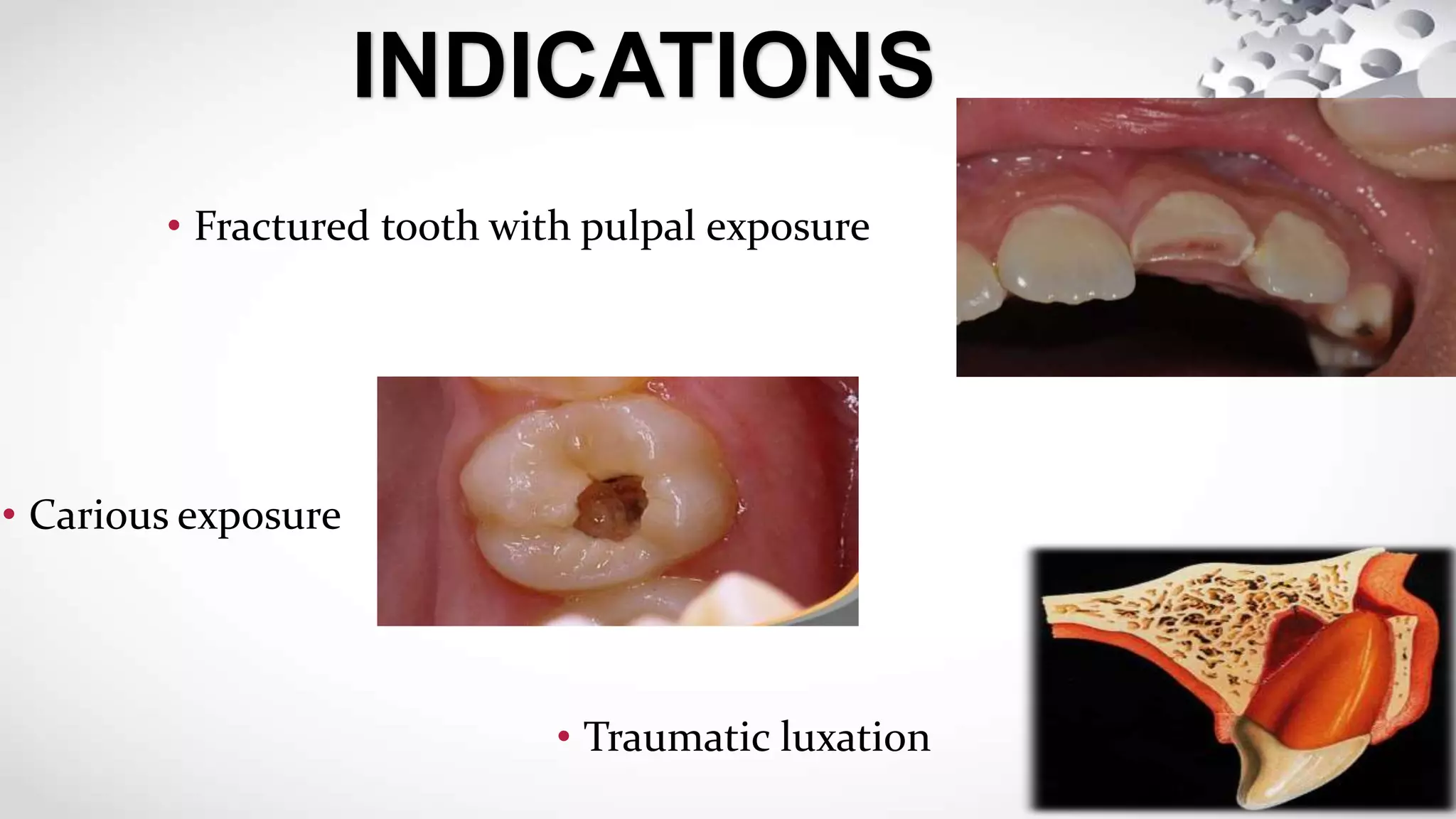
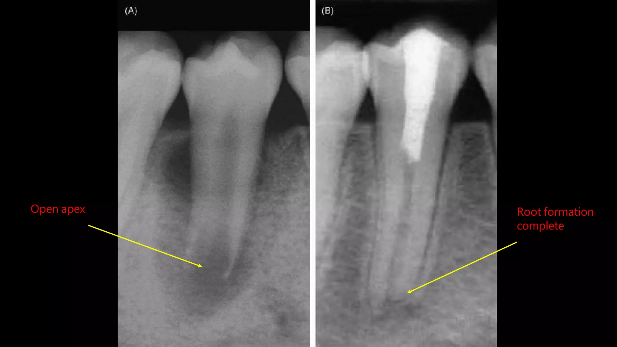
APEXOGENESIS
Rabina panta
1701938
INTRODUCTION
• Physiologic process
• Formation of apex in young, vital, immature permanent teeth
with appropriate pulp therapy
• Rootend Developmentoccurs in a tooth with a normal pulpand minimal
inflammation
• Pulp of immature teeth has significant reparativepotential
• Pulprevascularisation and repairoccurs moreefficiently in tooth
with an openapex
• Poor long term prognosisof an endodontically treated
immature teeth
Relativelythin dentine in obturated canalsof Immature rootsand open
apexareprone to fracture
RATIONALE
• Sustaining aviable Hertwig’ssheath tostimulate continues
development of root
• Toattain favourable crown:rootratio
• Toattain root endclosure
• Topreserve pulpvitality tosecure furtherroot development
and maturation
• Generating dentinal bridge at the site ofpulpotomy
GOALS
• Fractured tooth with pulpal exposure
INDICATIONS
• Carious exposure
• Traumatic luxation
• Severe crown-root fracture
which requires intra-radicular
retention forrestoration
CONTRAINDICATIONS
• Tooth with unfavourable
horizontal root fracture i.e.
close to gingival margin
• Necrotic or non vitalpulp
• Unrestorable carioustooth
• Direct pulp capping
When pulp chamber
is exposed
INVOLVES
• Indirect pulp capping When a thin dentin layer is present
between pulp and cavity
• Pulpotomy Extirpation of pulp is restricted
strictly to the coronal portion of
pulp chamber
• MTA (Mineral trioxide aggregrate)
MATERIALS USED
• Calcium hydroxide
• Formocresol (as an alternative to calcium hydroxide)
• Anesthesia application and rubberdam
isolation
• The instrument of choice for tissueremoval
is an abrasive diamond bur at slow speed
with adequatewater-cooling
PROCEDURE
• Access is gained into the pulpchamberand
infected dentin partlyremoved
• Peripheral carious lesion removed witha
spoon excavator
• Following coronal pulp amputation,the pulp
chamber is rinsed with sterile saline or sterile
water to remove all debris
• The excess liquid should then be carefully
removed viavacuumorsterile cotton pellets.
• Air should not be blown on the exposed pulp,
as this may cause desiccation and additional
tissuedamage.
• Once the pulpal bleeding is controlled, calciumhydroxide
paste is placed overtheamputation site
• Care must be taken to avoid placing the calcium
hydroxideon a blood clotand theentire pulpsurface
must becovered
• Once this is accomplished, a restorative base material
should be placed over the calcium hydroxide and then
allowed to setcompletely
• A coronal restoration should then be placed thatwill
ensure the maximum long-termseal
• The patient should be re-evaluated every three months for the first year, and
then every 6 months for 2 to 4 years to determine if successful root formation
is taking placeand that thereare no signs of pulp necrosis, rootresorption or
periradicularpathosis
Open apex Root formation
complete
Apexogenesis

Apexogenesis

  • 1.
  • 2.
    INTRODUCTION • Physiologic process •Formation of apex in young, vital, immature permanent teeth with appropriate pulp therapy
  • 3.
    • Rootend Developmentoccursin a tooth with a normal pulpand minimal inflammation • Pulp of immature teeth has significant reparativepotential • Pulprevascularisation and repairoccurs moreefficiently in tooth with an openapex • Poor long term prognosisof an endodontically treated immature teeth Relativelythin dentine in obturated canalsof Immature rootsand open apexareprone to fracture RATIONALE
  • 4.
    • Sustaining aviableHertwig’ssheath tostimulate continues development of root • Toattain favourable crown:rootratio • Toattain root endclosure • Topreserve pulpvitality tosecure furtherroot development and maturation • Generating dentinal bridge at the site ofpulpotomy GOALS
  • 5.
    • Fractured toothwith pulpal exposure INDICATIONS • Carious exposure • Traumatic luxation
  • 6.
    • Severe crown-rootfracture which requires intra-radicular retention forrestoration CONTRAINDICATIONS • Tooth with unfavourable horizontal root fracture i.e. close to gingival margin
  • 7.
    • Necrotic ornon vitalpulp • Unrestorable carioustooth
  • 8.
    • Direct pulpcapping When pulp chamber is exposed INVOLVES • Indirect pulp capping When a thin dentin layer is present between pulp and cavity • Pulpotomy Extirpation of pulp is restricted strictly to the coronal portion of pulp chamber
  • 9.
    • MTA (Mineraltrioxide aggregrate) MATERIALS USED • Calcium hydroxide • Formocresol (as an alternative to calcium hydroxide)
  • 10.
    • Anesthesia applicationand rubberdam isolation • The instrument of choice for tissueremoval is an abrasive diamond bur at slow speed with adequatewater-cooling PROCEDURE
  • 11.
    • Access isgained into the pulpchamberand infected dentin partlyremoved • Peripheral carious lesion removed witha spoon excavator
  • 12.
    • Following coronalpulp amputation,the pulp chamber is rinsed with sterile saline or sterile water to remove all debris • The excess liquid should then be carefully removed viavacuumorsterile cotton pellets. • Air should not be blown on the exposed pulp, as this may cause desiccation and additional tissuedamage.
  • 13.
    • Once thepulpal bleeding is controlled, calciumhydroxide paste is placed overtheamputation site • Care must be taken to avoid placing the calcium hydroxideon a blood clotand theentire pulpsurface must becovered • Once this is accomplished, a restorative base material should be placed over the calcium hydroxide and then allowed to setcompletely
  • 14.
    • A coronalrestoration should then be placed thatwill ensure the maximum long-termseal • The patient should be re-evaluated every three months for the first year, and then every 6 months for 2 to 4 years to determine if successful root formation is taking placeand that thereare no signs of pulp necrosis, rootresorption or periradicularpathosis
  • 15.
    Open apex Rootformation complete