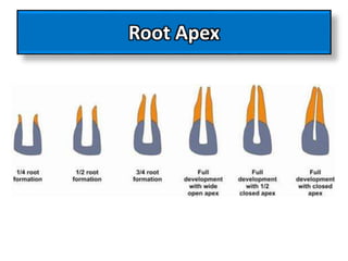
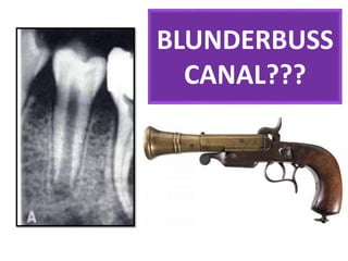
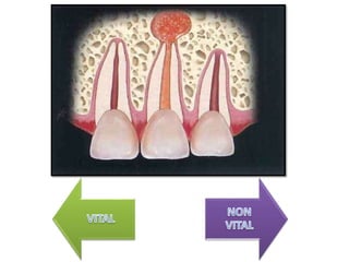
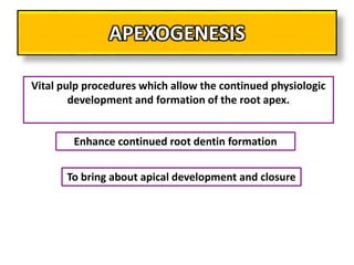
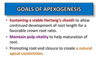
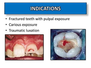
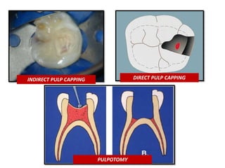
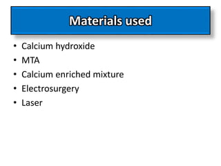
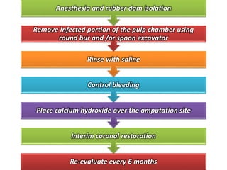
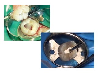
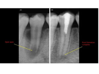
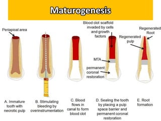
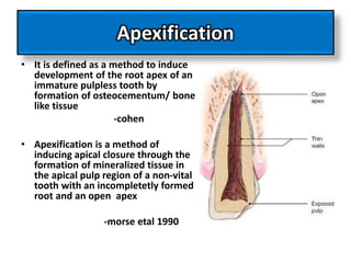
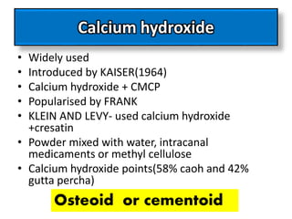
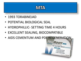
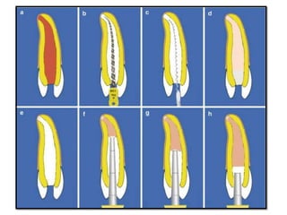
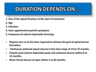
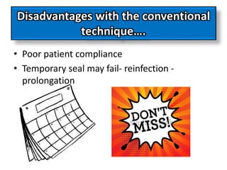
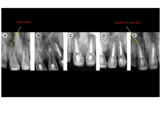
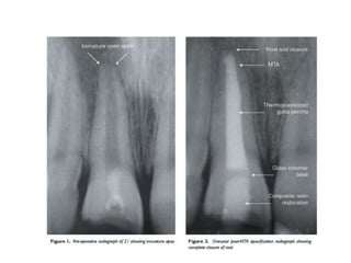
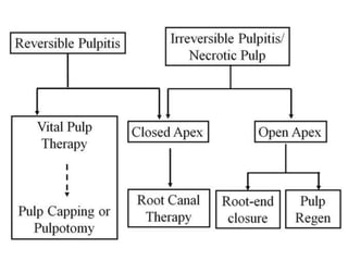
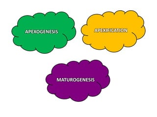
APEXOGENESIS AND
APEXIFICATION
Root Apex
BLUNDERBUSS
CANAL???
APEXOGENESIS
APEXOGENESIS
Vital pulp procedures which allow the continued physiologic
development and formation of the root apex.
To bring about apical development and closure
Enhance continued root dentin formation
RATIONALE FOR APEXOGENESIS
• Root end development- normal pulp and
minimal inflammation
• Pulp of immature teeth has significant
reparative potential
• Pulp revascularisation and repair occurs more
efficiently tooth with an open apex
GOALS OF APEXOGENESIS
• Sustaining a viable Hertwig’s sheath to allow
continued development of root length for a
favorable crown root ratio.
• Maintain pulp vitality to help maturation of
root.
• Promoting root end closure to create a natural
apical constriction.
INDICATIONS
• Fractured teeth with pulpal exposure
• Carious exposure
• Traumatic luxation
CONTRAINDICATIONS
INDIRECT PULP CAPPING DIRECT PULP CAPPING
PULPOTOMY
Materials used
• Calcium hydroxide
• MTA
• Calcium enriched mixture
• Electrosurgery
• Laser
PROCEDURE
Re-evaluate every 6 months
Interim coronal restoration
Place calcium hydroxide over the amputation site
Control bleeding
Rinse with saline
Remove Infected portion of the pulp chamber using
round bur and /or spoon excavator
Anesthesia and rubber dam isolation
APEXOGENESIS IN A YOUNG
NON - VITAL TEETH
Maturogenesis
Apexification
• It is defined as a method to induce
development of the root apex of an
immature pulpless tooth by
formation of osteocementum/ bone
like tissue
-cohen
• Apexification is a method of
inducing apical closure through the
formation of mineralized tissue in
the apical pulp region of a non-vital
tooth with an incompletetly formed
root and an open apex
-morse etal 1990
When ???
• Incomplete root development- caries, trauma
before root growth and development are
complete
• Extensive apical resorption due to trauma,
periapical pathosis, orthodontic treatment
Why cant we obturate it now???
Why should we wait till the apex
closes???
Why cant we obturate it now???
• No hard tissue stop against which gutta
percha can be packed
• Weak root dentin- fracture
Why apexification preferred over
RCT??
• Open apex
• Blunderbuss canals
• Thin and fragile canal walls
• Absolute dryness of canal difficult to achive
Objective
• To induce either closure of open apical third
of root canal or the formation of an apical
calcific barrier against which obturation can
be achieved
Materials used
• Calcium hydroxide
• Metacresylate- camphorated parachlorophenol
• Tricalcium phosphate+ beta tricalcium phosphate
• Resorbable tricalcium phosphate
• Collagen- calcium phosphate gel
• Mineral trioxide aggregate
• Dentine chips
• Hydroxyapatite
• Bone morphogenic proteins
• Platelet rich fibrin matrix
Calcium hydroxide
• Widely used
• Introduced by KAISER(1964)
• Calcium hydroxide + CMCP
• Popularised by FRANK
• KLEIN AND LEVY- used calcium hydroxide
+cresatin
• Powder mixed with water, intracanal
medicaments or methyl cellulose
• Calcium hydroxide points(58% caoh and 42%
gutta percha)
Osteoid or cementoid
MTA
• 1993 TORABINEJAD
• POTENTIAL BIOLOGICAL SEAL
• HYDROPHILLIC- SETTING TIME 4 HOURS
• EXCELLENT SEALING, BIOCOMPATIBLE
• AIDS CEMENTUM AND PDL REGENERATION
Procedure – apexification
FIRST VISIT
Rubberdam application following local anesthesia
Access opening
Removal of necrotic pulp
Working length determination
Removal of infected dentin from root canals
Canal dried
Calcium hydroxide placement
Seal with temporary restoration
2nd visit
Follow up for 6 months to 24 months
Radiographic evaluation for root apex
closure
Proceed for RCT
How is the barrier formed??
Follow up
• Absence of any fistula or sinus
• Absence or decrease in tooth mobility
• Formation of calcific bridge
• Continued apical development
• Absence of internal resorption or periapical
radiolucency
• Evidence of firm stop clinically or
radiographically
DURATION DEPENDS ON….
1. Size of the apical foramen at the start of treatment
2. Age
3. Infection
4. inter-appointment painful symptoms
5. frequency of calcium hydroxide dressings
• Reports vary as to the time required to achieve the goal of apical barrier
formation.
• Heithersay achieved apical closure in the time range of 14 to 75 months.
• Chawla used calcium hydroxide paste and achieved closure within 6 to
12 months.
• Kleier found closure of apex within 1 to 30 months.
Disadvantages with the conventional
technique….
• Poor patient compliance
• Temporary seal may fail- reinfection -
prolongation
One visit apexification
APEXOGENESIS
MATUROGENESIS
APEXIFICATION
Thank you

Apexogenesis and Apexification - pediatric dentistry

  • 2.
  • 3.
  • 4.
  • 7.
  • 8.
    APEXOGENESIS Vital pulp procedureswhich allow the continued physiologic development and formation of the root apex. To bring about apical development and closure Enhance continued root dentin formation
  • 9.
    RATIONALE FOR APEXOGENESIS •Root end development- normal pulp and minimal inflammation • Pulp of immature teeth has significant reparative potential • Pulp revascularisation and repair occurs more efficiently tooth with an open apex
  • 10.
    GOALS OF APEXOGENESIS •Sustaining a viable Hertwig’s sheath to allow continued development of root length for a favorable crown root ratio. • Maintain pulp vitality to help maturation of root. • Promoting root end closure to create a natural apical constriction.
  • 11.
    INDICATIONS • Fractured teethwith pulpal exposure • Carious exposure • Traumatic luxation
  • 12.
  • 14.
    INDIRECT PULP CAPPINGDIRECT PULP CAPPING PULPOTOMY
  • 15.
    Materials used • Calciumhydroxide • MTA • Calcium enriched mixture • Electrosurgery • Laser
  • 16.
  • 17.
    Re-evaluate every 6months Interim coronal restoration Place calcium hydroxide over the amputation site Control bleeding Rinse with saline Remove Infected portion of the pulp chamber using round bur and /or spoon excavator Anesthesia and rubber dam isolation
  • 20.
    APEXOGENESIS IN AYOUNG NON - VITAL TEETH
  • 21.
  • 22.
    Apexification • It isdefined as a method to induce development of the root apex of an immature pulpless tooth by formation of osteocementum/ bone like tissue -cohen • Apexification is a method of inducing apical closure through the formation of mineralized tissue in the apical pulp region of a non-vital tooth with an incompletetly formed root and an open apex -morse etal 1990
  • 23.
    When ??? • Incompleteroot development- caries, trauma before root growth and development are complete • Extensive apical resorption due to trauma, periapical pathosis, orthodontic treatment
  • 24.
    Why cant weobturate it now??? Why should we wait till the apex closes???
  • 25.
    Why cant weobturate it now??? • No hard tissue stop against which gutta percha can be packed • Weak root dentin- fracture
  • 26.
    Why apexification preferredover RCT?? • Open apex • Blunderbuss canals • Thin and fragile canal walls • Absolute dryness of canal difficult to achive
  • 27.
    Objective • To induceeither closure of open apical third of root canal or the formation of an apical calcific barrier against which obturation can be achieved
  • 28.
    Materials used • Calciumhydroxide • Metacresylate- camphorated parachlorophenol • Tricalcium phosphate+ beta tricalcium phosphate • Resorbable tricalcium phosphate • Collagen- calcium phosphate gel • Mineral trioxide aggregate • Dentine chips • Hydroxyapatite • Bone morphogenic proteins • Platelet rich fibrin matrix
  • 29.
    Calcium hydroxide • Widelyused • Introduced by KAISER(1964) • Calcium hydroxide + CMCP • Popularised by FRANK • KLEIN AND LEVY- used calcium hydroxide +cresatin • Powder mixed with water, intracanal medicaments or methyl cellulose • Calcium hydroxide points(58% caoh and 42% gutta percha) Osteoid or cementoid
  • 30.
    MTA • 1993 TORABINEJAD •POTENTIAL BIOLOGICAL SEAL • HYDROPHILLIC- SETTING TIME 4 HOURS • EXCELLENT SEALING, BIOCOMPATIBLE • AIDS CEMENTUM AND PDL REGENERATION
  • 31.
  • 33.
    FIRST VISIT Rubberdam applicationfollowing local anesthesia Access opening Removal of necrotic pulp Working length determination Removal of infected dentin from root canals Canal dried Calcium hydroxide placement Seal with temporary restoration
  • 34.
    2nd visit Follow upfor 6 months to 24 months Radiographic evaluation for root apex closure Proceed for RCT
  • 35.
    How is thebarrier formed??
  • 36.
    Follow up • Absenceof any fistula or sinus • Absence or decrease in tooth mobility • Formation of calcific bridge • Continued apical development • Absence of internal resorption or periapical radiolucency • Evidence of firm stop clinically or radiographically
  • 38.
    DURATION DEPENDS ON…. 1.Size of the apical foramen at the start of treatment 2. Age 3. Infection 4. inter-appointment painful symptoms 5. frequency of calcium hydroxide dressings • Reports vary as to the time required to achieve the goal of apical barrier formation. • Heithersay achieved apical closure in the time range of 14 to 75 months. • Chawla used calcium hydroxide paste and achieved closure within 6 to 12 months. • Kleier found closure of apex within 1 to 30 months.
  • 39.
    Disadvantages with theconventional technique…. • Poor patient compliance • Temporary seal may fail- reinfection - prolongation
  • 40.
  • 44.
  • 45.

Editor's Notes

  • #5 Dutch word: DONDERBUS- THUNDERGUN 18 CENTUARY WEAPON WITH SHORT AND WIDE BARELL Divergent and flaring root canals, funnel shape wider than coronal aspect of canal \
  • #13 Necrotic or non vital pulp unrestorable carious teeth Teeth with unfavourable horizontal root fracture Severe crown root fracture which requires intra radicular retention for restoration
  • #14 The dental pulp contains immune cells that allow it to mount a response against offending irritants. The pulp also contains odontoblasts, which are specialized to form dentin. In the absence of a vital pulp, the tooth structure is susceptible to infection, and dentin deposition is arrested. Maintenance of pulp vitality is imperative in an immature permanent tooth to allow continued root development. The pulp tissue is removed when pathologically inflamed or necrotic.
  • #15 Depending on the extent of inflammation, pulp capping, shallow pulpotomy, or conventional pulpotomy may be indicated. The dental pulp in young patients is more cellular and able to recover from injuries. Cvek et al (1) demonstrated that in teeth with complex crown fractures, the exposed pulp maintained its vitality for up to 7 days. Formocresol, gluteraldehyde, lasers, electrosurgery, feric sulfate, mta
  • #20 Do not blow air to avoid dessication
  • #25 ‘‘a method of inducing a calcified barrier in a root with an open apex or the continued apical development of an incompletely formed root in teeth with necrotic pulp’’
  • #36 Frank 1966 Calcium hydroxide and camphorated monochloro phenol to stimulate root end closure
  • #39 Apical development is monitored using pre and post operative radiographs
  • #40 frank
  • #41 1. Size of the apical foramen at the start of treatment - Teeth with apices < 2 mm in diameter has significant shorter time . 2. Age - Since less calcified material would be needed to occlude a narrow apex as compared to wide apex; it is understandable that the former would require shorter period for apexification . 3. Infection - some studies have reported that presence of periapical radiolucency at the start of treatment, increases the barrier formation time, whereas others have not . 4. Inter appointment painful symptoms - may delay time taken for apical healing . 5. Frequency of Ca(OH)2 dressings - there is no census on how frequently the dressing should be changed to induce apical healing. Some favour refilling every 3- 6 months, others favour refilling only if there is radiographic evidence of root resorption of paste or only after determining mechanically if the hard tissue barrier formed is adequate.
  • #42 Rationale is to establish an apical stop that would enable root canal to be filled immediately