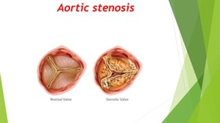
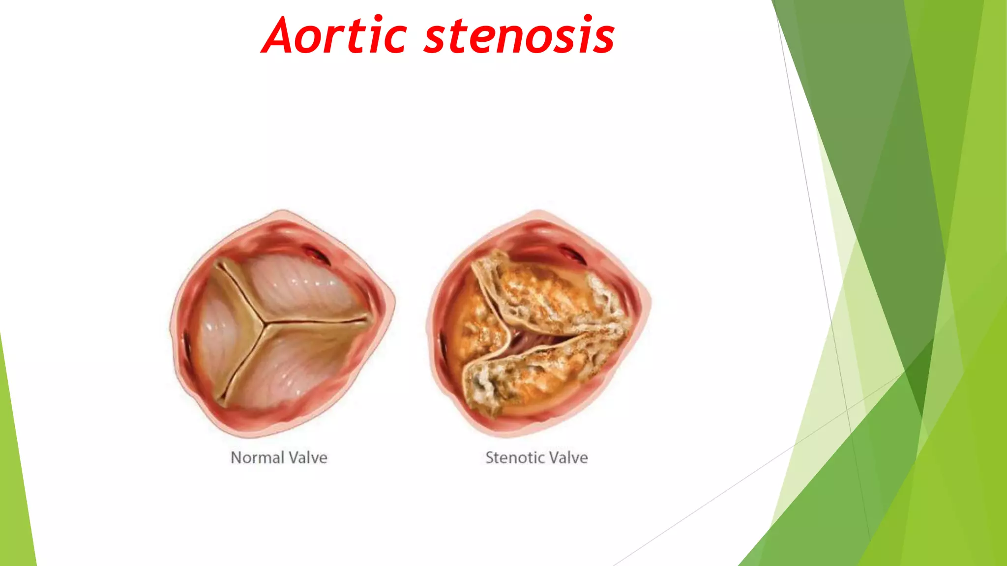
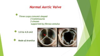
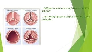
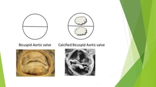
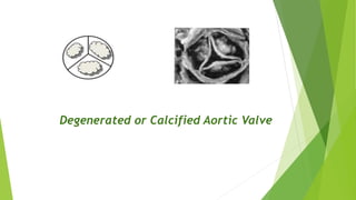
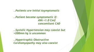
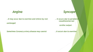
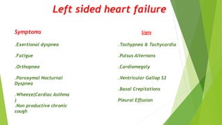
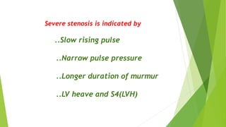
7. Bicuspid aortic Valve
.congenital abnormality.
.commonly emerge at the age of 50
to 65yrs.
Associated with Coarctation of
Aorta and Dilated ascending Aorta
8.
9. Degenerated or Calcified Aortic valve
25% of patients over age 65yrs have
echocardiographic evidence of AS
10 to 20% of them will develop
haemodynamically significant Aortic
stenosis.
Related with atherosclerotic vascular
disease.
14. Pathphysiology
Aortic Stenosis
Progressive decrease in the area of the
aortic valve
Decreased antegrade velocity when aortic area
decrease at least by half
Adaptation by hypertrophy
Early changes Late changes
Diastolic
dysfunction
Systolic
dysfunction
Decreased compliance
LV Diastolic pressure
Myocardial ischemia myocardial
fibrosis abnormal wall motion
Unchanged contractility
Normal stroke volume
Decreased contractility
Decreased stroke volume
Atrial
fibrillation
Mitral
Regurgitation
Heart
Failure
16. ..Patients are initial Asymptomatic
..Patient become symptomatic if:
AVA <1.0 Cm2
concomitant CAD
..Systolic Hypertension may coexist but
>200mm hg is uncommon
..Hypertrophic Obstructive
Cardiomyopathy may also coexist
18. Angina Syncope.
..It may occur due to exertion and relieve by rest .. It occurs due to peripheral
vasodilatation but
unchanged
cardiac output.
Sometimes Coronary artery disease may coexist ..It occurs due to exertion
21. Murmur
..Site---Aortic area (Right sec ICS)
..Radiation---To neck through carotid arteries
..Ejection---Systolic murmur
..Character—Harsh
..Position---Leaning forward held in expiration
22. Severe stenosis is indicated by
..Slow rising pulse
..Narrow pulse pressure
..Longer duration of murmur
..LV heave and S4(LVH)
26. Chest X ray
..In Early Stage
CXR may be normal or
Ascending aorta dilatation w/normal
heart size
..In Late Stage ,
Cardiac enlargement
Pulmonary Congestion
27. The ascending aorta
(yellow dotted
line)leading into the arch
is dilated , whereas the
distal arch and
descending aorta (red
dotted line )are normal
in size .
The left heart border
(blue dotted line)
28. The ascending aorta
(yellow dotted
line)leading into the
arch is dilated,
whereas the distal
arch and descending
aorta red dotted
line are normal in
size .
30. 3) Two Dimensional Echocardiography w/Doppler
Diagnostic of AS
Perform the following purposes
..Evaluate Aortic Valve morphology
..Calculate Aortic Valve area
..Evaluate Left ventricular walls thickness
..Calculate Ejection Fraction
..Estimate Aortic Valve gradient
31. Recommendations for Echocardiography in Aortic
Stenosis
Classification of Aortic Stenosis
Severity Mean Gradient(mmhg) Aortic Valve area
..Mild
..Moderate
..Severe
..Critical
..<25
..25-40
..>40
..>80
..>1.5
..1-1.5
..<1
..0.5
32. Anatomic evaluation
Combination of short and long axis images to identify
Number of leaflets
Describe leaf mobility, thickness,calcification
Combination of imaging doppler allows the
determination of the level of
Obstruction;Subvalvular,Valvular,Or Supravalvular.
TransEsophageal Echocardiography may be helpful
when image quality is suboptimal
33. Calcific Aortic Stenosis
Nodular calcific masses on aortic
side of cusps.
No commissural fusion
Free edges of cusps or not involved
Stellate –shaped systolic oriface
35. Bicuspid Aortic Valve
Fusion of the Right and Left coronary cusps (80%)
Fusion of the Right AND Left Non Coronary
cusps (20%)
36. Bicuspid Aortic Valve
Two cusps are seen in Systole with only two commissures framing an
elliptical Systolic oriface(the fish mouth appearance
Diastolic images may mimic a tricuspid valve when a raphe is
present
37. Bicuspid Aortic Valve
Parasternal long axis echocardiography may show
An Asymmetric closure line
Systolic doming
Diastolic prolapse of the cusps
In children valve may be stenotic without extensive
calcification.
In adult stenosis typically is due to calcific changes
,which often obscures the number of cusps ,making
determination of bicuspid VS tricuspid valve difficult.
38. Calcific Aortic Stenosis
Calcification of bicuspid valve,the severity can be graded semi quantitavely as
The degree of valve calcification is predictor of clinical outcome.
39. Aortic Sclerosis
Thickened calcified cusps with preserved mobility.
Typically associated with peak doppler velocity
of less than 2.5m/sec.
Rheumatic Aortic Stenosis
Characterized by
..Commissural fusion
..Triangular systolic orifice
..thickening &calcification
Accompanied by rheumatic mitral valve changes
40. Rheumatic Aortic Stenosis
Parasternal short axis view
showing commissural fusion
,leaflet thickening and
calcification ,small triangular
systolic orifice.
Subvalvular Aortic Stenosis
1) The discrete membrane
consisting of endocardial fold
and fibrous tissue.
2) A fibromuscular ridge.
3) Diffuse tunnel-like narrowing
of the LVOT.
4) Accessory of anomalous mitral
valve tissue.
41. Supravalvular Aortic Stenosis
Type 1- Thick fibrous ring above the
aortic valve with less mobility and has
the easily identifiable ‘hourglass’
appearance of the aorta.
Type 2- thin discrete fibrous membrane
located above the aortic valve.
The membrane usually mobile
and may demonstrate doming during
systole.
Type 3- diffuse narrowing
42. Asymptomatic patient murmur of AS heard
in physical exam
To dimensional echocardiography with
doppler study
Mild to moderate
aortic stenosis
Severe aortic
stenosis
Follow up the
patient
Exercise-testing
Normal Abnormal
Consider cardiac
catheterization and
Aortic Valve
replacement
43. Symptomatic patient
Two dimensional echocardiography w/doppler study
Mild to moderate
aortic stenosis
Severe aortic
stenosis
Cardiac catheterization
and plan for aortic
valve replacement
Work up for other
diseases w/h can
explain symptoms
44. 4) Cardiac catheterization
..Recommended for assessing concomitant CAD
..Recommended prior to aortic valve replacement
..If indications for Dobutamine test have been
met
45. 5) Emerging Role of BNP
..Recognized as a marker of EARLY LV failure
..Levels of >550pg/ml are associated with
poor outcomes
47. ..Following the onset of symptoms ,prognosis
without surgery is poor .
50% 03 years mortality rate
..Mean survival based on Symptoms :
1..Angina ->05 years.
2..Syncope ->03 years.
3..Heart failure ->02 years
..Sudden Cardiac death may occur in
asymptomatic individuals ~04%
49. Initial Treatment
..Depend upon the presenting complaints.
..If LV failure,
Symptom-improving drugs
Prognosis-improving drugs
..Treating hypertension normalizing lipid
profile and smoking cessation have the role .
50. Definitive treatment
Mechanical solution for mechanical problem
AORTIC VALVE REPLACEMENT
..There are two options for valve replacement .
..Surgical valve replacement.
..Transcatheter Aortic Valve Replacement
(TAVR)