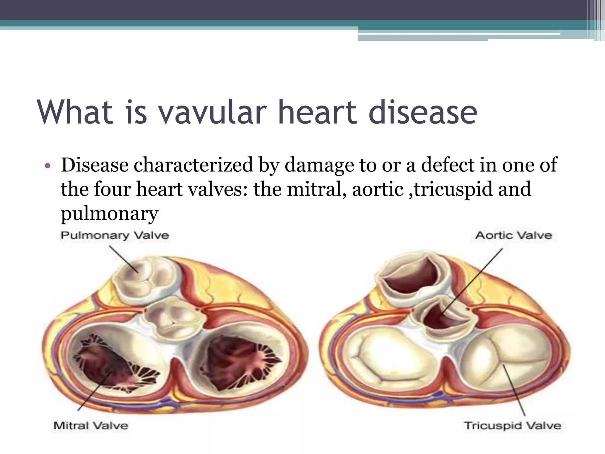
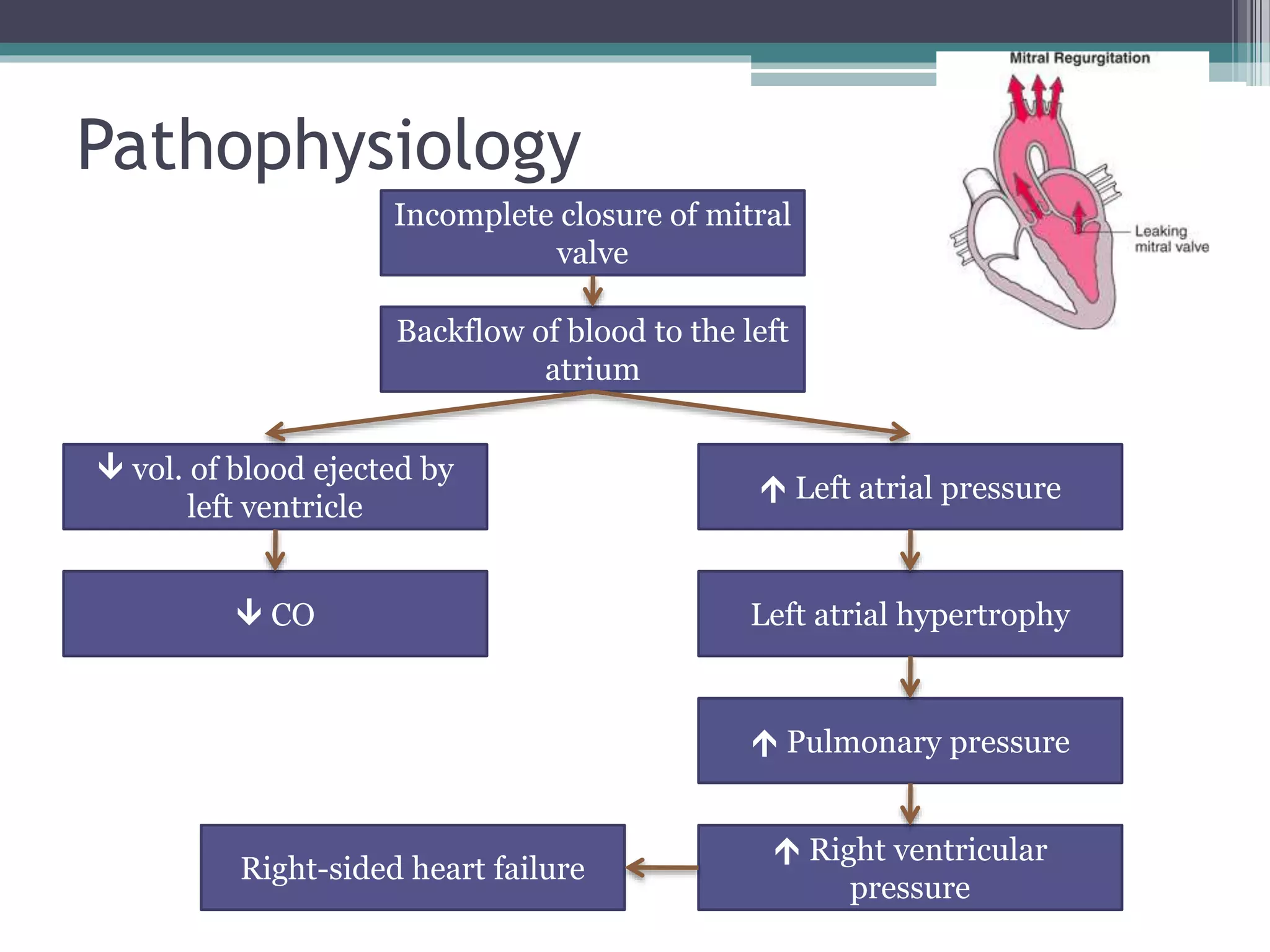
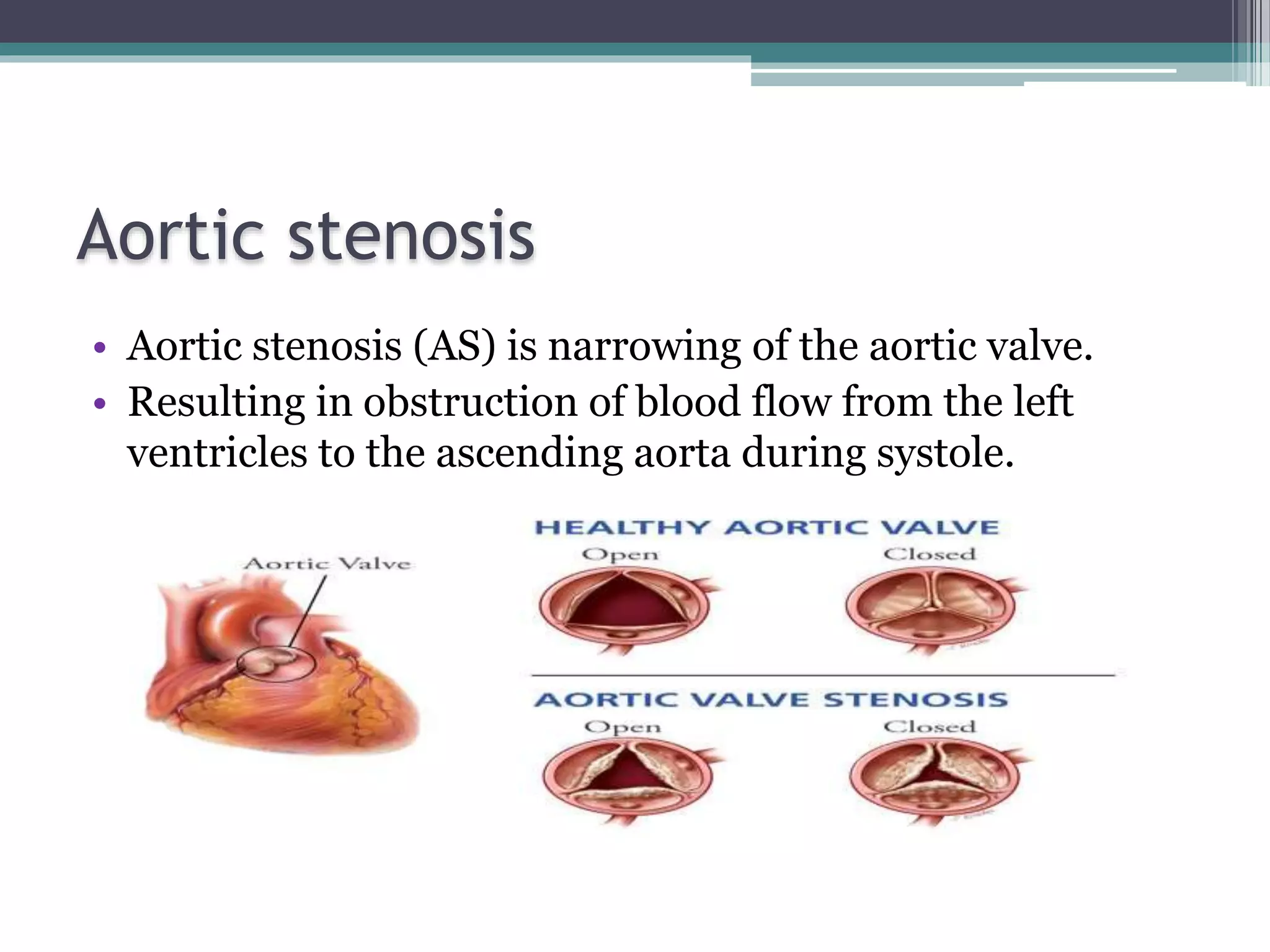
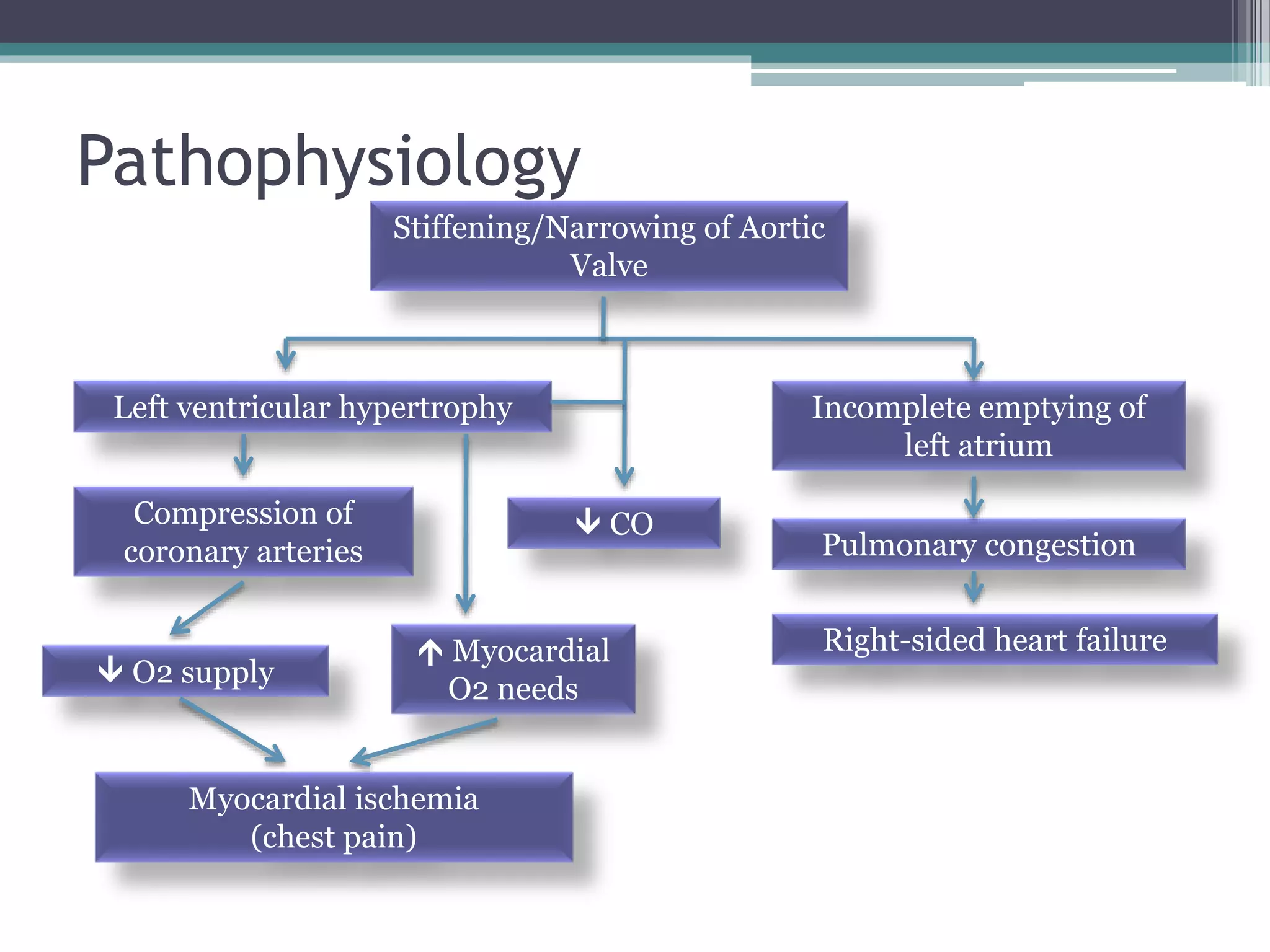
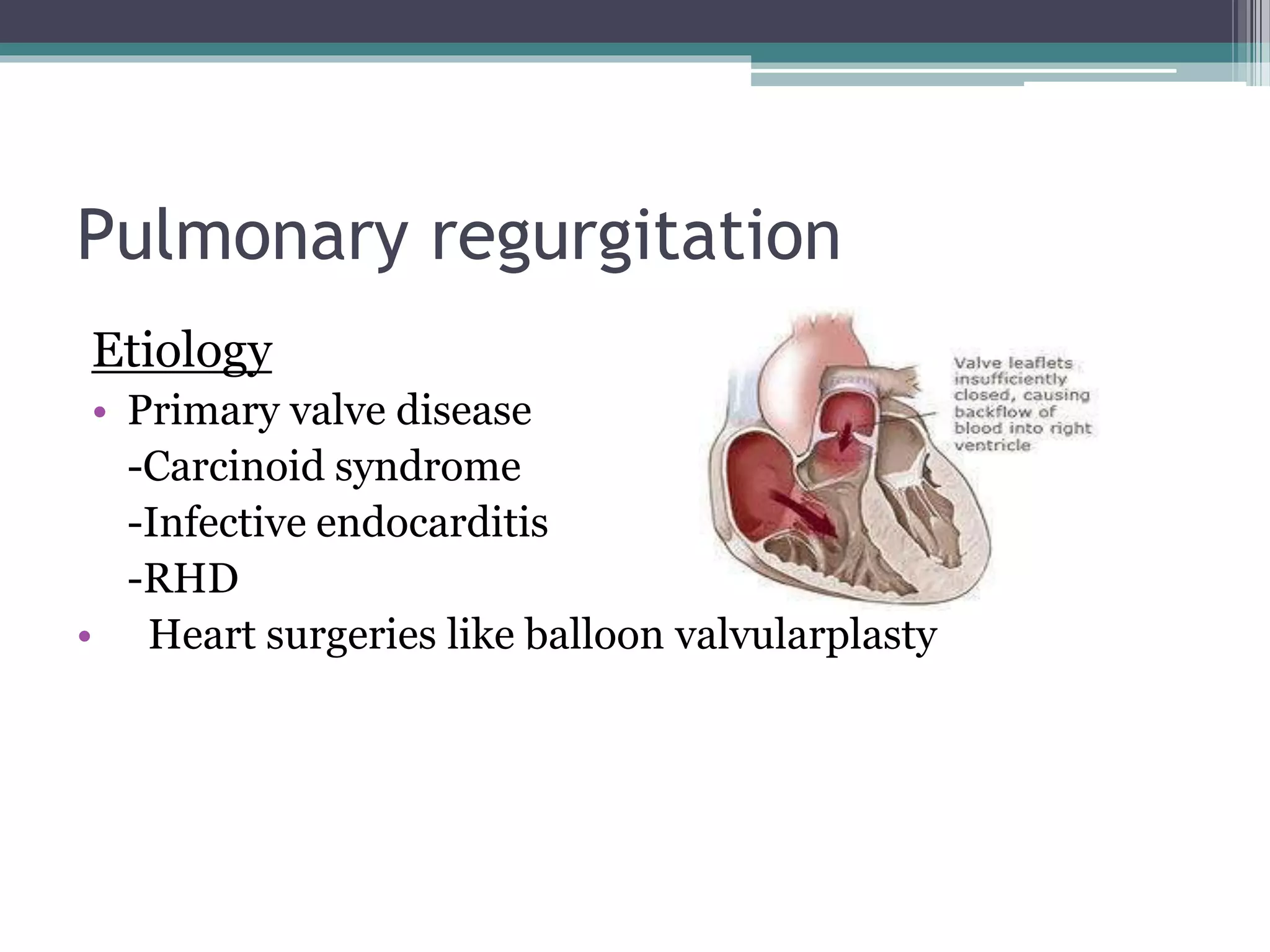
Valvular heart disease
Group members:
Osama Bin Khalid: 20170702
Shanil Samal Kumar: 20180241
Dipika Nandni: 20180274
Shiritika Keshni Ram: 20180093
What is vavular heart disease
• Disease characterized by damage to or a defect in one of
the four heart valves: the mitral, aortic ,tricuspid and
pulmonary
Types of valve disease
Principal Causes
Valve stenosis Valve regurgitation
• Congenital
• Rheumatic carditis
• Congenital
• Rheumatic carditis (acute or
chronic)
• Infective endocarditis
• Valve ring dilatation
(e.g. dilated cardiomyopathy)
• Syphilitic aortitis
• Traumatic valve rupture
• Damage to chordae and
papillary muscle (e.g. MI)
Valvular Heart Diseases
1. Mitral stenosis
2. Mitral regurgitation
3. Aortic stenosis
4. Aortic regurgitation
5. Tricuspid stenosis
6. Tricuspid regurgitation
7. Pulmonary stenosis
8. Pulmonary regurgitation
Mitral Stenosis
• Stenosis means abnormal narrowing of a passage
• Mitral stenosis is the result of structural changes that
usually include some loss of valvular substance,
contracture of the chordae tendineae, valve leaflets,
papillary muscles or fibrosis of mitral ring.
• It takes 10 or more years for the lesion to develop fully
Mitral stenosis
Etiology
• Almost always rheumatic in
origin
• Older people: can be caused by
heavy calcification of mitral
valve
• Congenital (rare)
• Systemic diseases: systemic
lupus erythematosus
Pathogenesis
Streptococcal
infection
Immune
response
macrophages engulf
Lymph
nodes
T- helper
cells
Stimulate
MHC1
Stimulate
plasma cells
Antivirus 1
antibodies
97% kills
antigen
3% goes to 4
different sites
neutrophils
endocytosis
Lysosomal
activities
Antigens released in
lymph nodes
Narrowing of mitral valve
 CO
O2/CO2 exchange
(fatigue, dyspnea,
orthopnea)
Hypotension
pulmonary
congestion
 pulmonary
pressure
 left atrial
pressure
Hypertrophy left
atrium
 blood flow to
left ventricle
Right-sided
heart failure
Fatigue
Pathophysiology
Clinical Features
Symptoms
• Breathlessness, cough (pulmonary congestion)
• Chest pain (pulmonary hypertension)
• Hemoptysis (pulmonary congestion or hypertension)
• Fatigue (low cardiac output)
• Oedema, ascites (right heart failure)
• Palpitation (atrial fibrillation)
• Thromboembolic complications
Signs
• Mitral faces (abnormal flushing of the cheeks that occurs from
cutaneous vasodilation in the setting of severe mitral valve stenosis)
• Auscultation - Loud first heart sound, opening snap
(created by forceful opening of mitral valve)
- Mid-diastolic murmur(apex)
• Crepitations, pulmonary edema, effusions (raised pulmonary
capillary pressure)
• RV heave
Investigation
• ECG: - right ventricular hypertrophy  tall R waves
• Chest x-ray: - enlarged LA & appendage
- signs of pulmonary venous congestion
• ECHO: - thickened immobile cusps
- reduced valve area
- enlarged LA
- reduced rate of diastolic filling of LV
• Doppler: - pressure gradient across mitral valve
• Cardiac catheterization: mitral stenosis and
regurgitation
Management
• Anticoagulant; to reduce the risk of systemic embolism
• Digoxin, beta blockers, or rate limiting calcium
antagonists; to control ventricular rate in atrial
fibrillation
• Afterload-reducing agents: angiotensin-converting
enzyme inhibitors or angiotensin receptor blockers (may
reduce the regurgitant volume and preserve left
ventricular function
• Diuretic; to control pulmonary congestion
• Mitral balloon valvuloplasty
• Valve replacement
Mitral regurgitation
Etiology
• Rheumatic disease
• Mitral valve prolapse
• Dilatation of the LV and mitral valve ring (e.g. coronary
artery disease, cardiomyopathy)
• Damage to valve cusps and chordae eg. endocarditis
• Ischemia or infarction of papillary muscle (MI)
Pathophysiology
Incomplete closure of mitral
valve
 vol. of blood ejected by
left ventricle
 Left atrial pressure
Right-sided heart failure
Left atrial hypertrophy CO
 Pulmonary pressure
Backflow of blood to the left
atrium
 Right ventricular
pressure
Clinical manifestation
Signs and Symptoms
• Fatigue & weakness – due to  CO – predominant complaint
• Exertional dyspnea & cough – pulmonary congestion
• Palpitations – due to atrial fibrillation (occur in 75% pts)
• Edema, ascites – Right-sided heart failure
• Atrial fibrillation
• Cardiomegaly
• Signs of pulmonary hypertension & right heart failure
• Apical pansystolic murmur +/- thrill
• Soft S1, apical S3
Investigations
• ECG: - left atrial hypertrophy
- left ventricular hypertrophy
• Chest x-ray: - enlarged LA,LV
- pulmonary venous congestion
- pulmonary oedema
• ECHO: - dilated LA,LV
- structural abnormalities of mitral valve (e.g.
prolapse)
• Doppler: - detects and quantifies regurgitation
• Cardiac catheterization: - dilated LA,LV
- mitral regurgitation
- coexisting coronary artery disease
Management
• Vasodilators (e.g. ACE inhibitors)
• Diuretics
• If atrial fibrillation presents,
▫ Anticoagulant
▫ Digoxin
• Mitral valve replacement
Aortic stenosis
• Aortic stenosis (AS) is narrowing of the aortic valve.
• Resulting in obstruction of blood flow from the left
ventricles to the ascending aorta during systole.
Etiology
INFANTS
 Congenital aortic stenosis
YOUNG ADULTS TO MIDDLE-AGED
 Calcification and fibrosis of congenitally bicuspid aortic valve
 Rheumatic aortic stenosis
MIDDLE-AGED TO ELDERLY
 Senile degenerative aortic stenosis
Pathophysiology
Pathophysiology
Stiffening/Narrowing of Aortic
Valve
Incomplete emptying of
left atrium
Left ventricular hypertrophy
Pulmonary congestion
Compression of
coronary arteries
Right-sided heart failure
 CO
 Myocardial
O2 needs
Myocardial ischemia
(chest pain)
 O2 supply
Clinical features
Symptoms
 Mild to moderate stenosis (asymptomatic)
 Exertional dyspnea
 Angina
 Exertional syncope
 Episodes of acute pulmonary edema
 Sudden death
Signs
 Ejection systolic murmur
 Slow-rising carotid pulse
 Thrusting apex beat (LV pressure overload)
 Narrow pulse pressure
 Signs of pulmonary venous congestion (e.g. crepititions)
Investigations
 ECG: - left ventricular hypertrophy - left bundle branch block
 Chest x-ray: - may be normal
- enlarged LV & dilated ascending aorta (PA view)
- calcified valve on lateral view
 ECHO: - calcified valve with restricted opening, hypertrophied LV
 Doppler: - measurement of severity of stenosis
- detection of associated aortic regurgitation
 Cardiac catheterization: - to identify coronary artery disease
- used to measure gradient between LV and aorta
Management
• Symptomatic severe aortic stenosis  valve
replacement.
• Congenital aortic stenosis  aortic balloon valvuloplasty
• Atrial fibrillation or post valve replacement with a
mechanical prosthesis  anticoagulant
• Prophylactic antibiotic to prevent endocarditis.
Aortic regurgitation
• Aortic regurgitation is the failure of aortic valve to close
tightly causing backflow of blood into the left ventricles.
Etiology
Congenital:
Bicuspid valve or disproportionate cusps
Acquired:
Rheumatic disease
Infective endocarditis
Trauma
Aortic dilatation (marfan’s syndrome, aneurysm,
dissection, syphilis)
Pathophysiology
Incomplete closure of the
aortic valve
Backflow of blood to Left
ventricle
Left ventricular
hypertrophy & dilation
 Left atrial pressure
Left-sided heart failure
(late stage)
Left atrium hypertrophy
 CO
 Pulmonary pressure
Right-sided heart failure
 Right ventricular
pressure
Clinical features
Symptoms
Mild or moderate aortic regurgitation:
• Usually asymptomatic
(because compensatory ventricular dilatation & hypertrophy
occur)
• Awareness of heartbeat, ‘palpitations’
particularly when lying on the left side, which results from
increased in stroke volume
• Severe aortic regurgitation:
Breathlessness
Angina
Clinical features
SIGNS
Pulses:
 Large volume or ‘collapsing’ pulse
 Low diastolic and increased pulse
pressure
 Bounding peripheral pulse
 Capillary pulsation in nail beds:
Quincke’s sign
 Femoral bruit(‘pistol shot’):
Duroziez’s sign
 Head nodding with pulse: de
Musset’s sign
Murmurs:
 Early diastolic murmur
 Austin Flint murmur (soft mid-
diastolic)
Other signs:
 Displaced, heaving apex beat
(volume overload)
 Crepitations
Investigation
• ECG: initially normal, later left ventricular hypertrophy & T-wave
inversion
• Chest x-ray: - cardiac dilatation, maybe aortic dilatation
- features of left heart failure
• ECHO: - dilated LV
- hyperdynamic LV
- fluttering anterior mitral leaflet
• Doppler: - detects reflux
• Cardiac catheterization: - dilated LV
- aortic regurgitation
- dilated aortic root
Management
• Treatment may be required for underlying conditions, such as
endocarditis or syphilis
• Aortic regurgitation with symptoms aortic valve
replacement (may be combined with aortic root replacement
and coronary bypass surgery)
• Asymptomatic patients  annually follow up with
echocardiography for evidence of increasing ventricular size
• Systolic BP should be controlled with vasodilating drugs, such
as Nifedipine or ACE inhibitors
Tricuspid stenosis
● Rheumatic in origin and is rare
● Occurs together with aortic or mitral stenosis
● May occur due to rheumatic heart disease (<5%)
● Decreased right ventricular output --> decrease in
cardiac output, left ventricular filling and increase in
systemic pressure
Clinical features
Symptoms Signs
● Symptoms of right heart failure
● hepatomegaly with presystolic
pulsation
● ascites
● peripheral oedema
● neck vein enlargement
● Decrease in co- fatigue,
hypotension
● Raised JVP
● Mid-diastolic murmur (best
heard at the lower left or right
sternal edge)
Diagnosis and Management
● The diagnosis can be confirmed by Doppler
echocardiography
Management
● Valve replacement
● Valvotomy
● Balloon valvuloplasty
Tricuspid Regurgitation
● Common and is most frequently ‘functional’ occurring
as a result of right ventricular dilatation
● Backflow to right atrium --> which causes venous
congestion--> decreased right ventricular output, and
decrease blood flow in lungs
Etiology
Primary
•Rheumatic heart disease
•Endocarditis, particularly in intravenous drug-users
•Ebstein’s congenital anomaly
Secondary
•Right ventricular failure
•Right ventricular infarction
•Pulmonary hypertension
Clinical features
Symptoms
● Symptoms are usually non-
specific
● Tiredness (reduced cardiac
output)
● Oedema
● Hepatic enlargement (due to
venous congestion)
Signs
● Raised JVP
● Pansystolic murmur (left
sternal edge)
● Pulsatile liver
Management
● Correction of the cause of right ventricular overload (if
TR is due to right ventricular dilation)
● Valve repair
● Tricuspid valve replacement
Pulmonary stenosis
Etiology
• Carcinoid syndrome
• Congenital (Tetralogy of
fallot)
• Genetic conditions
(Noonans syndrome)
• Mechanical stress
Pathophysiology
Hard to open and push blood pass heart valve
Pressure in RV increases
Thickening of the heart muscle
Concentric right ventricular hypertrophy
Heart struggles to get enough blood
Right sided heart failure
Signs and symptoms
Symptoms Signs
• Swelling of ankles and feet
• Hepatomegaly
• Cyanosis, dyspnea on exertion
and fatigue
• Poor weight gain or failure to
thrive in infants
• Ejection systolic murmur
(loudest at the left upper
sternum & radiating towards
the left shoulder)
• Murmur often preceded by an
ejection sound (click)
• May be wide splitting of
second heart sound (delay in
ventricular ejection
• May be a thrill (best felt when
patient leans forward and
breathes out)
• Distended neck veins
Investigations
• Doppler echocardiography (definitive investigation)
• ECG: - right ventricular hypertrophy
• Chest x-ray: - post-stenotic dilatation in the pulmonary
artery
Management
• Mild to moderate isolated pulmonary stenosis is
relatively common and does not usually progress or
require treatment
• Severe pulmonary stenosis  percutaneous pulmonary
balloon valvuloplasty or surgical valvotomy
Pulmonary regurgitation
Etiology
• Primary valve disease
-Carcinoid syndrome
-Infective endocarditis
-RHD
• Heart surgeries like balloon valvularplasty
Pathophysiology
Backflow increases blood volume in the RV
Increases workload of RV during systole
eccentric ventricular hypertrophy
right sided heart failure
Signs and symptoms
Symptoms Signs
• fatigue
• Dyspnoea
• Graham steell Murmur
• Prominent JVP
Investigation
• ECG
-right ventricle enlargement
-right axis deviation
• X-ray
-right atrium enlargement
-right ventricle hypertrophy
• Echocardiogram
Management
• Usually asymptomatic
• Reduce pulmonary artery pressure
• Valve replacement (rare)
References
• Ralston SH, Penman ID, Strachan MWJ & Hobson RP,
Davidson’s Principles and Practice of Medicine, 22rd
edition, China, Elsevier, 2018, pg. 616-625.

Valvular heart disease

  • 1.
    Valvular heart disease Groupmembers: Osama Bin Khalid: 20170702 Shanil Samal Kumar: 20180241 Dipika Nandni: 20180274 Shiritika Keshni Ram: 20180093
  • 2.
    What is vavularheart disease • Disease characterized by damage to or a defect in one of the four heart valves: the mitral, aortic ,tricuspid and pulmonary
  • 3.
  • 4.
    Principal Causes Valve stenosisValve regurgitation • Congenital • Rheumatic carditis • Congenital • Rheumatic carditis (acute or chronic) • Infective endocarditis • Valve ring dilatation (e.g. dilated cardiomyopathy) • Syphilitic aortitis • Traumatic valve rupture • Damage to chordae and papillary muscle (e.g. MI)
  • 5.
    Valvular Heart Diseases 1.Mitral stenosis 2. Mitral regurgitation 3. Aortic stenosis 4. Aortic regurgitation 5. Tricuspid stenosis 6. Tricuspid regurgitation 7. Pulmonary stenosis 8. Pulmonary regurgitation
  • 6.
    Mitral Stenosis • Stenosismeans abnormal narrowing of a passage • Mitral stenosis is the result of structural changes that usually include some loss of valvular substance, contracture of the chordae tendineae, valve leaflets, papillary muscles or fibrosis of mitral ring. • It takes 10 or more years for the lesion to develop fully
  • 7.
    Mitral stenosis Etiology • Almostalways rheumatic in origin • Older people: can be caused by heavy calcification of mitral valve • Congenital (rare) • Systemic diseases: systemic lupus erythematosus
  • 8.
    Pathogenesis Streptococcal infection Immune response macrophages engulf Lymph nodes T- helper cells Stimulate MHC1 Stimulate plasmacells Antivirus 1 antibodies 97% kills antigen 3% goes to 4 different sites neutrophils endocytosis Lysosomal activities Antigens released in lymph nodes
  • 9.
    Narrowing of mitralvalve  CO O2/CO2 exchange (fatigue, dyspnea, orthopnea) Hypotension pulmonary congestion  pulmonary pressure  left atrial pressure Hypertrophy left atrium  blood flow to left ventricle Right-sided heart failure Fatigue Pathophysiology
  • 10.
    Clinical Features Symptoms • Breathlessness,cough (pulmonary congestion) • Chest pain (pulmonary hypertension) • Hemoptysis (pulmonary congestion or hypertension) • Fatigue (low cardiac output) • Oedema, ascites (right heart failure) • Palpitation (atrial fibrillation) • Thromboembolic complications
  • 11.
    Signs • Mitral faces(abnormal flushing of the cheeks that occurs from cutaneous vasodilation in the setting of severe mitral valve stenosis) • Auscultation - Loud first heart sound, opening snap (created by forceful opening of mitral valve) - Mid-diastolic murmur(apex) • Crepitations, pulmonary edema, effusions (raised pulmonary capillary pressure) • RV heave
  • 12.
    Investigation • ECG: -right ventricular hypertrophy  tall R waves • Chest x-ray: - enlarged LA & appendage - signs of pulmonary venous congestion • ECHO: - thickened immobile cusps - reduced valve area - enlarged LA - reduced rate of diastolic filling of LV • Doppler: - pressure gradient across mitral valve • Cardiac catheterization: mitral stenosis and regurgitation
  • 13.
    Management • Anticoagulant; toreduce the risk of systemic embolism • Digoxin, beta blockers, or rate limiting calcium antagonists; to control ventricular rate in atrial fibrillation • Afterload-reducing agents: angiotensin-converting enzyme inhibitors or angiotensin receptor blockers (may reduce the regurgitant volume and preserve left ventricular function • Diuretic; to control pulmonary congestion • Mitral balloon valvuloplasty • Valve replacement
  • 14.
    Mitral regurgitation Etiology • Rheumaticdisease • Mitral valve prolapse • Dilatation of the LV and mitral valve ring (e.g. coronary artery disease, cardiomyopathy) • Damage to valve cusps and chordae eg. endocarditis • Ischemia or infarction of papillary muscle (MI)
  • 15.
    Pathophysiology Incomplete closure ofmitral valve  vol. of blood ejected by left ventricle  Left atrial pressure Right-sided heart failure Left atrial hypertrophy CO  Pulmonary pressure Backflow of blood to the left atrium  Right ventricular pressure
  • 16.
    Clinical manifestation Signs andSymptoms • Fatigue & weakness – due to  CO – predominant complaint • Exertional dyspnea & cough – pulmonary congestion • Palpitations – due to atrial fibrillation (occur in 75% pts) • Edema, ascites – Right-sided heart failure • Atrial fibrillation • Cardiomegaly • Signs of pulmonary hypertension & right heart failure • Apical pansystolic murmur +/- thrill • Soft S1, apical S3
  • 17.
    Investigations • ECG: -left atrial hypertrophy - left ventricular hypertrophy • Chest x-ray: - enlarged LA,LV - pulmonary venous congestion - pulmonary oedema • ECHO: - dilated LA,LV - structural abnormalities of mitral valve (e.g. prolapse) • Doppler: - detects and quantifies regurgitation • Cardiac catheterization: - dilated LA,LV - mitral regurgitation - coexisting coronary artery disease
  • 18.
    Management • Vasodilators (e.g.ACE inhibitors) • Diuretics • If atrial fibrillation presents, ▫ Anticoagulant ▫ Digoxin • Mitral valve replacement
  • 19.
    Aortic stenosis • Aorticstenosis (AS) is narrowing of the aortic valve. • Resulting in obstruction of blood flow from the left ventricles to the ascending aorta during systole.
  • 20.
    Etiology INFANTS  Congenital aorticstenosis YOUNG ADULTS TO MIDDLE-AGED  Calcification and fibrosis of congenitally bicuspid aortic valve  Rheumatic aortic stenosis MIDDLE-AGED TO ELDERLY  Senile degenerative aortic stenosis
  • 21.
  • 22.
    Pathophysiology Stiffening/Narrowing of Aortic Valve Incompleteemptying of left atrium Left ventricular hypertrophy Pulmonary congestion Compression of coronary arteries Right-sided heart failure  CO  Myocardial O2 needs Myocardial ischemia (chest pain)  O2 supply
  • 23.
    Clinical features Symptoms  Mildto moderate stenosis (asymptomatic)  Exertional dyspnea  Angina  Exertional syncope  Episodes of acute pulmonary edema  Sudden death Signs  Ejection systolic murmur  Slow-rising carotid pulse  Thrusting apex beat (LV pressure overload)  Narrow pulse pressure  Signs of pulmonary venous congestion (e.g. crepititions)
  • 24.
    Investigations  ECG: -left ventricular hypertrophy - left bundle branch block  Chest x-ray: - may be normal - enlarged LV & dilated ascending aorta (PA view) - calcified valve on lateral view  ECHO: - calcified valve with restricted opening, hypertrophied LV  Doppler: - measurement of severity of stenosis - detection of associated aortic regurgitation  Cardiac catheterization: - to identify coronary artery disease - used to measure gradient between LV and aorta
  • 25.
    Management • Symptomatic severeaortic stenosis  valve replacement. • Congenital aortic stenosis  aortic balloon valvuloplasty • Atrial fibrillation or post valve replacement with a mechanical prosthesis  anticoagulant • Prophylactic antibiotic to prevent endocarditis.
  • 26.
    Aortic regurgitation • Aorticregurgitation is the failure of aortic valve to close tightly causing backflow of blood into the left ventricles.
  • 27.
    Etiology Congenital: Bicuspid valve ordisproportionate cusps Acquired: Rheumatic disease Infective endocarditis Trauma Aortic dilatation (marfan’s syndrome, aneurysm, dissection, syphilis)
  • 28.
  • 29.
    Incomplete closure ofthe aortic valve Backflow of blood to Left ventricle Left ventricular hypertrophy & dilation  Left atrial pressure Left-sided heart failure (late stage) Left atrium hypertrophy  CO  Pulmonary pressure Right-sided heart failure  Right ventricular pressure
  • 30.
    Clinical features Symptoms Mild ormoderate aortic regurgitation: • Usually asymptomatic (because compensatory ventricular dilatation & hypertrophy occur) • Awareness of heartbeat, ‘palpitations’ particularly when lying on the left side, which results from increased in stroke volume • Severe aortic regurgitation: Breathlessness Angina
  • 31.
    Clinical features SIGNS Pulses:  Largevolume or ‘collapsing’ pulse  Low diastolic and increased pulse pressure  Bounding peripheral pulse  Capillary pulsation in nail beds: Quincke’s sign  Femoral bruit(‘pistol shot’): Duroziez’s sign  Head nodding with pulse: de Musset’s sign Murmurs:  Early diastolic murmur  Austin Flint murmur (soft mid- diastolic) Other signs:  Displaced, heaving apex beat (volume overload)  Crepitations
  • 32.
    Investigation • ECG: initiallynormal, later left ventricular hypertrophy & T-wave inversion • Chest x-ray: - cardiac dilatation, maybe aortic dilatation - features of left heart failure • ECHO: - dilated LV - hyperdynamic LV - fluttering anterior mitral leaflet • Doppler: - detects reflux • Cardiac catheterization: - dilated LV - aortic regurgitation - dilated aortic root
  • 33.
    Management • Treatment maybe required for underlying conditions, such as endocarditis or syphilis • Aortic regurgitation with symptoms aortic valve replacement (may be combined with aortic root replacement and coronary bypass surgery) • Asymptomatic patients  annually follow up with echocardiography for evidence of increasing ventricular size • Systolic BP should be controlled with vasodilating drugs, such as Nifedipine or ACE inhibitors
  • 34.
    Tricuspid stenosis ● Rheumaticin origin and is rare ● Occurs together with aortic or mitral stenosis ● May occur due to rheumatic heart disease (<5%) ● Decreased right ventricular output --> decrease in cardiac output, left ventricular filling and increase in systemic pressure
  • 35.
    Clinical features Symptoms Signs ●Symptoms of right heart failure ● hepatomegaly with presystolic pulsation ● ascites ● peripheral oedema ● neck vein enlargement ● Decrease in co- fatigue, hypotension ● Raised JVP ● Mid-diastolic murmur (best heard at the lower left or right sternal edge)
  • 36.
    Diagnosis and Management ●The diagnosis can be confirmed by Doppler echocardiography Management ● Valve replacement ● Valvotomy ● Balloon valvuloplasty
  • 37.
    Tricuspid Regurgitation ● Commonand is most frequently ‘functional’ occurring as a result of right ventricular dilatation ● Backflow to right atrium --> which causes venous congestion--> decreased right ventricular output, and decrease blood flow in lungs
  • 38.
    Etiology Primary •Rheumatic heart disease •Endocarditis,particularly in intravenous drug-users •Ebstein’s congenital anomaly Secondary •Right ventricular failure •Right ventricular infarction •Pulmonary hypertension
  • 39.
    Clinical features Symptoms ● Symptomsare usually non- specific ● Tiredness (reduced cardiac output) ● Oedema ● Hepatic enlargement (due to venous congestion) Signs ● Raised JVP ● Pansystolic murmur (left sternal edge) ● Pulsatile liver
  • 40.
    Management ● Correction ofthe cause of right ventricular overload (if TR is due to right ventricular dilation) ● Valve repair ● Tricuspid valve replacement
  • 41.
    Pulmonary stenosis Etiology • Carcinoidsyndrome • Congenital (Tetralogy of fallot) • Genetic conditions (Noonans syndrome) • Mechanical stress
  • 42.
    Pathophysiology Hard to openand push blood pass heart valve Pressure in RV increases Thickening of the heart muscle Concentric right ventricular hypertrophy Heart struggles to get enough blood Right sided heart failure
  • 43.
    Signs and symptoms SymptomsSigns • Swelling of ankles and feet • Hepatomegaly • Cyanosis, dyspnea on exertion and fatigue • Poor weight gain or failure to thrive in infants • Ejection systolic murmur (loudest at the left upper sternum & radiating towards the left shoulder) • Murmur often preceded by an ejection sound (click) • May be wide splitting of second heart sound (delay in ventricular ejection • May be a thrill (best felt when patient leans forward and breathes out) • Distended neck veins
  • 44.
    Investigations • Doppler echocardiography(definitive investigation) • ECG: - right ventricular hypertrophy • Chest x-ray: - post-stenotic dilatation in the pulmonary artery
  • 45.
    Management • Mild tomoderate isolated pulmonary stenosis is relatively common and does not usually progress or require treatment • Severe pulmonary stenosis  percutaneous pulmonary balloon valvuloplasty or surgical valvotomy
  • 46.
    Pulmonary regurgitation Etiology • Primaryvalve disease -Carcinoid syndrome -Infective endocarditis -RHD • Heart surgeries like balloon valvularplasty
  • 47.
    Pathophysiology Backflow increases bloodvolume in the RV Increases workload of RV during systole eccentric ventricular hypertrophy right sided heart failure
  • 48.
    Signs and symptoms SymptomsSigns • fatigue • Dyspnoea • Graham steell Murmur • Prominent JVP
  • 49.
    Investigation • ECG -right ventricleenlargement -right axis deviation • X-ray -right atrium enlargement -right ventricle hypertrophy • Echocardiogram
  • 50.
    Management • Usually asymptomatic •Reduce pulmonary artery pressure • Valve replacement (rare)
  • 51.
    References • Ralston SH,Penman ID, Strachan MWJ & Hobson RP, Davidson’s Principles and Practice of Medicine, 22rd edition, China, Elsevier, 2018, pg. 616-625.

Editor's Notes

  • #9 3% goes to 4 different sites because of the m protein in the bacteria that has similar structure as this 4 : Heart- troponin, actin, myosin Brain- basal ganglia- gangliosides Joints- vermertin Skin- keratin
  • #10 Significant mitral stenosis results in increased pressure and enlargement and hypertrophy of the left atrium, pulmonary venous hypertension, increased pulmonary vascular resistance, and pulmonary hypertension.
  • #32 Collapsing pulse is also know as water hammer pulse