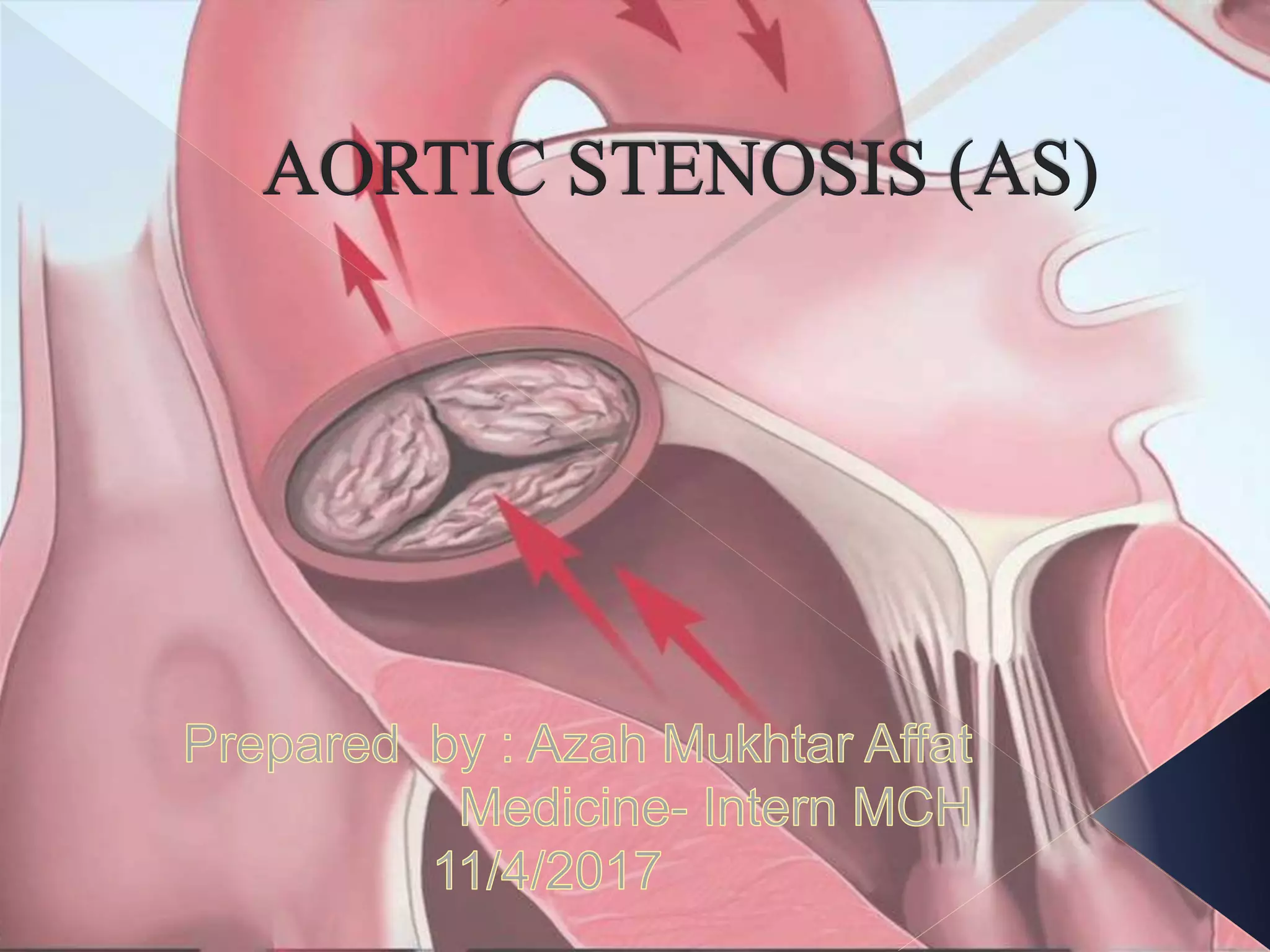
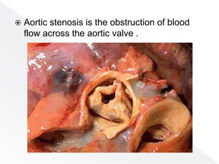
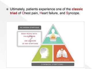
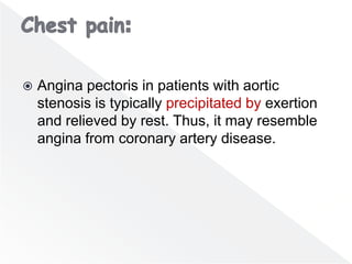
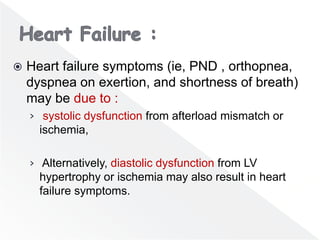
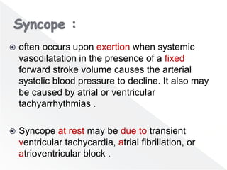
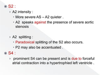
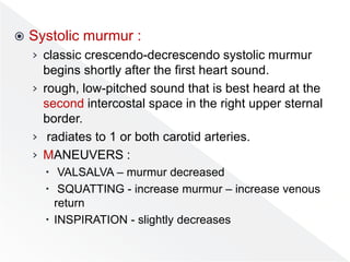
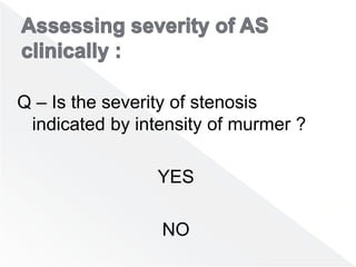
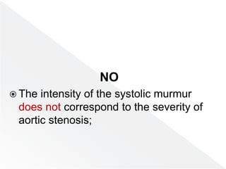
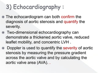
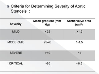
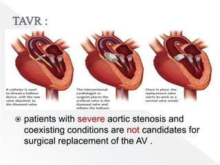
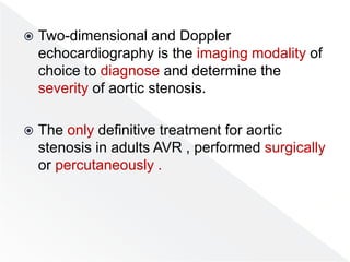
๏‚ž Overview
๏‚ž Approach to the patient
๏‚ž Diagnostic Studies
๏‚ž Treatment And Management
๏‚ž Conclussion
๏‚ž Aortic stenosis is the obstruction of blood
flow across the aortic valve .
๏‚ž Most cases of aortic stenosis are due to the
obstruction at the valvular level .
๏‚ž Valvular aortic stenosis can be either congenital or
acquired.
๏‚ž Congenital valvular aortic stenosis :
โ€บ Congenitally unicuspid, bicuspid, or even quadricuspid
valves may cause aortic stenosis .
โ€บ In neonates and infants younger than 1 year, a
unicuspid valve can produce severe obstruction .
โ€บ In adults who develop symptoms from congenital aortic
stenosis, the problem is usually a bicuspid valve .
๏‚ž Acquired valvular aortic stenosis :
1) Degenerative calcification , (senile calcific
aortic stenosis) .
โ€บ Risk factors include :
๏‚– advanced age,
๏‚– hypertension
๏‚– hypercholesterolemia,
๏‚– diabetes mellitus,
๏‚– and smoking.
2) Rheumatic heart disease :
โ€บ progressive fibrosis of the valve leaflets with
varying degrees of commissural fusion, often with
calcification .
โ€บ Coexistent mitral valve disease is common.
3) Infrequent causes of aortic stenosis include
:
โ€ข Obstructive vegetations
โ€ข Homozygous type II hypercholesterolemia
โ€ข Paget disease . SLE
โ€ข Irradiation .
๏‚ž Most patients with a congenitally bicuspid
aortic valve do not develop symptoms until
middle age or later.
๏‚ž Patients with rheumatic aortic stenosis
typically present with symptoms after the
sixth decade of life .
๏‚ž Aortic sclerosis increases in incidence with
age and is present in 29% of individuals older
than 65 years and in 37% of individuals older
than 75 years.
๏‚ž In elderly persons, the prevalence of AS is
between 2% and 9%.
๏‚ž Degenerative calcific aortic stenosis usually
manifests in individuals older than 75 years
and occurs most frequently in males.
๏‚ž Asymptomatic patients, even with critical aortic
stenosis, have an excellent prognosis for
survival, with an expected death rate of less
than 1% per year; only 4% of sudden cardiac
deaths in severe AS occur in asymptomatic
patients.
๏‚ž Among symptomatic patients with medically
treated, moderate-to-severe AS , mortality
rates from the onset of symptoms are
approximately 25% at 1 year and 50% at 2
years. More than 50% of deaths are sudden.
๏‚ž Aortic stenosis usually has an asymptomatic
latent period of 10-20 years .
๏‚ž Symptoms develop gradually :
โ€บ Exertional dyspnea is the most common initial
complaint .
โ€บ In addition, patients may develop exertional chest
pain, effort dizziness or lightheadedness, easy
fatigability, and progressive inability to exercise .
๏‚ž Ultimately, patients experience one of the classic
triad of Chest pain, Heart failure, and Syncope.
๏‚ž Angina pectoris in patients with aortic
stenosis is typically precipitated by exertion
and relieved by rest. Thus, it may resemble
angina from coronary artery disease.
๏‚ž Heart failure symptoms (ie, PND , orthopnea,
dyspnea on exertion, and shortness of breath)
may be due to :
โ€บ systolic dysfunction from afterload mismatch or
ischemia,
โ€บ Alternatively, diastolic dysfunction from LV
hypertrophy or ischemia may also result in heart
failure symptoms.
๏‚ž often occurs upon exertion when systemic
vasodilatation in the presence of a fixed
forward stroke volume causes the arterial
systolic blood pressure to decline. It also may
be caused by atrial or ventricular
tachyarrhythmias .
๏‚ž Syncope at rest may be due to transient
ventricular tachycardia, atrial fibrillation, or
atrioventricular block .
๏‚ž Syncope may be accompanied by
convulsions.
๏‚ž Patients with AS may have a higher
incidence of nitroglycerin-induced syncope
than does the general population.
๏‚ž Always consider AS as a possible etiology
for a patient who demonstrates particular
hemodynamic sensitivity to nitrates.
๏‚ž GI bleeding due to angiodysplasia (Heyde
syndrome ) .
๏‚ž Patients may present with manifestations
of infectiveendocarditis ( fever,
fatigue,anorexia, back pain, and weight loss).
The risk of infective endocarditis is higher in
younger patients with mild valvular deformity
than in older patients with degenerated
calcified aortic valves.
๏‚ž BP :
โ€บ SBP normal โ€“ in absence of HTN or AR
โ€บ Advanced AS, pulse pressure .
โ€บ Systemic HTN in elderly .
๏‚ž Carotid :
โ€บ Hallmark โ€“ slow rising and weak (parvus), delayed
peak (tardus), small-volume .
๏‚ž JVP :
โ€บ JVP elevated if biventricular HF - A more
prominent (rare) .
๏‚ž Precordial :
โ€บ Sustained with no displacement apex beat
(displace if LV dilated )
โ€บ LV heave.
๏‚ž S1 :
โ€บ usually normal or soft.
๏‚ž Aortic Ejection Click :
โ€บ After S1 .
โ€บ loudest at apex
โ€บ Click DOES NOT vary with resp (as does PS).
โ€บ rare in elderly individuals with acquired calcific
aortic stenosis .
๏‚ž S2 :
โ€บ A2 intensity :
๏‚– More severe AS โ€“ A2 quieter .
๏‚– A2 speaks against the presence of severe aortic
stenosis
โ€บ A2 splitting :
๏‚– Paradoxical splitting of the S2 also occurs.
๏‚– P2 may also be accentuated .
๏‚ž S4 :
โ€บ prominent S4 can be present and is due to forceful
atrial contraction into a hypertrophied left ventricle .
๏‚ž Systolic murmur :
โ€บ classic crescendo-decrescendo systolic murmur
begins shortly after the first heart sound.
โ€บ rough, low-pitched sound that is best heard at the
second intercostal space in the right upper sternal
border.
โ€บ radiates to 1 or both carotid arteries.
โ€บ MANEUVERS :
๏‚– VALSALVA โ€“ murmur decreased
๏‚– SQUATTING - increase murmur โ€“ increase venous
return
๏‚– INSPIRATION - slightly decreases
Q โ€“ Is the severity of stenosis
indicated by intensity of murmer ?
YES
NO
NO
๏‚ž The intensity of the systolic murmur
does not correspond to the severity of
aortic stenosis;
๏‚ž rather, the timing of the peak and the
duration of the murmur corresponds to the
severity of aortic stenosis.
๏‚ž Diagnostic studies in the emergency
department should include:
โ€บ electrocardiography (ECG),
โ€บ chest radiography .
โ€บ serum electrolyte levels.
โ€บ cardiac biomarkers, and a complete blood count
(CBC).
โ€บ ABG are generally not necessary but may be
obtained if hypoxemia or a mixed respiratory
disease state is suspected.
๏‚ž Two-dimensional and Doppler
echocardiography is the imaging modality of
choice to diagnose and determine the severity
of aortic stenosis.
๏‚ž The echocardiogram can both confirm the
diagnosis of aortic stenosis and quantify the
severity.
๏‚ž Two-dimensional echocardiography can
demonstrate a thickened aortic valve, reduced
leaflet mobility, and concentric LVH .
๏‚ž Doppler is used to quantify the severity of aortic
stenosis by measuring the pressure gradient
across the aortic valve and by calculating the
aortic valve area (AVA). .
๏‚ž Criteria for Determining Severity of Aortic
Stenosis :
Aortic valve area
(cm2)
Mean gradient (mm
Hg)
Severity
>1.5
<25
MILD
1-1.5
25-40
MODERATE
<1
>40
SEVERE
<0.5
>80
CRITICAL
๏‚ž Indicated if :
โ€บ clinical findings are not consistent with Doppler
echocardiogram results, for further hemodynamic
assessment.
โ€บ the angina of aortic stenosis may be due to coexistent
coronary disease .
๏‚ž Measuring the LV ED and systolic volume and
calculating the EF .
๏‚ž coronary angiography is important in all patients
older than 35 years who are being considered for
valve surgery.
๏‚ž Exercise stress testing is contraindicated in
symptomatic patients with severe aortic
stenosis, but it may be considered in
asymptomatic patients with severe aortic
stenosis.
๏‚ž (BNP) may provide incremental prognostic
information in predicting symptom onset in
asymptomatic patients with severe aortic
stenosis.
๏‚ž Preoperative BNP provides prognostic
information on postoperative outcome .
๏‚ž evaluate myocardial perfusion at rest and during exertion and
exercise may be considered as part of the complete workup of
AS.
๏‚ž may provide information on LV function, including LVEF, ESV,
and EDV.
๏‚ž The only definitive treatment for aortic stenosis in
adults is aortic valve replacement, performed
surgically or percutaneously.
๏‚ž For patients who are not candidates for aortic
replacement, percutaneous aortic balloon
valvuloplasty may provide some symptom relief .
๏‚ž (ACC) recommends considering (PCI) in all
patients with significant proximal coronary
stenosis in major coronary arteries before
transcatheter aortic valve replacement (TAVR) .
๏‚ž focused on acute exacerbations of the symptoms of
aortic stenosis.
๏‚ž ABC
๏‚ž cardiopulmonary arrest resuscitation
๏‚ž hospital admission, ICU admission, and cardiology
consultation all should be considered .
๏‚ž heart failure oxygen, cardiac and oximetry
monitoring, IV access, loop diuretics, nitrates ,
morphine (as needed and tolerated), and
noninvasive or invasive ventilatory support
๏‚ž Angina pectoris monitoring and studies as
listed above. relieve chest discomfort. This may
include the administration of nitrates, oxygen,
and morphine.
๏‚ž Syncope treated as in any patient
presenting with a syncopal episode.
๏‚ž Atrial fibrillation considered a medical
emergency, and sinus rhythm should be
restored urgently in patients who are
hemodynamically unstable
๏‚ž used as a palliative measure in critically ill adult
patients who are not surgical candidates .
๏‚ž The high rate of restenosis preclude its use as a
definitive treatment method in adults with severe
aortic stenosis.
๏‚ž In most adults with symptomatic, severe aortic
stenosis, aortic valve replacement is the surgical
treatment of choice.
๏‚ž If concomitant coronary disease is present, aortic
valve replacement and coronary artery bypass graft
(CABG) should be performed simultaneously.
๏‚ž Valve replacement options :
โ€บ Mechanical valves
๏‚– Anticoagulation (INR 2-3)
๏‚– < 65 years-old
โ€บ Biological valves
๏‚– No anticoagulation
๏‚– > 65 years-old, woman in child-bearing years .
๏‚– Porcine or bovine pericardial tissue
๏‚– life expectancy 10-15 years, < 10 years in young patients
โ€บ Homograft
๏‚– Cadaveric valves
๏‚– For patients with active endocarditis
๏‚– No anticoagulation
๏‚– Durability not better than a tissue valve
โ€บ Ross procedure
๏‚– Pulmonary valve transplanted in aortic position +
homograft in pulmonary position
๏‚ž patients with severe aortic stenosis and
coexisting conditions are not candidates for
surgical replacement of the AV .
๏‚ž In a study comparing TAVR (via a transfemoral
or a transapical approach) and surgical
replacement in patients who were candidates
for valve replacement but considered to be
high risk, survival at 1 year was similar for both
procedures.
๏‚ž However, important differences in major
vascular complications and stroke were more
frequent with TAVR, whereas major bleeding
and new-onset AF were more frequent with
surgical valve replacement.
๏‚ž Limited in symptomatic patients with aortic
stenosis who are not candidates for surgery.
๏‚ž In patients with pulmonary congestion ,
cautious use of digitalis, diuretics, and (ACE)
inhibitors might attempted.
๏‚ž Beta-blockers might be used if the predominant
symptom is angina.
๏‚ž In any case, excessive decrease in preload or
systemic arterial blood pressure should be
avoided .
๏‚ž Endocarditis prophylaxis :
โ€บ Antibiotic prophylaxis for the prevention of bacterial
endocarditis is no longer recommended in patients
with valvular aortic stenosis.
๏‚ž Activity :
โ€บ Patients with mild aortic stenosis can lead a normal
life. In cases of moderate aortic stenosis, moderate
to severe physical exertion and competitive sports
should be avoided.
๏‚ž randomized trials have failed to show a benefit
for patients with AS treated with statins, and
there is a lack of randomized trials evaluating
whether optimal treatment of hypertension or
diabetes reduces incident severe AS.
๏‚ž Possible complications of aortic stenosis
include the following:
โ€บ Sudden cardiac death
โ€บ Heart failure
โ€บ Conduction defects
โ€บ Calcific embolization
๏‚ž mild aortic stenosis yearly H & E and an
echocardiogram every 3-5 years .
๏‚ž moderate aortic stenosis echo every 2 years
๏‚ž asymptomatic patients with severe AS
yearly echocardiograms are recommended .
๏‚ž Patients with mechanical valves should receive
lifelong anticoagulation with warfarin and should
undergo periodic screening of their anticoagulation
status.
๏‚ž (senile calcific aortic stenosis) is the most
common cause of AS
๏‚ž Patients with severe aortic stenosis may be
asymptomatic for many years despite the
presence of severe LV outflow tract
obstruction (LVOTO).
๏‚ž classic triad of Chest pain, Heart failure,
and Syncope .
๏‚ž ECG is not a reliable test for aortic stenosis .
๏‚ž Two-dimensional and Doppler
echocardiography is the imaging modality of
choice to diagnose and determine the
severity of aortic stenosis.
๏‚ž The only definitive treatment for aortic
stenosis in adults AVR , performed surgically
or percutaneously .

Aortic stenosis 11 4

  • 2.
    ๏‚ž Overview ๏‚ž Approachto the patient ๏‚ž Diagnostic Studies ๏‚ž Treatment And Management ๏‚ž Conclussion
  • 5.
    ๏‚ž Aortic stenosisis the obstruction of blood flow across the aortic valve .
  • 7.
    ๏‚ž Most casesof aortic stenosis are due to the obstruction at the valvular level . ๏‚ž Valvular aortic stenosis can be either congenital or acquired. ๏‚ž Congenital valvular aortic stenosis : โ€บ Congenitally unicuspid, bicuspid, or even quadricuspid valves may cause aortic stenosis . โ€บ In neonates and infants younger than 1 year, a unicuspid valve can produce severe obstruction . โ€บ In adults who develop symptoms from congenital aortic stenosis, the problem is usually a bicuspid valve .
  • 8.
    ๏‚ž Acquired valvularaortic stenosis : 1) Degenerative calcification , (senile calcific aortic stenosis) . โ€บ Risk factors include : ๏‚– advanced age, ๏‚– hypertension ๏‚– hypercholesterolemia, ๏‚– diabetes mellitus, ๏‚– and smoking.
  • 9.
    2) Rheumatic heartdisease : โ€บ progressive fibrosis of the valve leaflets with varying degrees of commissural fusion, often with calcification . โ€บ Coexistent mitral valve disease is common.
  • 10.
    3) Infrequent causesof aortic stenosis include : โ€ข Obstructive vegetations โ€ข Homozygous type II hypercholesterolemia โ€ข Paget disease . SLE โ€ข Irradiation .
  • 11.
    ๏‚ž Most patientswith a congenitally bicuspid aortic valve do not develop symptoms until middle age or later. ๏‚ž Patients with rheumatic aortic stenosis typically present with symptoms after the sixth decade of life . ๏‚ž Aortic sclerosis increases in incidence with age and is present in 29% of individuals older than 65 years and in 37% of individuals older than 75 years.
  • 12.
    ๏‚ž In elderlypersons, the prevalence of AS is between 2% and 9%. ๏‚ž Degenerative calcific aortic stenosis usually manifests in individuals older than 75 years and occurs most frequently in males.
  • 13.
    ๏‚ž Asymptomatic patients,even with critical aortic stenosis, have an excellent prognosis for survival, with an expected death rate of less than 1% per year; only 4% of sudden cardiac deaths in severe AS occur in asymptomatic patients. ๏‚ž Among symptomatic patients with medically treated, moderate-to-severe AS , mortality rates from the onset of symptoms are approximately 25% at 1 year and 50% at 2 years. More than 50% of deaths are sudden.
  • 15.
    ๏‚ž Aortic stenosisusually has an asymptomatic latent period of 10-20 years . ๏‚ž Symptoms develop gradually : โ€บ Exertional dyspnea is the most common initial complaint . โ€บ In addition, patients may develop exertional chest pain, effort dizziness or lightheadedness, easy fatigability, and progressive inability to exercise .
  • 16.
    ๏‚ž Ultimately, patientsexperience one of the classic triad of Chest pain, Heart failure, and Syncope.
  • 17.
    ๏‚ž Angina pectorisin patients with aortic stenosis is typically precipitated by exertion and relieved by rest. Thus, it may resemble angina from coronary artery disease.
  • 18.
    ๏‚ž Heart failuresymptoms (ie, PND , orthopnea, dyspnea on exertion, and shortness of breath) may be due to : โ€บ systolic dysfunction from afterload mismatch or ischemia, โ€บ Alternatively, diastolic dysfunction from LV hypertrophy or ischemia may also result in heart failure symptoms.
  • 19.
    ๏‚ž often occursupon exertion when systemic vasodilatation in the presence of a fixed forward stroke volume causes the arterial systolic blood pressure to decline. It also may be caused by atrial or ventricular tachyarrhythmias . ๏‚ž Syncope at rest may be due to transient ventricular tachycardia, atrial fibrillation, or atrioventricular block .
  • 20.
    ๏‚ž Syncope maybe accompanied by convulsions. ๏‚ž Patients with AS may have a higher incidence of nitroglycerin-induced syncope than does the general population. ๏‚ž Always consider AS as a possible etiology for a patient who demonstrates particular hemodynamic sensitivity to nitrates.
  • 21.
    ๏‚ž GI bleedingdue to angiodysplasia (Heyde syndrome ) . ๏‚ž Patients may present with manifestations of infectiveendocarditis ( fever, fatigue,anorexia, back pain, and weight loss). The risk of infective endocarditis is higher in younger patients with mild valvular deformity than in older patients with degenerated calcified aortic valves.
  • 22.
    ๏‚ž BP : โ€บSBP normal โ€“ in absence of HTN or AR โ€บ Advanced AS, pulse pressure . โ€บ Systemic HTN in elderly . ๏‚ž Carotid : โ€บ Hallmark โ€“ slow rising and weak (parvus), delayed peak (tardus), small-volume . ๏‚ž JVP : โ€บ JVP elevated if biventricular HF - A more prominent (rare) .
  • 23.
    ๏‚ž Precordial : โ€บSustained with no displacement apex beat (displace if LV dilated ) โ€บ LV heave. ๏‚ž S1 : โ€บ usually normal or soft. ๏‚ž Aortic Ejection Click : โ€บ After S1 . โ€บ loudest at apex โ€บ Click DOES NOT vary with resp (as does PS). โ€บ rare in elderly individuals with acquired calcific aortic stenosis .
  • 24.
    ๏‚ž S2 : โ€บA2 intensity : ๏‚– More severe AS โ€“ A2 quieter . ๏‚– A2 speaks against the presence of severe aortic stenosis โ€บ A2 splitting : ๏‚– Paradoxical splitting of the S2 also occurs. ๏‚– P2 may also be accentuated . ๏‚ž S4 : โ€บ prominent S4 can be present and is due to forceful atrial contraction into a hypertrophied left ventricle .
  • 25.
    ๏‚ž Systolic murmur: โ€บ classic crescendo-decrescendo systolic murmur begins shortly after the first heart sound. โ€บ rough, low-pitched sound that is best heard at the second intercostal space in the right upper sternal border. โ€บ radiates to 1 or both carotid arteries. โ€บ MANEUVERS : ๏‚– VALSALVA โ€“ murmur decreased ๏‚– SQUATTING - increase murmur โ€“ increase venous return ๏‚– INSPIRATION - slightly decreases
  • 26.
    Q โ€“ Isthe severity of stenosis indicated by intensity of murmer ? YES NO
  • 27.
    NO ๏‚ž The intensityof the systolic murmur does not correspond to the severity of aortic stenosis;
  • 28.
    ๏‚ž rather, thetiming of the peak and the duration of the murmur corresponds to the severity of aortic stenosis.
  • 30.
    ๏‚ž Diagnostic studiesin the emergency department should include: โ€บ electrocardiography (ECG), โ€บ chest radiography . โ€บ serum electrolyte levels. โ€บ cardiac biomarkers, and a complete blood count (CBC). โ€บ ABG are generally not necessary but may be obtained if hypoxemia or a mixed respiratory disease state is suspected. ๏‚ž Two-dimensional and Doppler echocardiography is the imaging modality of choice to diagnose and determine the severity of aortic stenosis.
  • 34.
    ๏‚ž The echocardiogramcan both confirm the diagnosis of aortic stenosis and quantify the severity. ๏‚ž Two-dimensional echocardiography can demonstrate a thickened aortic valve, reduced leaflet mobility, and concentric LVH . ๏‚ž Doppler is used to quantify the severity of aortic stenosis by measuring the pressure gradient across the aortic valve and by calculating the aortic valve area (AVA). .
  • 35.
    ๏‚ž Criteria forDetermining Severity of Aortic Stenosis : Aortic valve area (cm2) Mean gradient (mm Hg) Severity >1.5 <25 MILD 1-1.5 25-40 MODERATE <1 >40 SEVERE <0.5 >80 CRITICAL
  • 36.
    ๏‚ž Indicated if: โ€บ clinical findings are not consistent with Doppler echocardiogram results, for further hemodynamic assessment. โ€บ the angina of aortic stenosis may be due to coexistent coronary disease . ๏‚ž Measuring the LV ED and systolic volume and calculating the EF . ๏‚ž coronary angiography is important in all patients older than 35 years who are being considered for valve surgery.
  • 37.
    ๏‚ž Exercise stresstesting is contraindicated in symptomatic patients with severe aortic stenosis, but it may be considered in asymptomatic patients with severe aortic stenosis.
  • 38.
    ๏‚ž (BNP) mayprovide incremental prognostic information in predicting symptom onset in asymptomatic patients with severe aortic stenosis. ๏‚ž Preoperative BNP provides prognostic information on postoperative outcome .
  • 39.
    ๏‚ž evaluate myocardialperfusion at rest and during exertion and exercise may be considered as part of the complete workup of AS. ๏‚ž may provide information on LV function, including LVEF, ESV, and EDV.
  • 41.
    ๏‚ž The onlydefinitive treatment for aortic stenosis in adults is aortic valve replacement, performed surgically or percutaneously. ๏‚ž For patients who are not candidates for aortic replacement, percutaneous aortic balloon valvuloplasty may provide some symptom relief . ๏‚ž (ACC) recommends considering (PCI) in all patients with significant proximal coronary stenosis in major coronary arteries before transcatheter aortic valve replacement (TAVR) .
  • 42.
    ๏‚ž focused onacute exacerbations of the symptoms of aortic stenosis. ๏‚ž ABC ๏‚ž cardiopulmonary arrest resuscitation ๏‚ž hospital admission, ICU admission, and cardiology consultation all should be considered . ๏‚ž heart failure oxygen, cardiac and oximetry monitoring, IV access, loop diuretics, nitrates , morphine (as needed and tolerated), and noninvasive or invasive ventilatory support
  • 43.
    ๏‚ž Angina pectorismonitoring and studies as listed above. relieve chest discomfort. This may include the administration of nitrates, oxygen, and morphine. ๏‚ž Syncope treated as in any patient presenting with a syncopal episode. ๏‚ž Atrial fibrillation considered a medical emergency, and sinus rhythm should be restored urgently in patients who are hemodynamically unstable
  • 44.
    ๏‚ž used asa palliative measure in critically ill adult patients who are not surgical candidates . ๏‚ž The high rate of restenosis preclude its use as a definitive treatment method in adults with severe aortic stenosis.
  • 45.
    ๏‚ž In mostadults with symptomatic, severe aortic stenosis, aortic valve replacement is the surgical treatment of choice. ๏‚ž If concomitant coronary disease is present, aortic valve replacement and coronary artery bypass graft (CABG) should be performed simultaneously.
  • 46.
    ๏‚ž Valve replacementoptions : โ€บ Mechanical valves ๏‚– Anticoagulation (INR 2-3) ๏‚– < 65 years-old โ€บ Biological valves ๏‚– No anticoagulation ๏‚– > 65 years-old, woman in child-bearing years . ๏‚– Porcine or bovine pericardial tissue ๏‚– life expectancy 10-15 years, < 10 years in young patients โ€บ Homograft ๏‚– Cadaveric valves ๏‚– For patients with active endocarditis ๏‚– No anticoagulation ๏‚– Durability not better than a tissue valve โ€บ Ross procedure ๏‚– Pulmonary valve transplanted in aortic position + homograft in pulmonary position
  • 47.
    ๏‚ž patients withsevere aortic stenosis and coexisting conditions are not candidates for surgical replacement of the AV .
  • 48.
    ๏‚ž In astudy comparing TAVR (via a transfemoral or a transapical approach) and surgical replacement in patients who were candidates for valve replacement but considered to be high risk, survival at 1 year was similar for both procedures. ๏‚ž However, important differences in major vascular complications and stroke were more frequent with TAVR, whereas major bleeding and new-onset AF were more frequent with surgical valve replacement.
  • 49.
    ๏‚ž Limited insymptomatic patients with aortic stenosis who are not candidates for surgery. ๏‚ž In patients with pulmonary congestion , cautious use of digitalis, diuretics, and (ACE) inhibitors might attempted. ๏‚ž Beta-blockers might be used if the predominant symptom is angina. ๏‚ž In any case, excessive decrease in preload or systemic arterial blood pressure should be avoided .
  • 50.
    ๏‚ž Endocarditis prophylaxis: โ€บ Antibiotic prophylaxis for the prevention of bacterial endocarditis is no longer recommended in patients with valvular aortic stenosis. ๏‚ž Activity : โ€บ Patients with mild aortic stenosis can lead a normal life. In cases of moderate aortic stenosis, moderate to severe physical exertion and competitive sports should be avoided.
  • 51.
    ๏‚ž randomized trialshave failed to show a benefit for patients with AS treated with statins, and there is a lack of randomized trials evaluating whether optimal treatment of hypertension or diabetes reduces incident severe AS.
  • 52.
    ๏‚ž Possible complicationsof aortic stenosis include the following: โ€บ Sudden cardiac death โ€บ Heart failure โ€บ Conduction defects โ€บ Calcific embolization
  • 53.
    ๏‚ž mild aorticstenosis yearly H & E and an echocardiogram every 3-5 years . ๏‚ž moderate aortic stenosis echo every 2 years ๏‚ž asymptomatic patients with severe AS yearly echocardiograms are recommended . ๏‚ž Patients with mechanical valves should receive lifelong anticoagulation with warfarin and should undergo periodic screening of their anticoagulation status.
  • 55.
    ๏‚ž (senile calcificaortic stenosis) is the most common cause of AS ๏‚ž Patients with severe aortic stenosis may be asymptomatic for many years despite the presence of severe LV outflow tract obstruction (LVOTO). ๏‚ž classic triad of Chest pain, Heart failure, and Syncope . ๏‚ž ECG is not a reliable test for aortic stenosis .
  • 56.
    ๏‚ž Two-dimensional andDoppler echocardiography is the imaging modality of choice to diagnose and determine the severity of aortic stenosis. ๏‚ž The only definitive treatment for aortic stenosis in adults AVR , performed surgically or percutaneously .