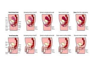
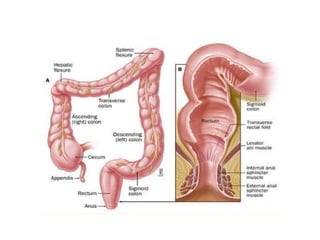
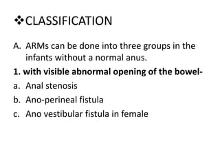
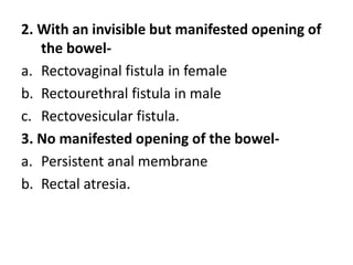
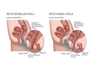
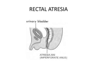
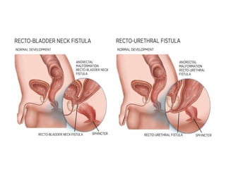
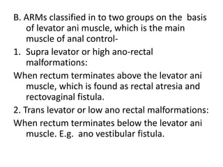
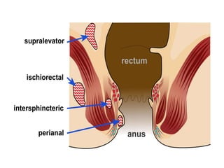
ANORECTAL MALFORMATION
DEFINITION
Anorectal malformations are developmental
deformities of the lower end of the alimentary
tract. i.e. anorectal canal.
CAUSES
• The Exact cause of these malformation is not
known.
• Due to arrest in embryonic development of
the anus, lower rectum and urogenital tract at
the 8th week of embryonic life.
CLASSIFICATION
A. ARMs can be done into three groups in the
infants without a normal anus.
1. with visible abnormal opening of the bowel-
a. Anal stenosis
b. Ano-perineal fistula
c. Ano vestibular fistula in female
2. With an invisible but manifested opening of
the bowel-
a. Rectovaginal fistula in female
b. Rectourethral fistula in male
c. Rectovesicular fistula.
3. No manifested opening of the bowel-
a. Persistent anal membrane
b. Rectal atresia.
ANO PERINEAL FISTULA
RECTAL ATRESIA
B. ARMs classified in to two groups on the basis
of levator ani muscle, which is the main
muscle of anal control-
1. Supra levator or high ano-rectal
malformations:
When rectum terminates above the levator ani
muscle, which is found as rectal atresia and
rectovaginal fistula.
2. Trans levator or low ano rectal malformations:
When rectum terminates below the levator ani
muscle. E.g. ano vestibular fistula.
 CLINICAL MANIFESTATION
• ARMs usually diagnosed after birth immediately or within
the hour by care giver.
• The important presenting feature are abnormally formed
or no anal opening and absence of meconium or presence
of fistula with passage of stool through fistula.
• In female baby the fistula may present between rectum
and vagina or perineum.
• In male baby the fistula is commonly found between
rectum and urinary tract or perineum.
The specific feature for specific anomalies are as
follow:
• Imperforate anal membrane-
Infant fails to pass meconium.
Greenish bulging membrane.
• Anal stenosis-
The baby may pass ribbon like stool with
difficulty as the anal opening is very small.
• Anal agenesis-
It presents with only anal dimple.
Usually fistula found to the perineum or urethra
in male and perineum and vulva in female.
• Rectoperineal fistula-
It is found as small orifice in the perineum,
usually anterior to the center of the external
sphincter
In male baby it is found to the scrotum and in
female to the vulva.
• Rectovaginal fistula-
It present with a communication between
rectum and vagina.
 DIAGNOSIS:
• Physical examination
• USG for diagnose the rectal pouch.
• X ray
• Urine examination for fistula diagnosis
• Micturating cystourethrogram for urinary
tractabnormalities.
 MANAGEMENT:
• The reconstructive surgery
• In low ARMs rectal anoplasty in male.
• In female perineal anoplasty
• In high ARMs, initial colostomy is done in the
neonatal period (PSARP) at age of 10 to 12
months. Or infant is having 7 to 9 kg body
weight.
Colostomy closure is done after 10 to 12 week of
successful definitive surgery.
THANK YOU

Anorectal malformation

  • 1.
  • 2.
    DEFINITION Anorectal malformations aredevelopmental deformities of the lower end of the alimentary tract. i.e. anorectal canal.
  • 3.
    CAUSES • The Exactcause of these malformation is not known. • Due to arrest in embryonic development of the anus, lower rectum and urogenital tract at the 8th week of embryonic life.
  • 6.
    CLASSIFICATION A. ARMs canbe done into three groups in the infants without a normal anus. 1. with visible abnormal opening of the bowel- a. Anal stenosis b. Ano-perineal fistula c. Ano vestibular fistula in female
  • 7.
    2. With aninvisible but manifested opening of the bowel- a. Rectovaginal fistula in female b. Rectourethral fistula in male c. Rectovesicular fistula. 3. No manifested opening of the bowel- a. Persistent anal membrane b. Rectal atresia.
  • 11.
  • 12.
  • 14.
    B. ARMs classifiedin to two groups on the basis of levator ani muscle, which is the main muscle of anal control- 1. Supra levator or high ano-rectal malformations: When rectum terminates above the levator ani muscle, which is found as rectal atresia and rectovaginal fistula. 2. Trans levator or low ano rectal malformations: When rectum terminates below the levator ani muscle. E.g. ano vestibular fistula.
  • 16.
     CLINICAL MANIFESTATION •ARMs usually diagnosed after birth immediately or within the hour by care giver. • The important presenting feature are abnormally formed or no anal opening and absence of meconium or presence of fistula with passage of stool through fistula. • In female baby the fistula may present between rectum and vagina or perineum. • In male baby the fistula is commonly found between rectum and urinary tract or perineum.
  • 17.
    The specific featurefor specific anomalies are as follow: • Imperforate anal membrane- Infant fails to pass meconium. Greenish bulging membrane. • Anal stenosis- The baby may pass ribbon like stool with difficulty as the anal opening is very small.
  • 18.
    • Anal agenesis- Itpresents with only anal dimple. Usually fistula found to the perineum or urethra in male and perineum and vulva in female.
  • 19.
    • Rectoperineal fistula- Itis found as small orifice in the perineum, usually anterior to the center of the external sphincter In male baby it is found to the scrotum and in female to the vulva. • Rectovaginal fistula- It present with a communication between rectum and vagina.
  • 20.
     DIAGNOSIS: • Physicalexamination • USG for diagnose the rectal pouch. • X ray • Urine examination for fistula diagnosis • Micturating cystourethrogram for urinary tractabnormalities.
  • 22.
     MANAGEMENT: • Thereconstructive surgery • In low ARMs rectal anoplasty in male. • In female perineal anoplasty • In high ARMs, initial colostomy is done in the neonatal period (PSARP) at age of 10 to 12 months. Or infant is having 7 to 9 kg body weight.
  • 23.
    Colostomy closure isdone after 10 to 12 week of successful definitive surgery.
  • 24.