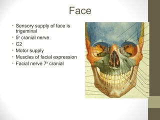
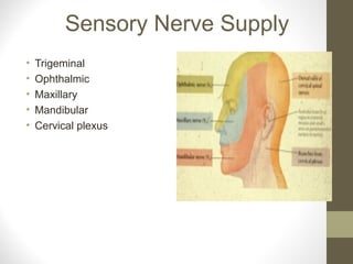
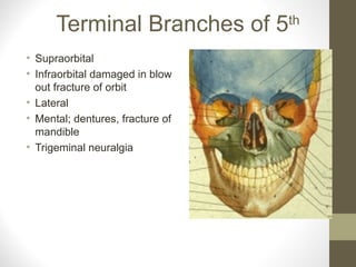
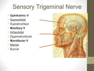
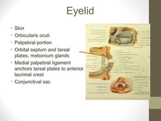
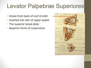
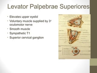
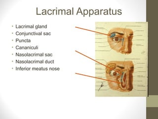
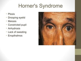
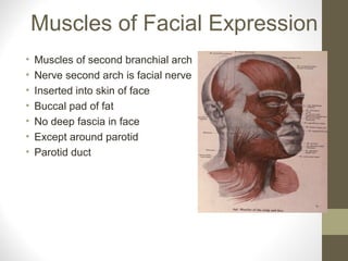
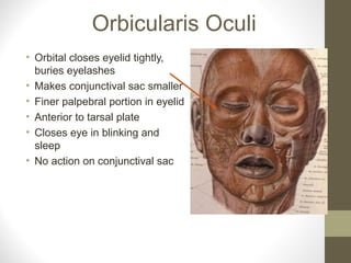
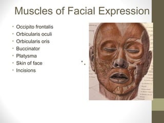
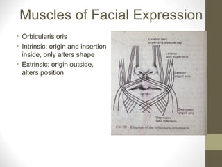
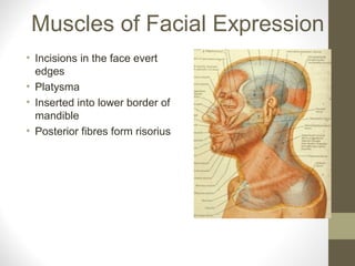
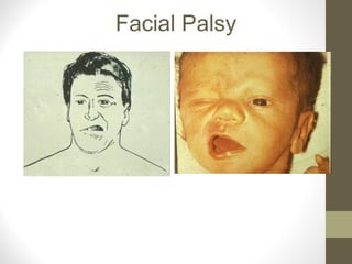
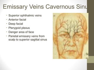
By- Dr. Armaan singh
Anatomy of the Face
(complete Info)
Face
• Sensory supply of face is
trigeminal
• 5th
cranial nerve
• C2
• Motor supply
• Muscles of facial expression
• Facial nerve 7th
cranial
Sensory Nerve Supply
• Trigeminal
• Ophthalmic
• Maxillary
• Mandibular
• Cervical plexus
Terminal Branches of 5th
• Supraorbital
• Infraorbital damaged in blow
out fracture of orbit
• Lateral
• Mental; dentures, fracture of
mandible
• Trigeminal neuralgia
Sensory Trigeminal Nerve
• Ophthalmic V
• Supraorbital
• Supratrochlear
• Maxillary V
• Infraorbital
• Zygomaticofacial
• Mandibular V
• Mental
• Buccal
Fractured Zygoma
• View from above shows
depression
• Lateral X-ray view
should be done
Great Auricular
• C2 from cervical plexus
• Skin over angle of mandible
Scalp
• Skin
• Subcutaneous
• Epicranial aponeurosis
• Three fused together
• Loose areolar tissue
• Periosteum
• Occipito-frontalis
• Attached posteriorly
• No anterior attachment
Scalp
• Occipital portion of occipito-frontals
and epicranial aponeurosis
attached to bone posterior
• Frontal no bony attachment
• Scalp wounds cause black eyes
• Occipital belly
• Posterior auricular of facial nerve
• Frontal belly by temporal of facial
nerve
Blood and Nerve Supply of Scalp
• Supraorbital vessels and nerve
• Scalp as far as vertex
• Posterior scalp
• Greater occipital C2
• Occipital and posterior
auricular vessels
• Zygomatico temporal
• Greater auricular
Eyelid
• Skin
• Orbicularis oculi
• Palpebral portion
• Orbital septum and tarsal
plates, mebonium glands
• Medial palpebral ligament
anchors tarsal plates to anterior
lacrimal crest
• Conjunctival sac
Levator Palpebrae Superiores
• Arises from back of roof of orbit
• Inserted into skin of upper eyelid
• The superior tarsal plate
• Superior fornix of conjunctiva
Levator Palpebrae Superiores
• Elevates upper eyelid
• Voluntary muscle supplied by 3rd
oculomotor nerve
• Smooth muscle
• Sympathetic T1
• Superior cervical ganglion
Lacrimal Apparatus
• Lacrimal gland
• Conjunctival sac
• Puncta
• Cananiculi
• Nasolacrimal sac
• Nasolacrimal duct
• Inferior meatus nose
Horner's Syndrome
• Ptosis
• Drooping eyelid
• Meiosis
• Constricted pupil
• Anhydrosis
• Lack of sweating
• Enopthalmos
Muscles of Facial Expression
• Muscles of second branchial arch
• Nerve second arch is facial nerve
• Inserted into skin of face
• Buccal pad of fat
• No deep fascia in face
• Except around parotid
• Parotid duct
Orbicularis Oculi
• Orbital closes eyelid tightly,
buries eyelashes
• Makes conjunctival sac smaller
• Finer palpebral portion in eyelid
• Anterior to tarsal plate
• Closes eye in blinking and
sleep
• No action on conjunctival sac
Muscles of Facial Expression
• Occipito frontalis
• Orbicularis oculi
• Orbicularis oris
• Buccinator
• Platysma
• Skin of face
• Incisions
Muscles of Facial Expression
• Orbicularis oris
• Intrinsic: origin and insertion
inside, only alters shape
• Extrinsic: origin outside,
alters position
Muscles of Facial Expression
• Incisions in the face evert
edges
• Platysma
• Inserted into lower border of
mandible
• Posterior fibres form risorius
Buccinator
• Buccinator 7th
nerve
• Accessory muscle of mastication
• Outer aspect of maxilla and
mandible
• Related to molar teeth
• Pterygomandibular raphe
• Action keeps food out of
vestibule
• Sucking
• Blowing trumpet
Facial Nerve
Motor nerve to:
• Posterior auricular to occipital belly
• Posterior belly of digastric
Enters parotid and divides into
branches
• Temporal
• Zygomatic
• Buccal
• Mandibular
• Cervical
Facial Nerve
• Stroke
• Upper motor neurone of facial
• Forehead is spared
• Can wrinkle forehead
• Bilaterally innervated
• All muscles of one side
paralysed if it is a lower motor
neurone e.g. Bell’s palsy
Bell’s Palsy
• Lower motor neurone of facial
nerve
• Forehead and all the
muscles on the side of lesion
are affected
Facial Palsy
Corneal Reflex
• Afferent (sensory) cilary branches
nasocilary of ophthalmic of 5th
• Efferent (motor)
• Zygomatic branch of facial 7th
• Last reflex to go
Blood Supply and Pulses in Face
Internal carotid
• Supraorbital artery
• Branch of ophthalmic
External carotid
• Superficial temporal artery
• Facial artery
Venous Drainage of Face
• Supraorbital and anterior facial
• Communicate superior
ophthalmic veins which drain
into cavernous sinus
• Deep facial vein
• Superficial temporal
• Retromandibular
• Common facial
Emissary Veins Cavernous Sinus
• Superior ophthalmic veins
• Anterior facial
• Deep facial
• Pterygoid plexus
• Danger area of face
• Parietal emissary veins from
scalp to superior sagittal sinus
Lymph Glands
• Submental
• Submandibular
• Buccal and mandibular
• Preauricural
• Occipital

Anatomy of the_face

  • 1.
    By- Dr. Armaansingh Anatomy of the Face (complete Info)
  • 2.
    Face • Sensory supplyof face is trigeminal • 5th cranial nerve • C2 • Motor supply • Muscles of facial expression • Facial nerve 7th cranial
  • 3.
    Sensory Nerve Supply •Trigeminal • Ophthalmic • Maxillary • Mandibular • Cervical plexus
  • 4.
    Terminal Branches of5th • Supraorbital • Infraorbital damaged in blow out fracture of orbit • Lateral • Mental; dentures, fracture of mandible • Trigeminal neuralgia
  • 5.
    Sensory Trigeminal Nerve •Ophthalmic V • Supraorbital • Supratrochlear • Maxillary V • Infraorbital • Zygomaticofacial • Mandibular V • Mental • Buccal
  • 6.
    Fractured Zygoma • Viewfrom above shows depression • Lateral X-ray view should be done
  • 7.
    Great Auricular • C2from cervical plexus • Skin over angle of mandible
  • 8.
    Scalp • Skin • Subcutaneous •Epicranial aponeurosis • Three fused together • Loose areolar tissue • Periosteum • Occipito-frontalis • Attached posteriorly • No anterior attachment
  • 9.
    Scalp • Occipital portionof occipito-frontals and epicranial aponeurosis attached to bone posterior • Frontal no bony attachment • Scalp wounds cause black eyes • Occipital belly • Posterior auricular of facial nerve • Frontal belly by temporal of facial nerve
  • 10.
    Blood and NerveSupply of Scalp • Supraorbital vessels and nerve • Scalp as far as vertex • Posterior scalp • Greater occipital C2 • Occipital and posterior auricular vessels • Zygomatico temporal • Greater auricular
  • 11.
    Eyelid • Skin • Orbicularisoculi • Palpebral portion • Orbital septum and tarsal plates, mebonium glands • Medial palpebral ligament anchors tarsal plates to anterior lacrimal crest • Conjunctival sac
  • 12.
    Levator Palpebrae Superiores •Arises from back of roof of orbit • Inserted into skin of upper eyelid • The superior tarsal plate • Superior fornix of conjunctiva
  • 13.
    Levator Palpebrae Superiores •Elevates upper eyelid • Voluntary muscle supplied by 3rd oculomotor nerve • Smooth muscle • Sympathetic T1 • Superior cervical ganglion
  • 14.
    Lacrimal Apparatus • Lacrimalgland • Conjunctival sac • Puncta • Cananiculi • Nasolacrimal sac • Nasolacrimal duct • Inferior meatus nose
  • 15.
    Horner's Syndrome • Ptosis •Drooping eyelid • Meiosis • Constricted pupil • Anhydrosis • Lack of sweating • Enopthalmos
  • 16.
    Muscles of FacialExpression • Muscles of second branchial arch • Nerve second arch is facial nerve • Inserted into skin of face • Buccal pad of fat • No deep fascia in face • Except around parotid • Parotid duct
  • 17.
    Orbicularis Oculi • Orbitalcloses eyelid tightly, buries eyelashes • Makes conjunctival sac smaller • Finer palpebral portion in eyelid • Anterior to tarsal plate • Closes eye in blinking and sleep • No action on conjunctival sac
  • 18.
    Muscles of FacialExpression • Occipito frontalis • Orbicularis oculi • Orbicularis oris • Buccinator • Platysma • Skin of face • Incisions
  • 19.
    Muscles of FacialExpression • Orbicularis oris • Intrinsic: origin and insertion inside, only alters shape • Extrinsic: origin outside, alters position
  • 20.
    Muscles of FacialExpression • Incisions in the face evert edges • Platysma • Inserted into lower border of mandible • Posterior fibres form risorius
  • 21.
    Buccinator • Buccinator 7th nerve •Accessory muscle of mastication • Outer aspect of maxilla and mandible • Related to molar teeth • Pterygomandibular raphe • Action keeps food out of vestibule • Sucking • Blowing trumpet
  • 22.
    Facial Nerve Motor nerveto: • Posterior auricular to occipital belly • Posterior belly of digastric Enters parotid and divides into branches • Temporal • Zygomatic • Buccal • Mandibular • Cervical
  • 23.
    Facial Nerve • Stroke •Upper motor neurone of facial • Forehead is spared • Can wrinkle forehead • Bilaterally innervated • All muscles of one side paralysed if it is a lower motor neurone e.g. Bell’s palsy
  • 24.
    Bell’s Palsy • Lowermotor neurone of facial nerve • Forehead and all the muscles on the side of lesion are affected
  • 25.
  • 26.
    Corneal Reflex • Afferent(sensory) cilary branches nasocilary of ophthalmic of 5th • Efferent (motor) • Zygomatic branch of facial 7th • Last reflex to go
  • 27.
    Blood Supply andPulses in Face Internal carotid • Supraorbital artery • Branch of ophthalmic External carotid • Superficial temporal artery • Facial artery
  • 28.
    Venous Drainage ofFace • Supraorbital and anterior facial • Communicate superior ophthalmic veins which drain into cavernous sinus • Deep facial vein • Superficial temporal • Retromandibular • Common facial
  • 29.
    Emissary Veins CavernousSinus • Superior ophthalmic veins • Anterior facial • Deep facial • Pterygoid plexus • Danger area of face • Parietal emissary veins from scalp to superior sagittal sinus
  • 30.
    Lymph Glands • Submental •Submandibular • Buccal and mandibular • Preauricural • Occipital