Downloaded 197 times


This document provides an overview of the anatomy of the scalp, face, and temporal region. It describes the layers of the scalp including the skin, connective tissue, aponeurosis, and pericranium. It details the muscles of the scalp including the occipitofrontalis. It discusses the sensory and vascular supply as well as lymph drainage of the scalp. It then describes the bones, muscles, nerves, vessels, and lymph drainage of the face. Finally, it briefly mentions that the temporal region is located on the side of the head and contains the temporal lobe of the brain.
Introduction to the anatomy of the skull, face, and related fossae, presented by Dr. Noor B. Najjar.
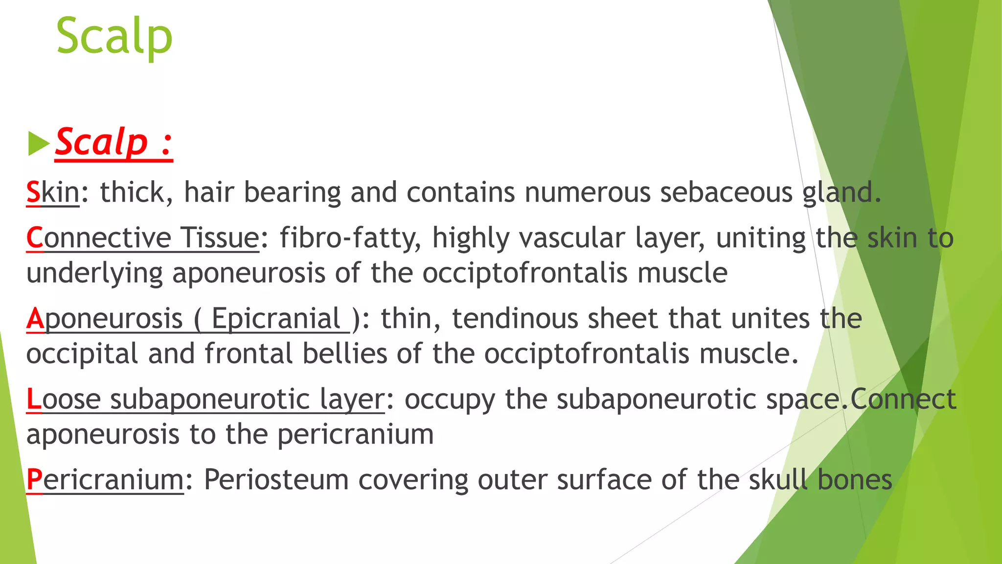
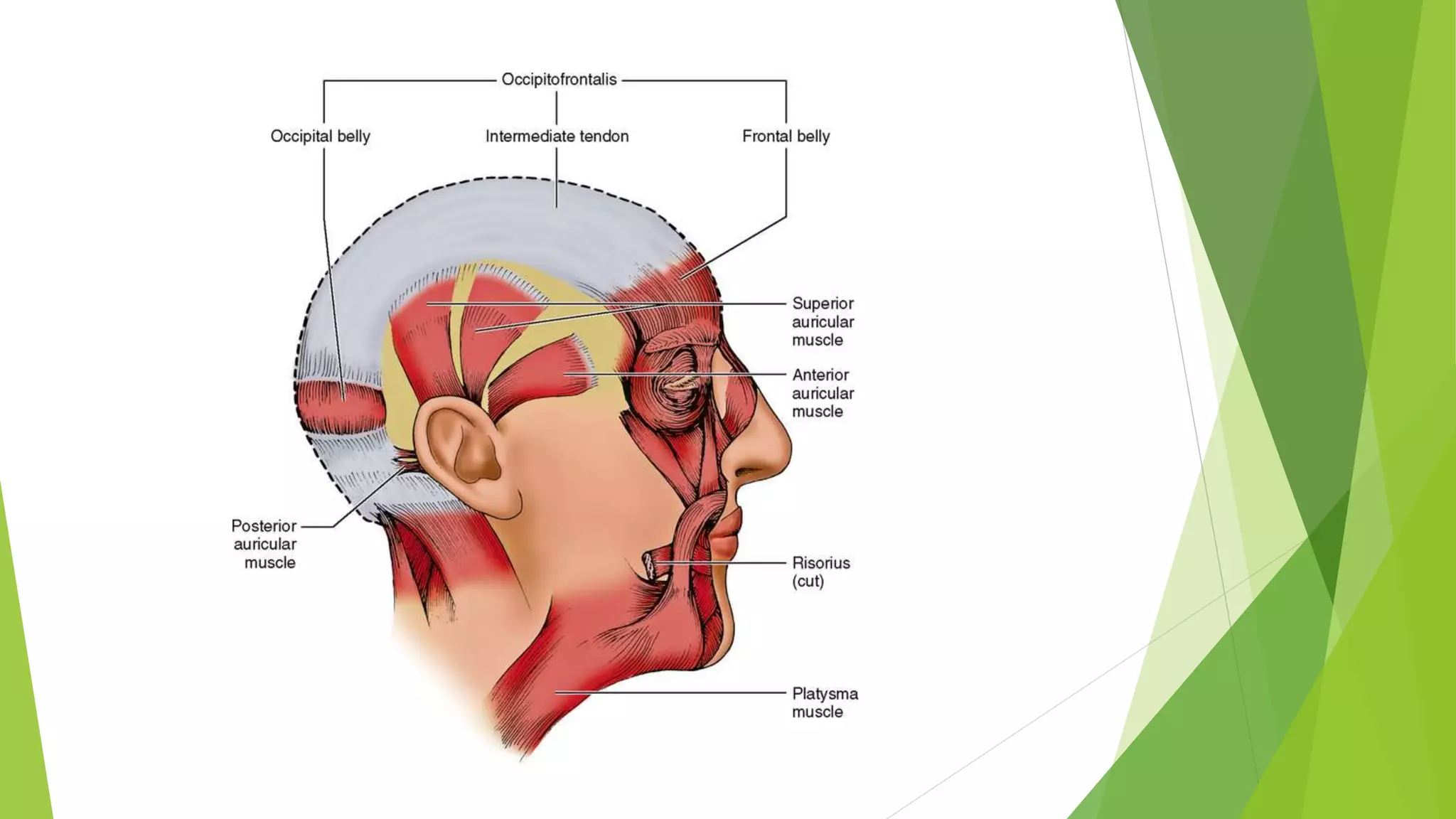
Description of scalp anatomy, including skin, connective tissue, pericranium, and layers associated with the occiptofrontalis muscle.
Details on occiptofrontalis muscle, its origin, insertion, action, and nerve supply through facial nerve branches.
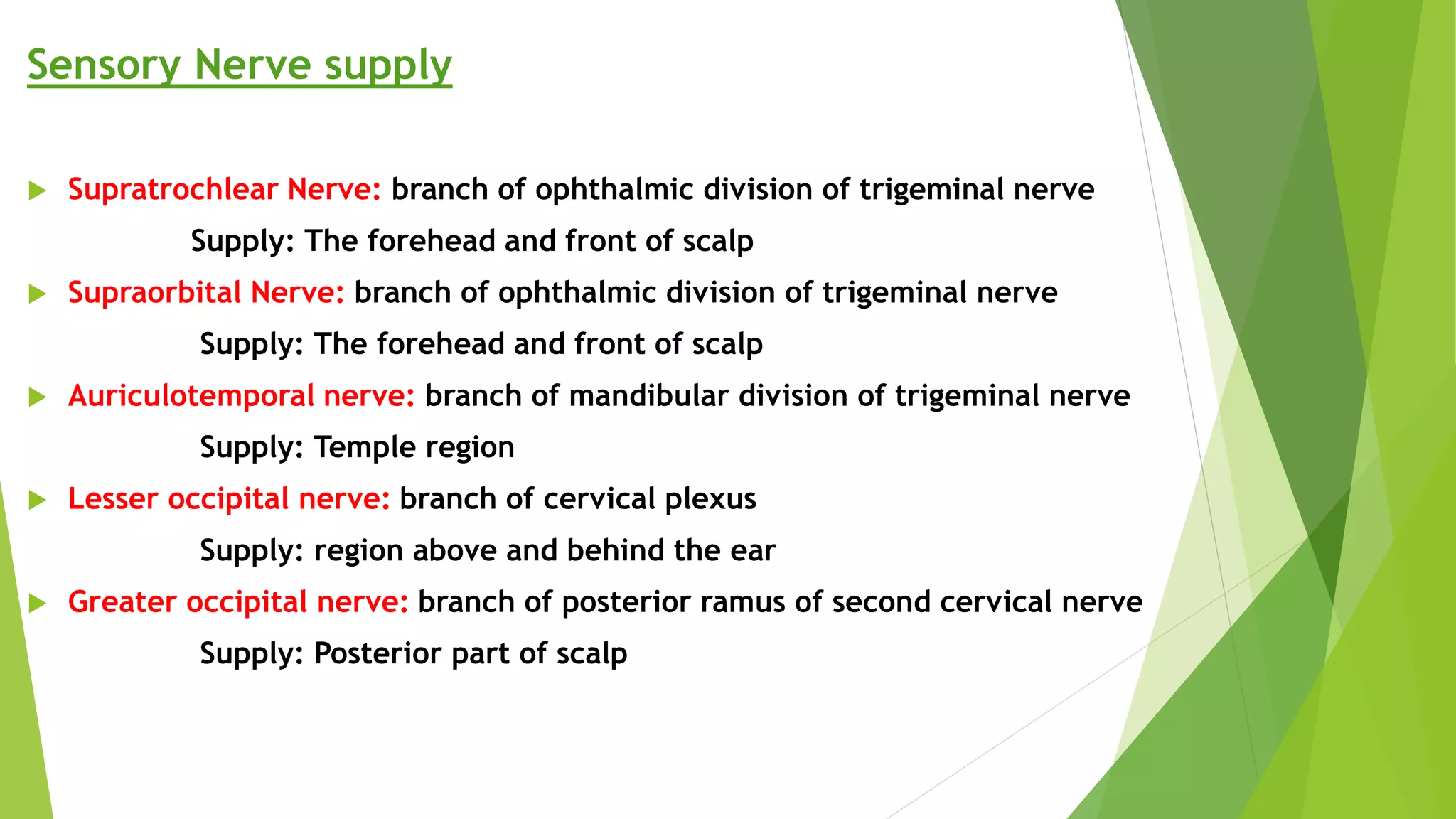
Sensory innervation of the scalp including nerves like supratrochlear, supraorbital, auriculotemporal.
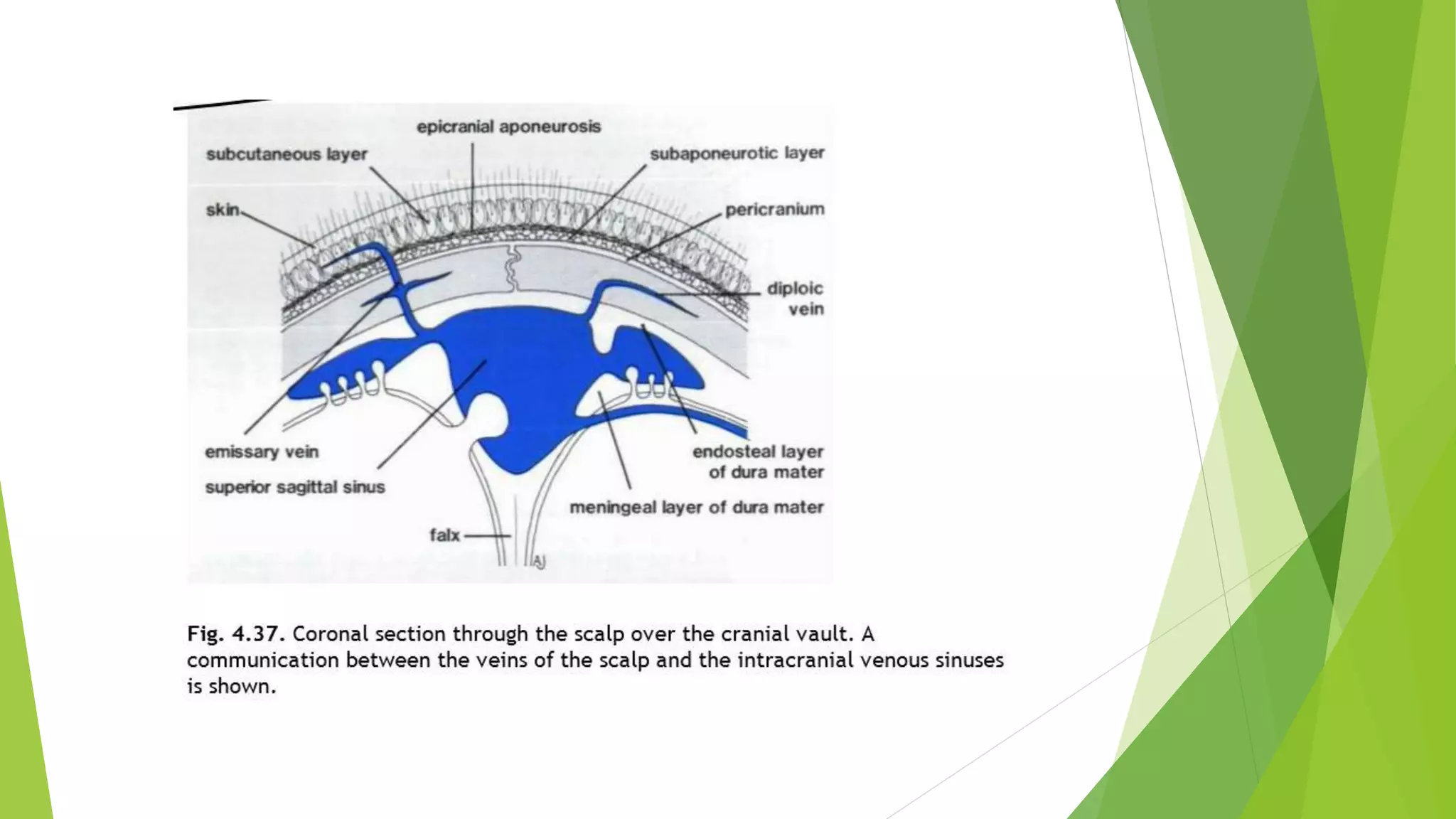
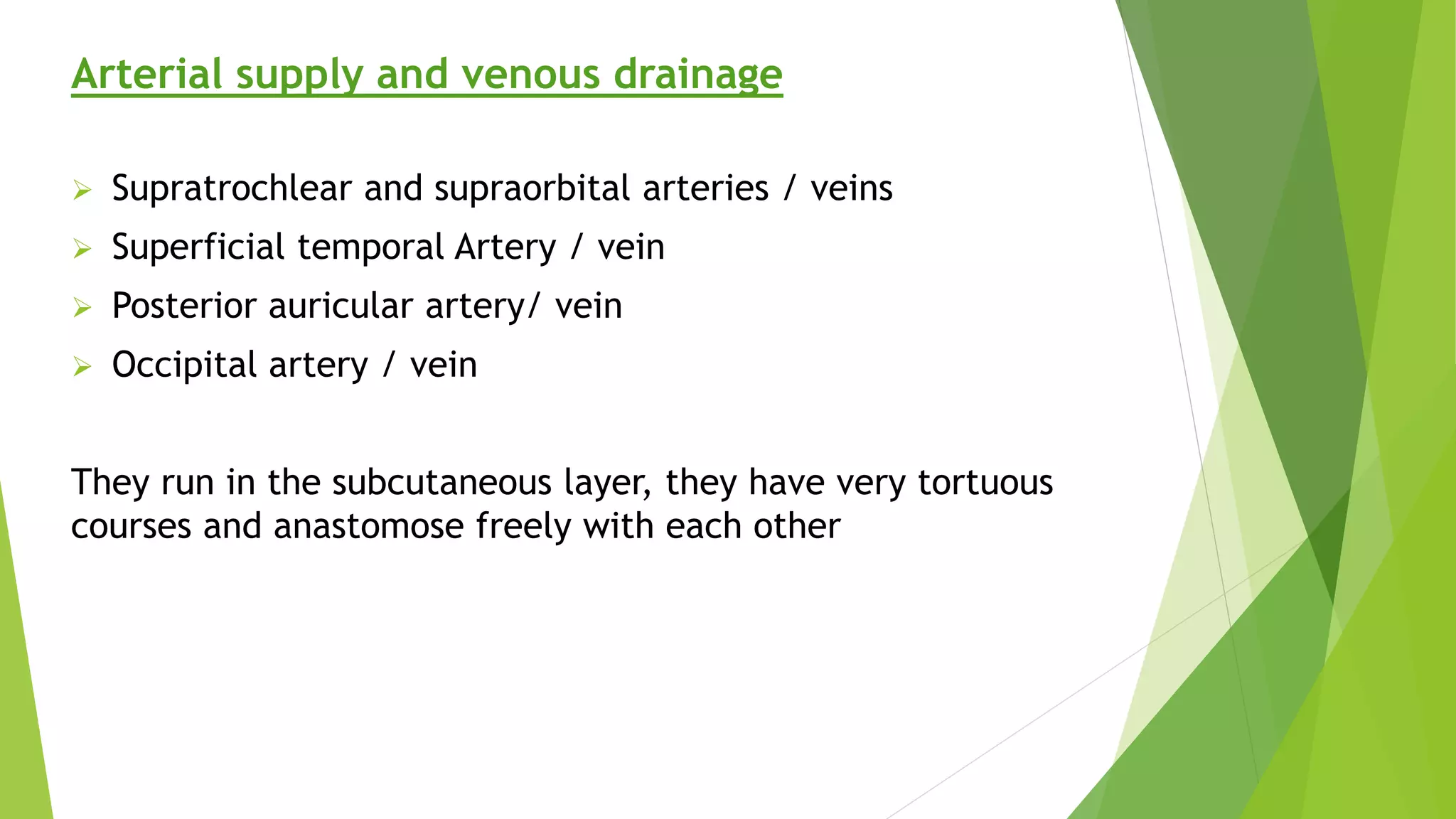
Arterial supply and venous drainage of scalping features major arteries/veins and their courses.
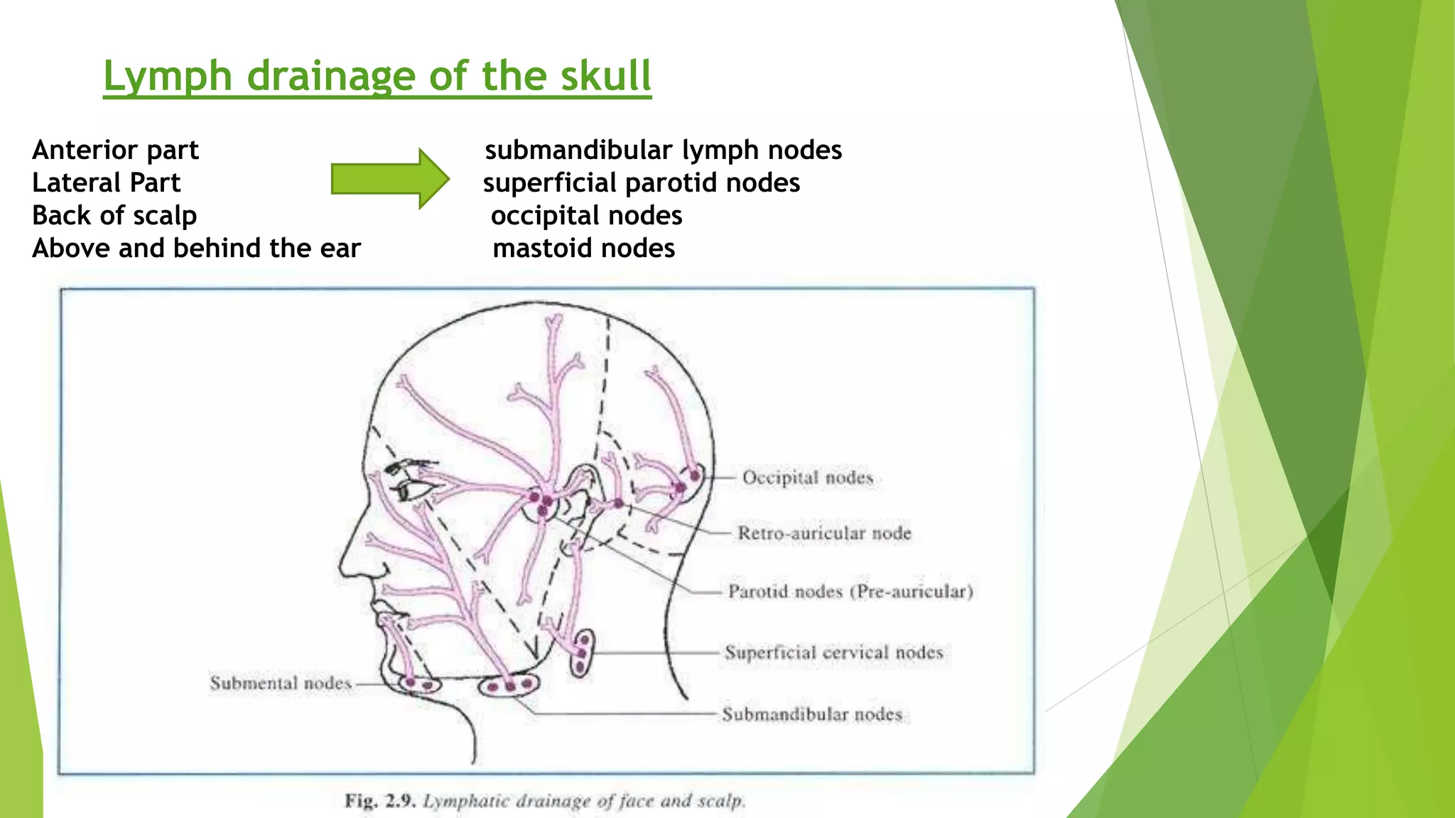
Details on lymphatic drainage regions for the skull and scalp into various lymph nodes.
Facial sensory innervation by trigeminal nerve branches with specific areas covered by each.
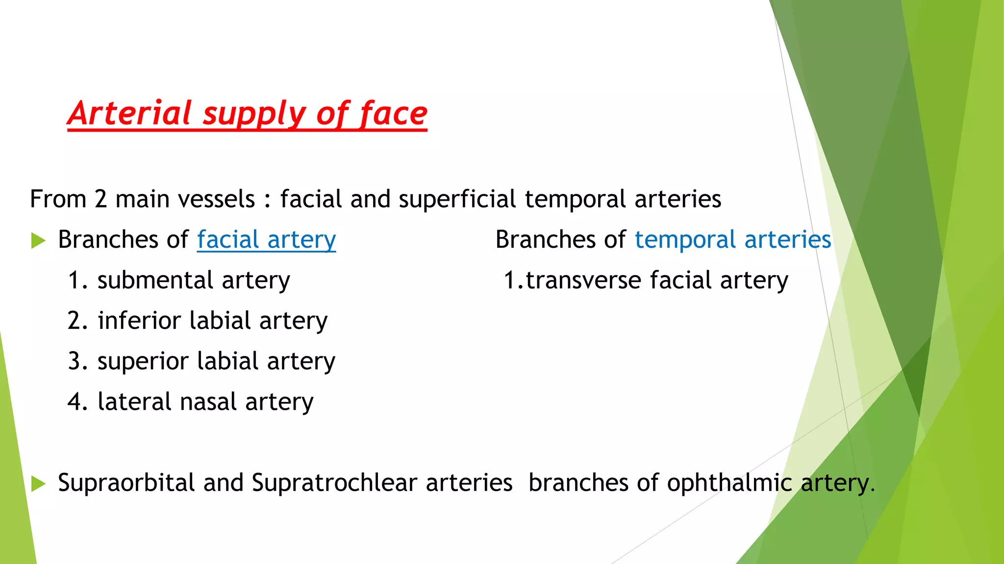
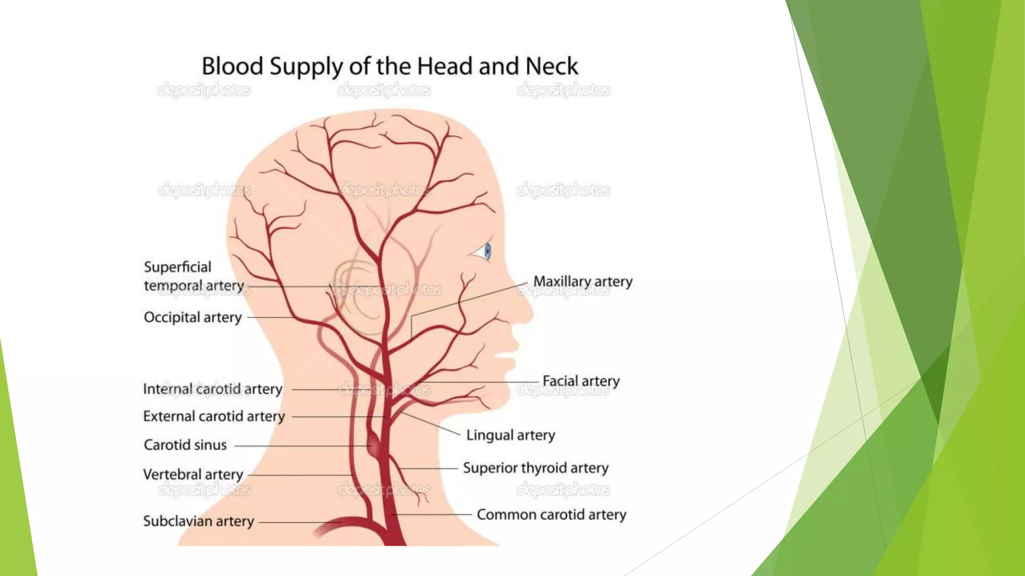
Arterial supply to the face from facial and superficial temporal arteries along with specific branches.
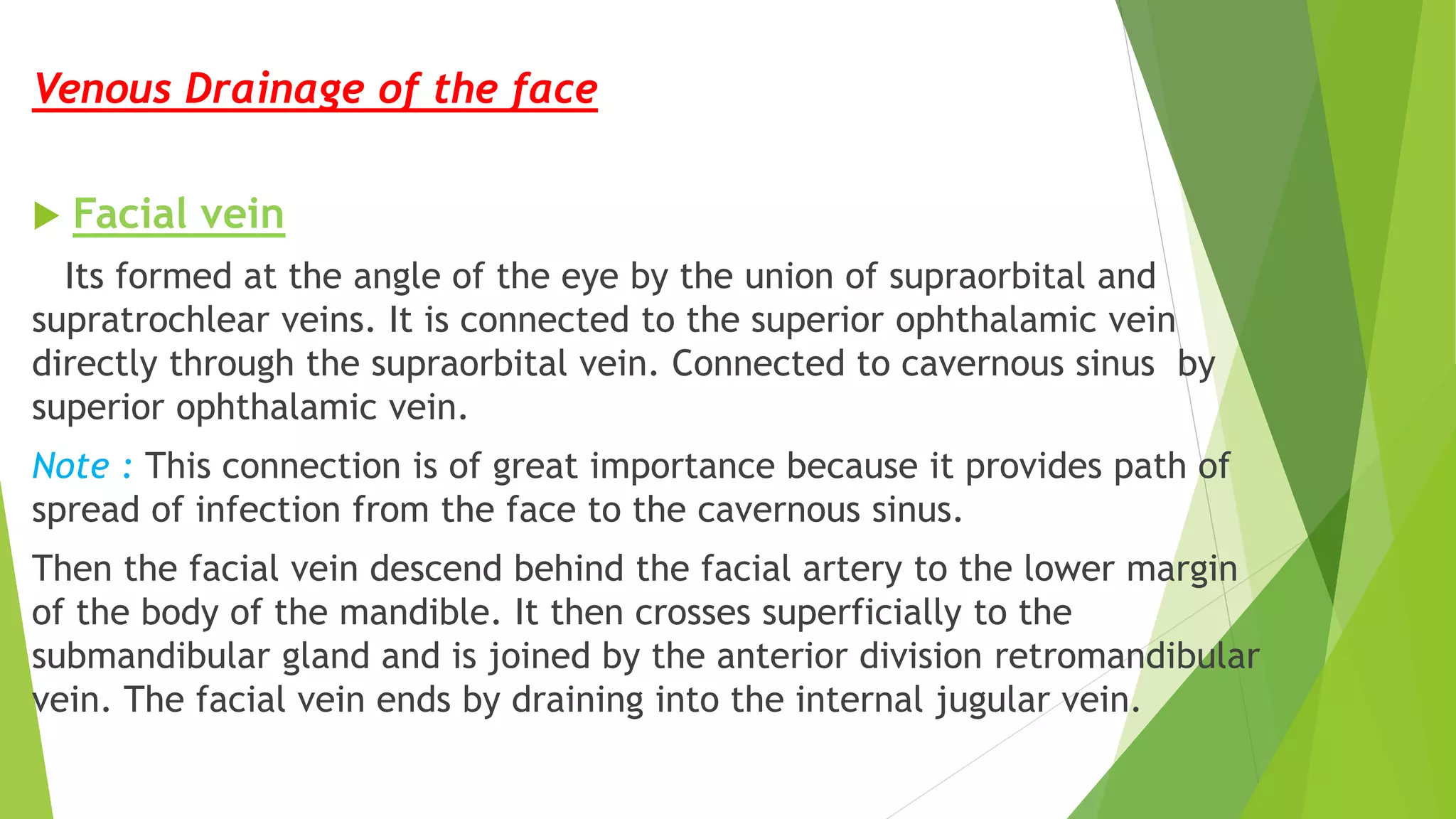
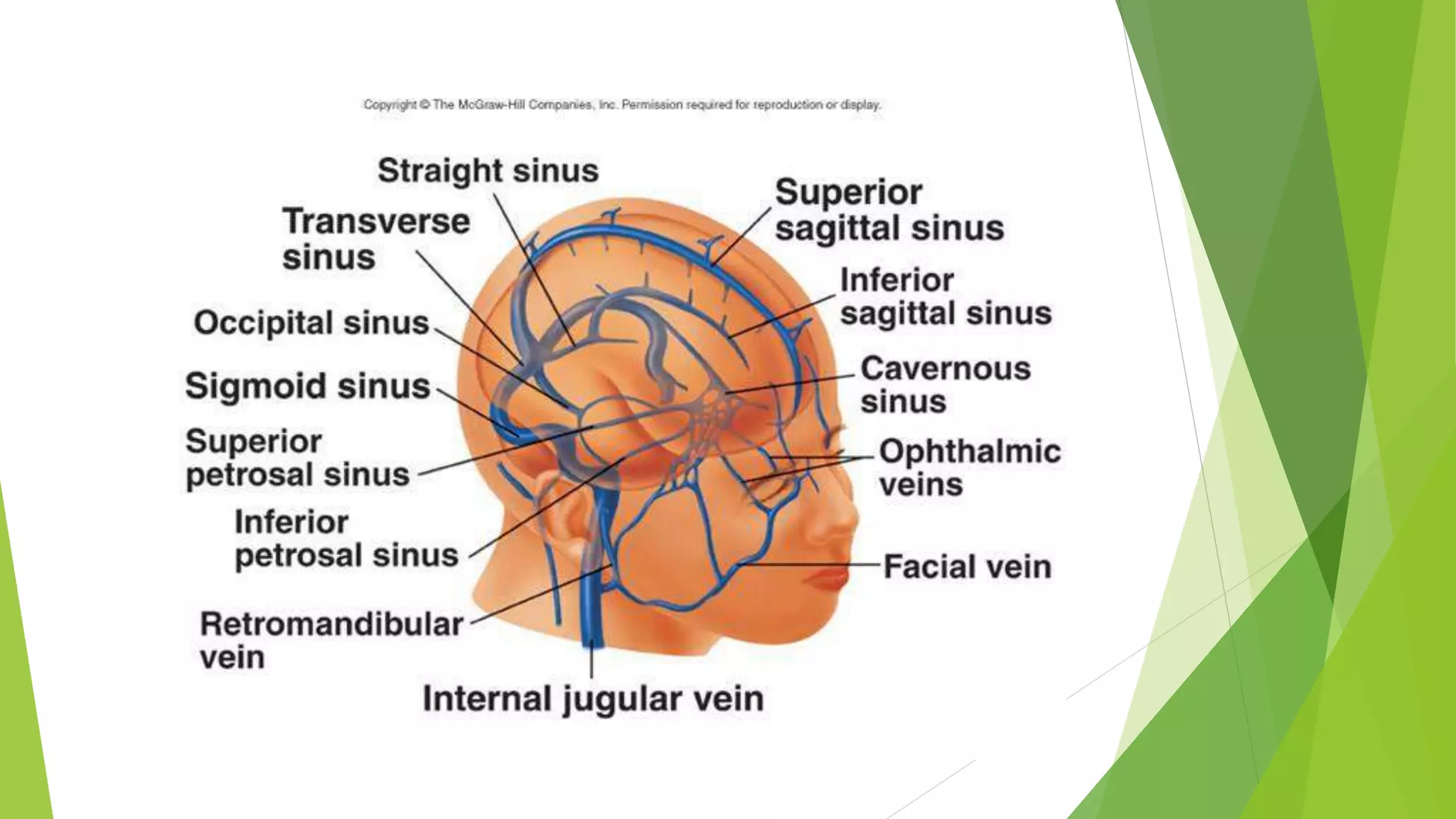
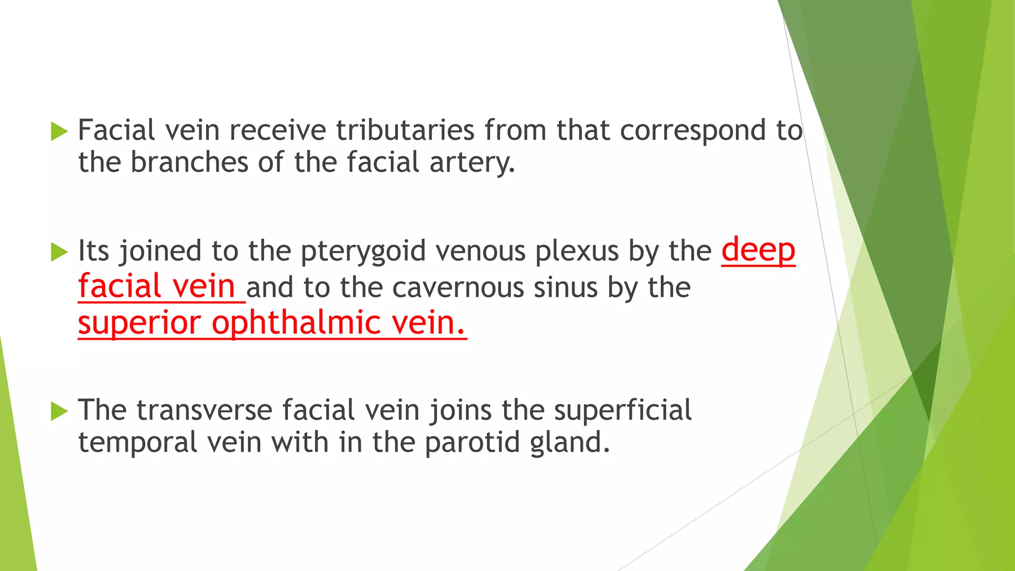
Facial vein structure, formation, tributaries, and its connections enabling potential infection paths.
Detailing tributaries to the facial vein including connections to the pterygoid venous plexus.
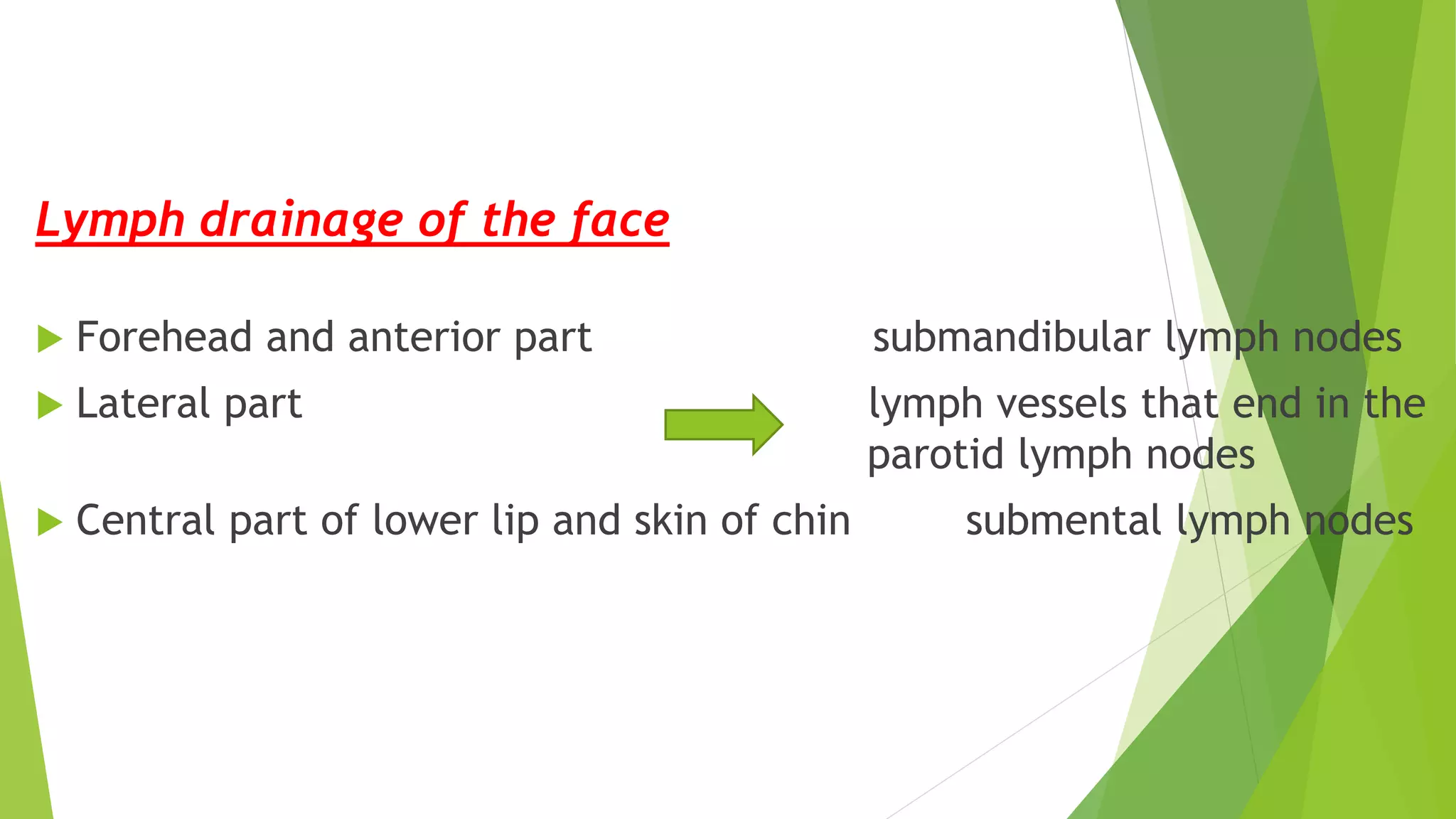
Describing lymphatic drainage pathways for forehead, lateral face, and lower lip skin.
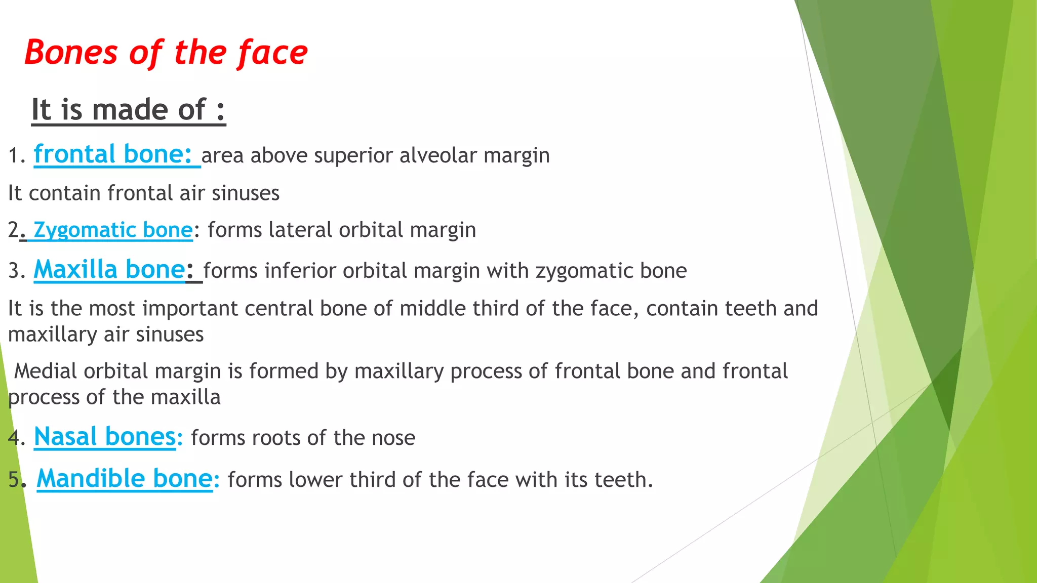
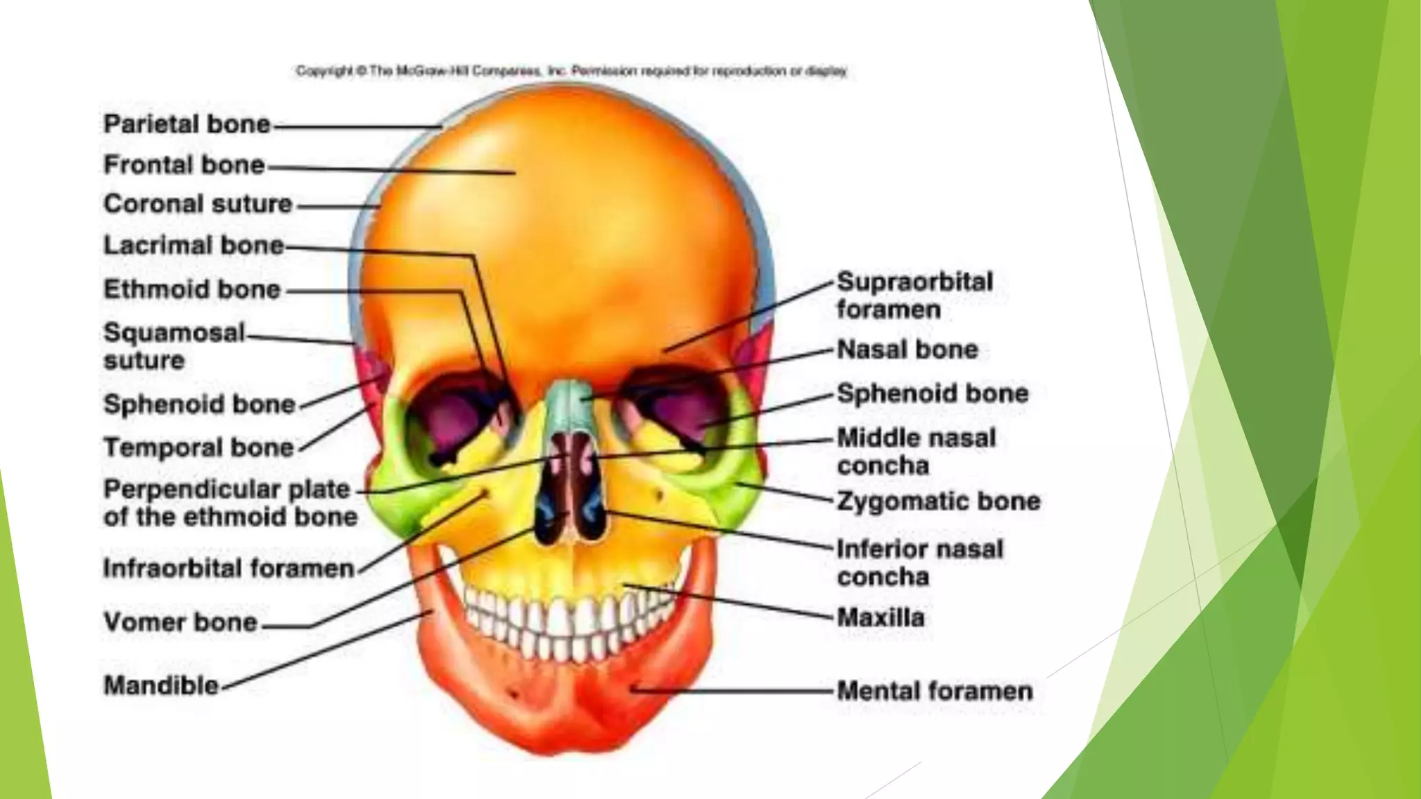
Overview of facial bones including frontal, zygomatic, maxilla, nasal bones, and mandible.
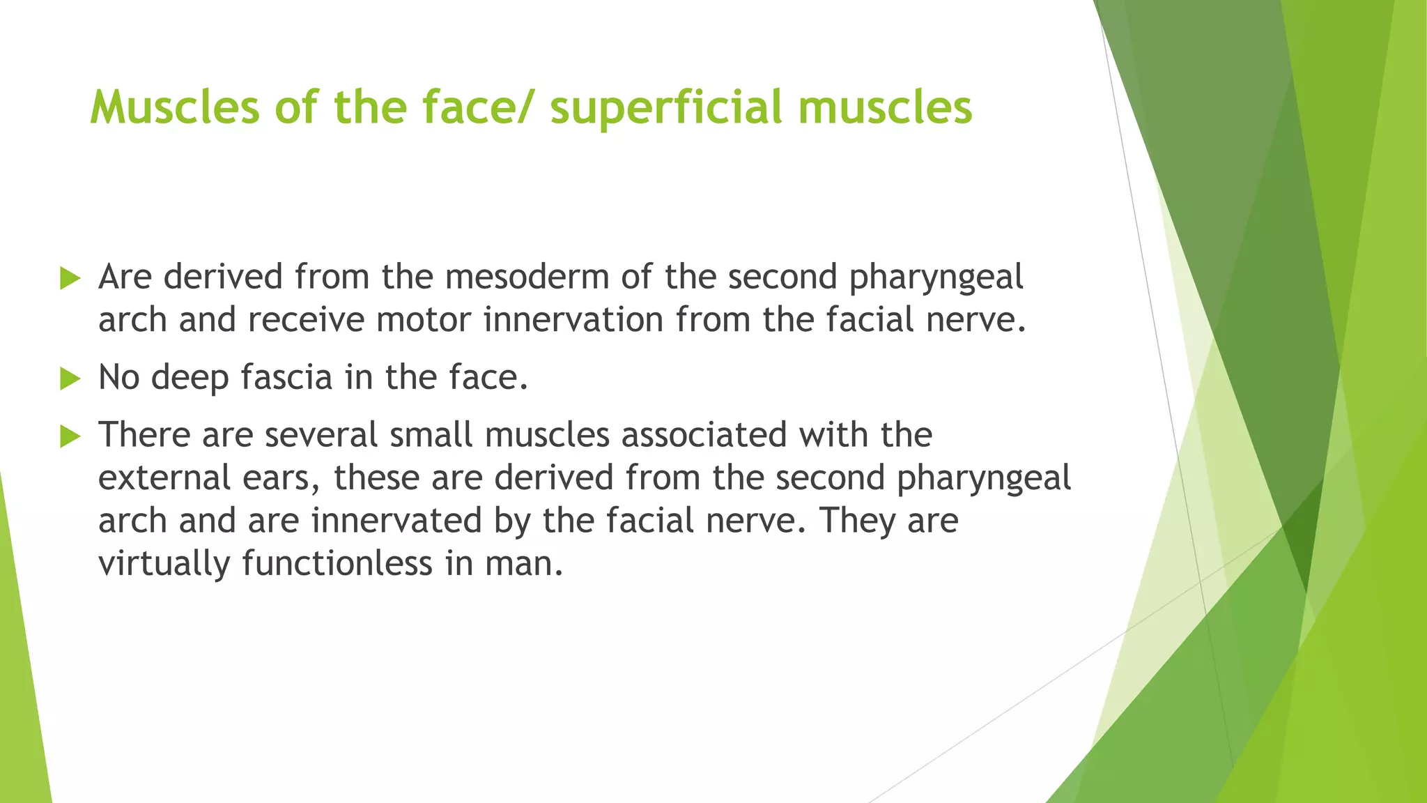
Description of superficial muscles of the face derived from the second pharyngeal arch.
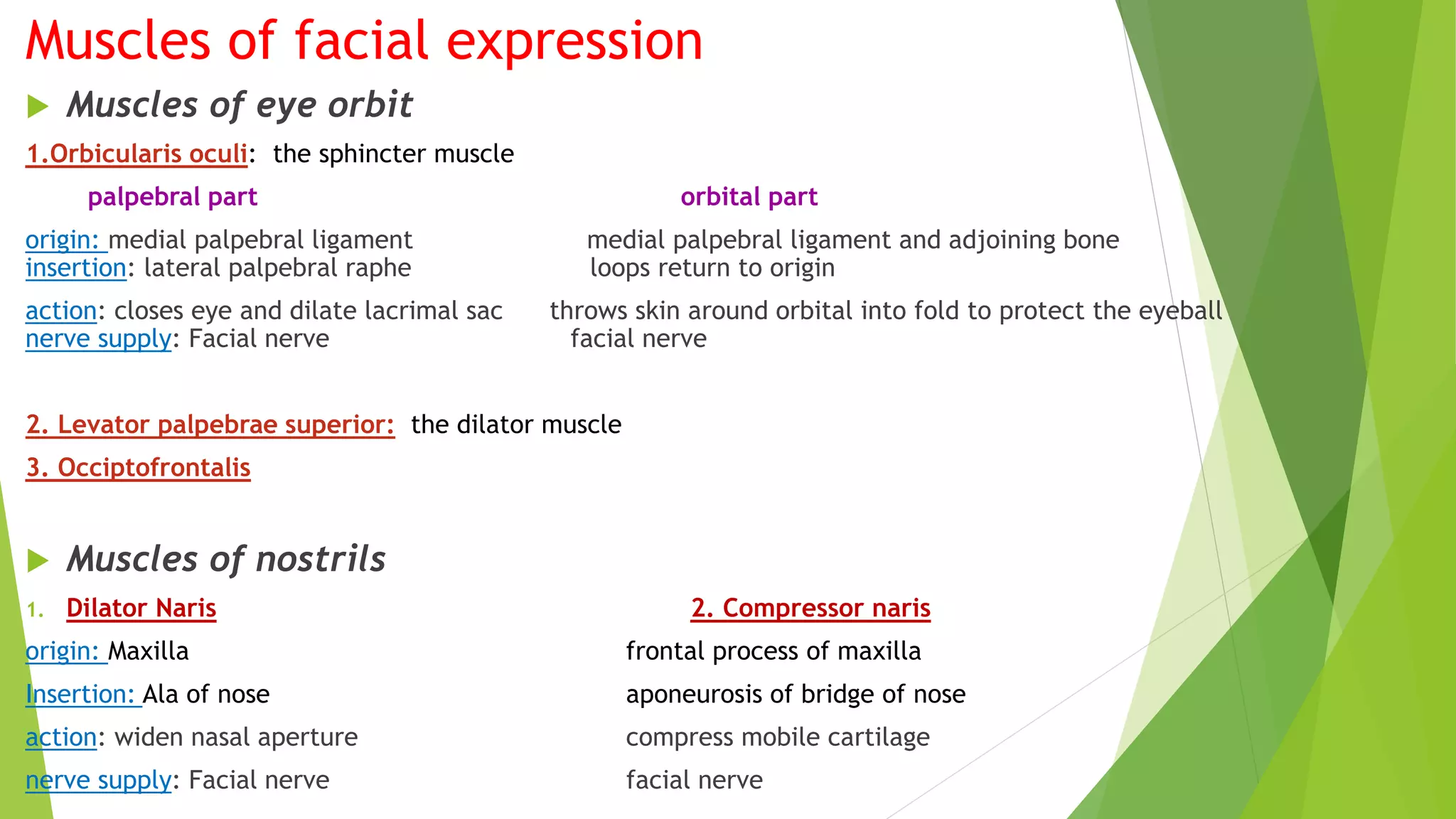
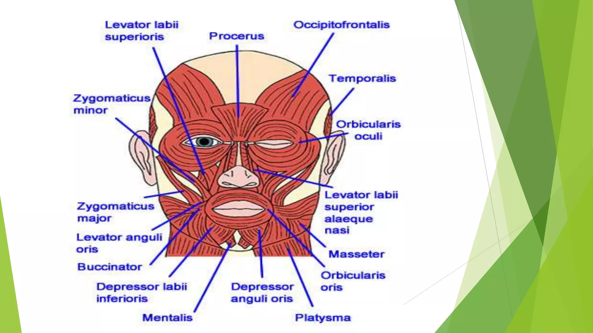
Muscles acting on facial expressions including orbicularis oculi, their origins, insertions, and innervations.
Details on the orbicularis oris and other dilator muscles of the lips, their functions, and nerve supplies.
Description of buccinator muscle origins and function in compressing cheeks during blowing.
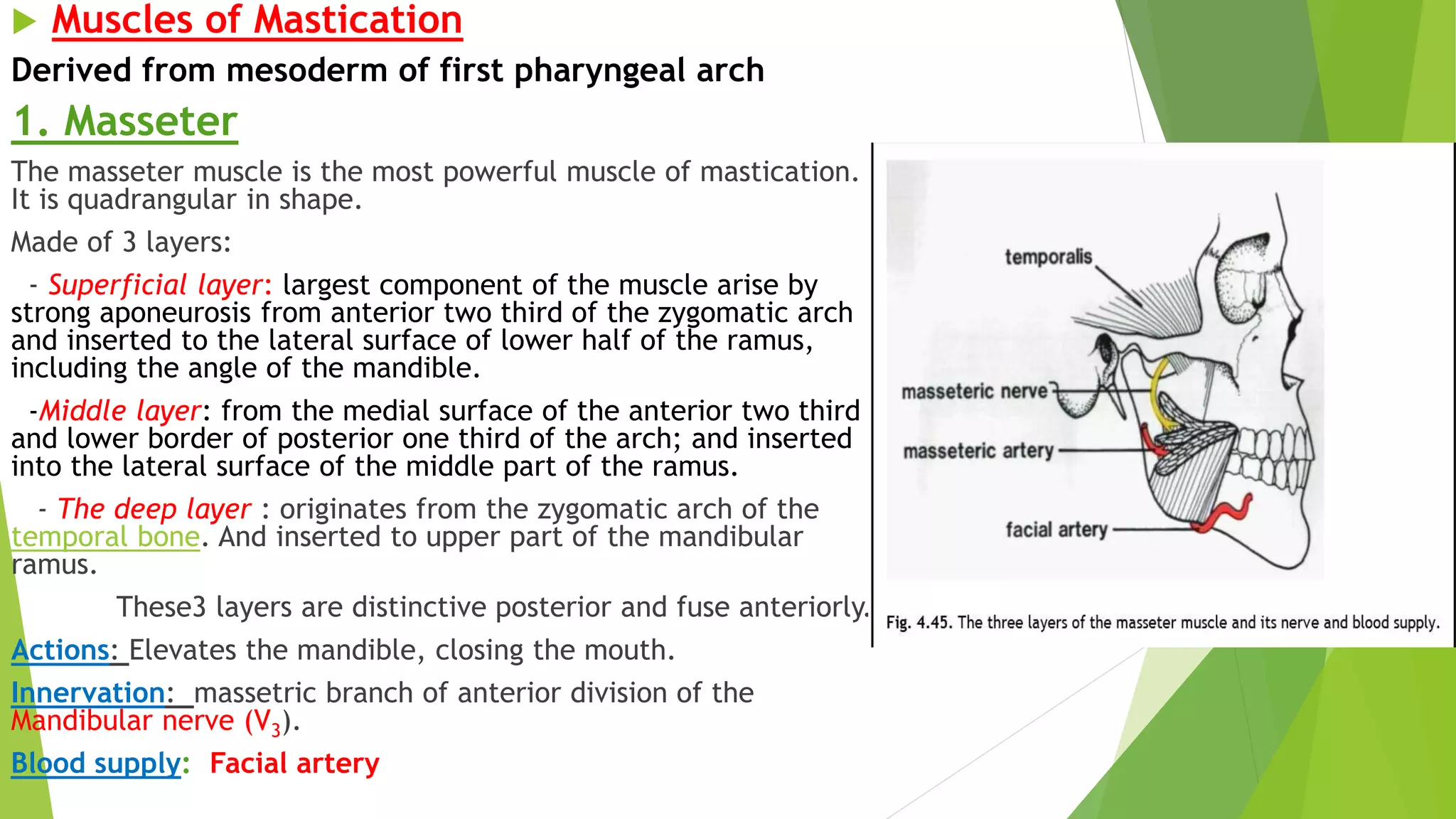
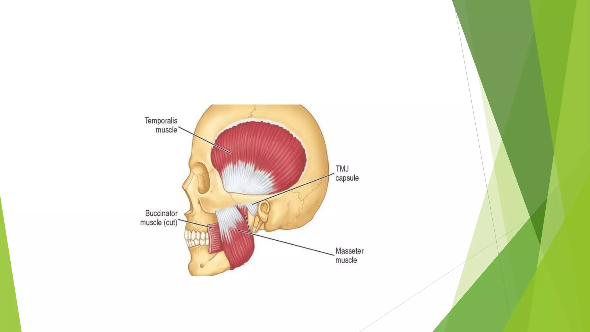
Masseter muscle overview including its layers, function, and innervation required for chewing.
Origin, insertion, actions, and innervation of the temporalis muscle in mastication.
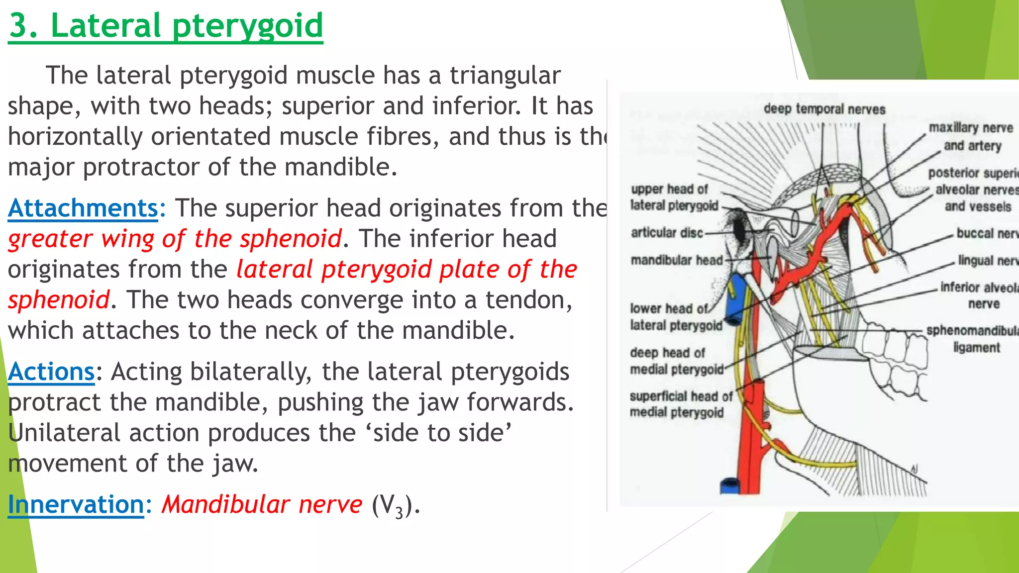
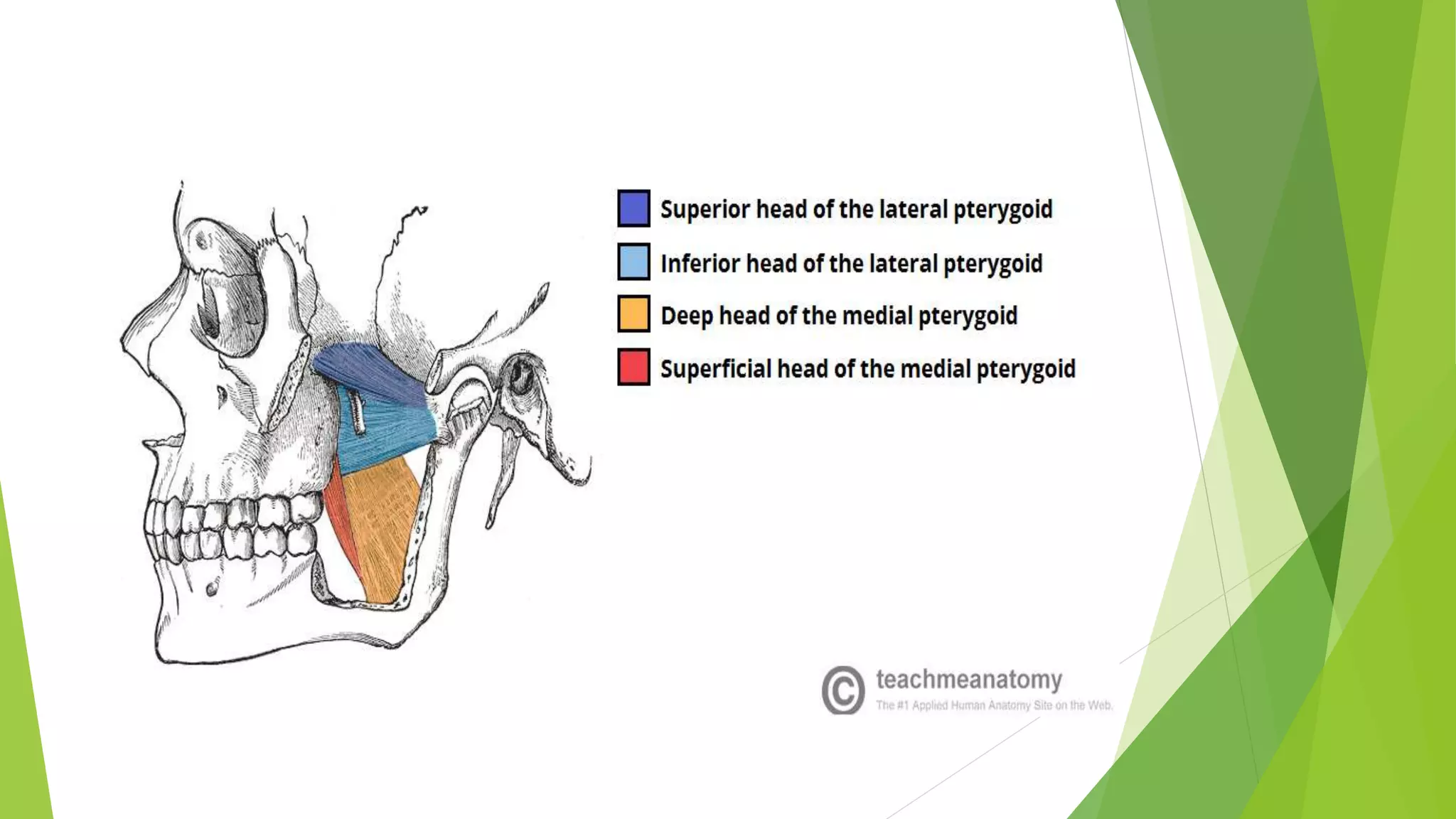
Function and anatomy of the lateral pterygoid muscle, including its action in jaw movement.
Characteristics and functions of the medial pterygoid muscle related to chewing.
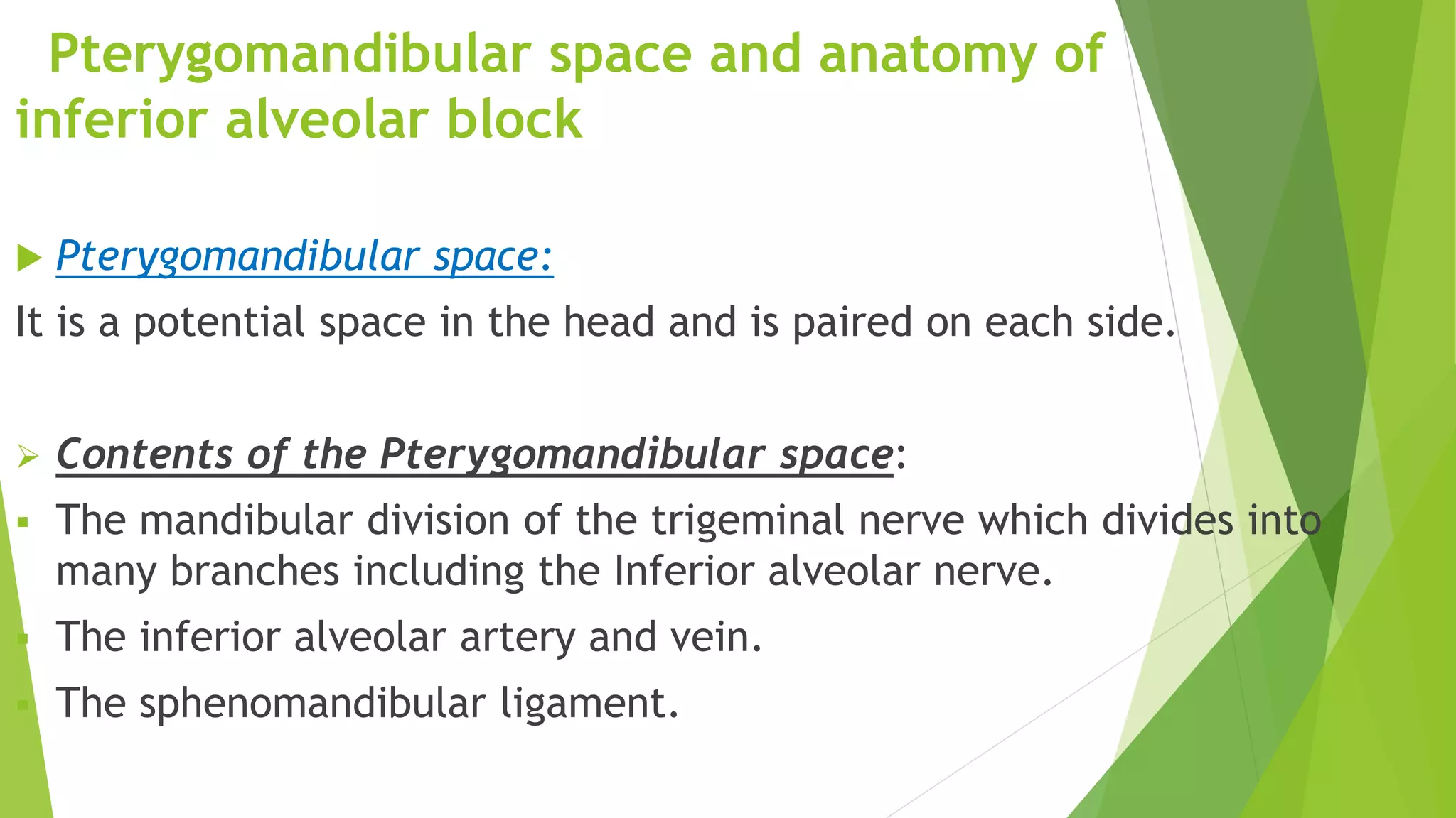
Description of the pterygomandibular space including its contents and their relevance.
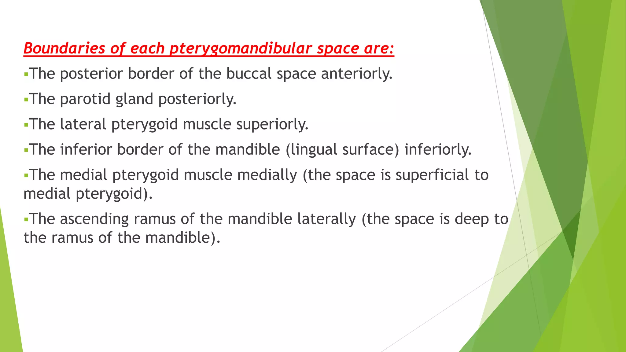
Anatomical boundaries of the pterygomandibular space providing contextual anatomical relevance.
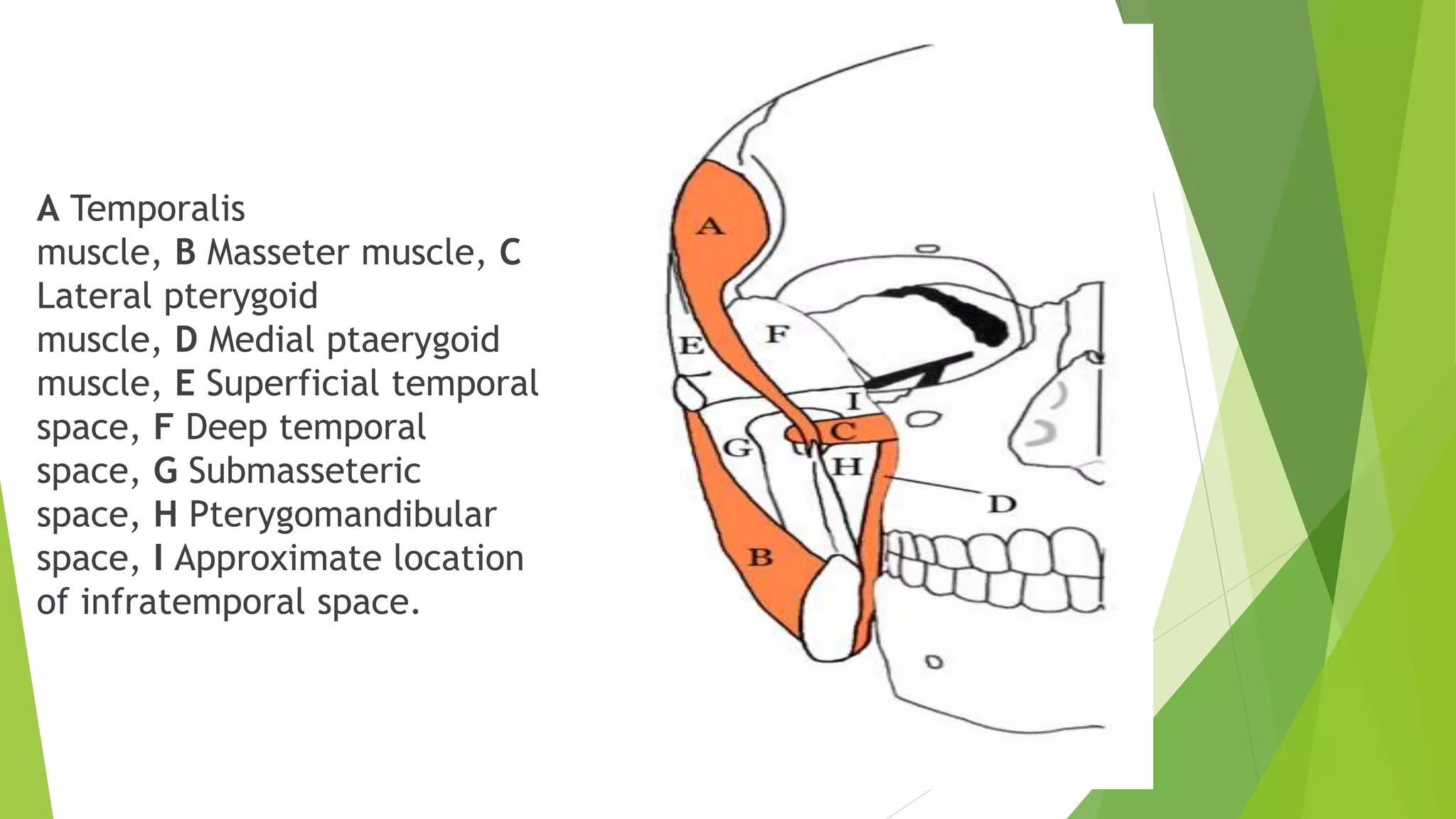
Illustration detailing multiple muscles in association with pterygomandibular space.
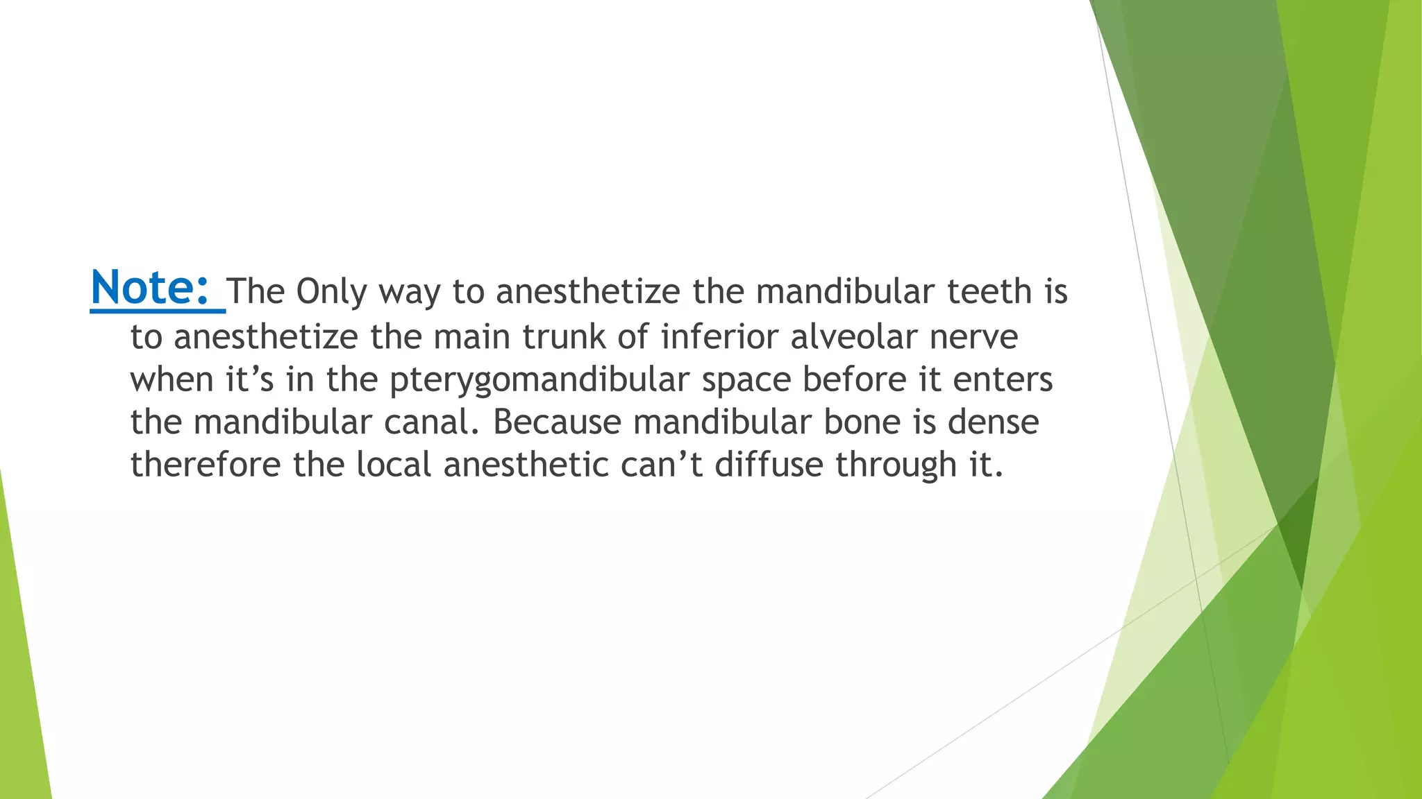
Explains the technique for administering anesthesia to mandibular teeth through pterygomandibular space.
Describes the ligament and muscles associated with the styloid process and hyoid bone.
Overview of muscles originating from the styloid process, including their origins and actions.
Anatomy of the sphenomandibular ligament and its clinical relevance relating to jaw movement.
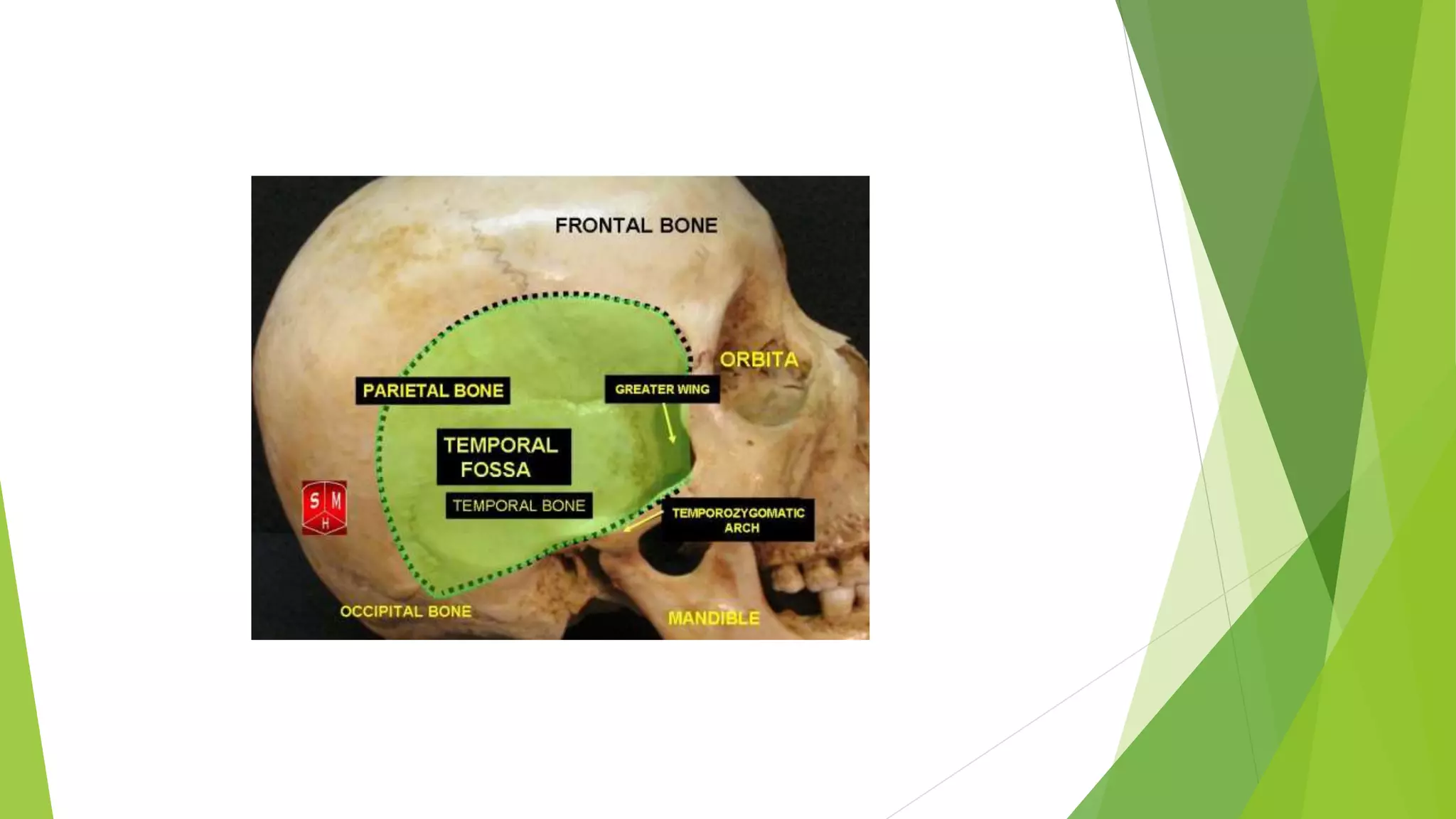
Description of the temporal region's anatomy, related structures, and the temporalis muscle.
Detailed examination of the parotid gland's location, structure, and duct pathways.
Review of non-glandular structures traversing the parotid gland and their functions.
Explains the parasympathetic and sympathetic innervation to parotid gland affecting saliva secretion.
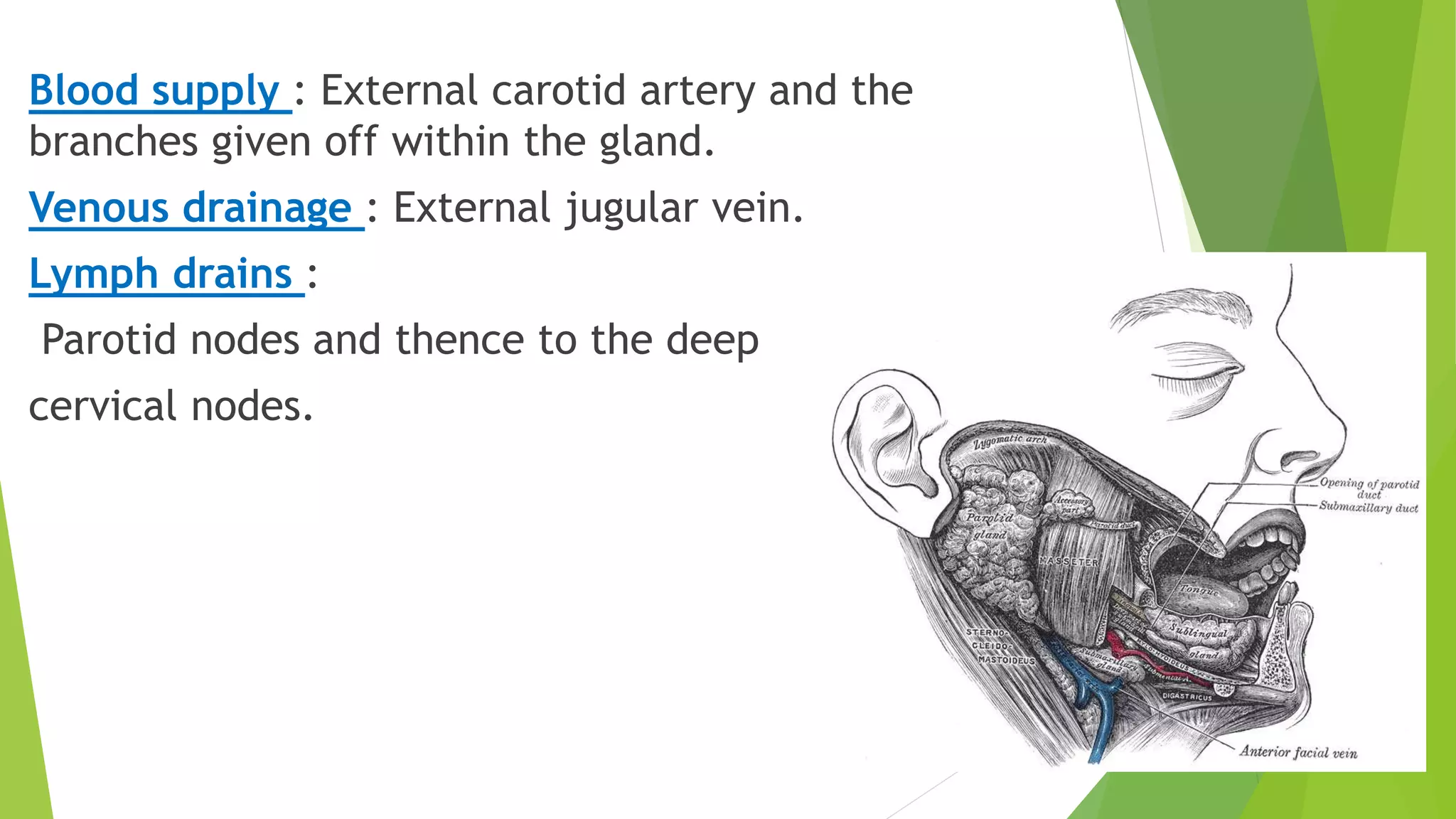
Arterial and venous blood supply along with lymph drainage patterns related to the gland.
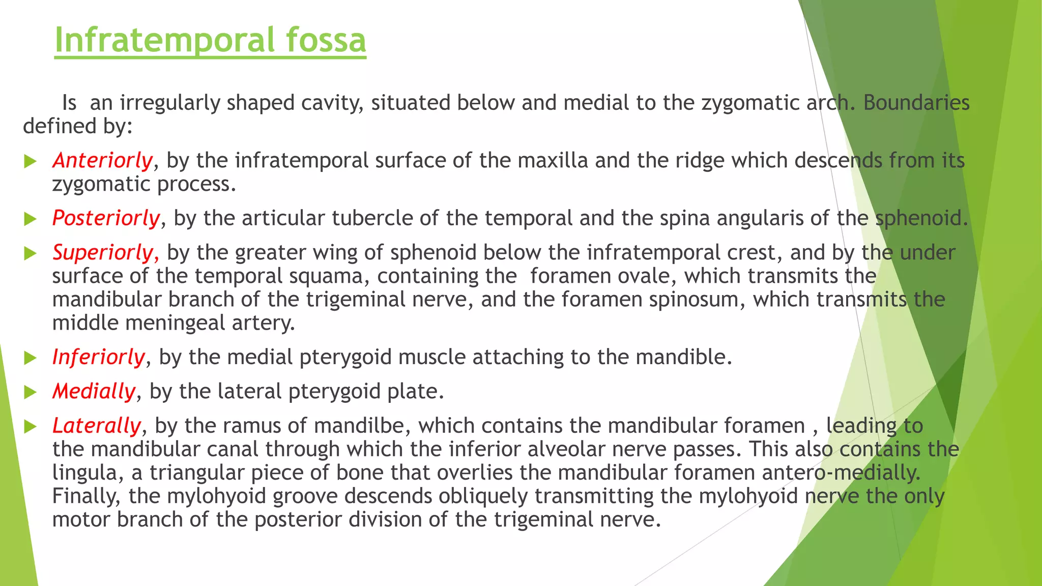
Defining boundaries, contents, and structures within the infratemporal fossa.
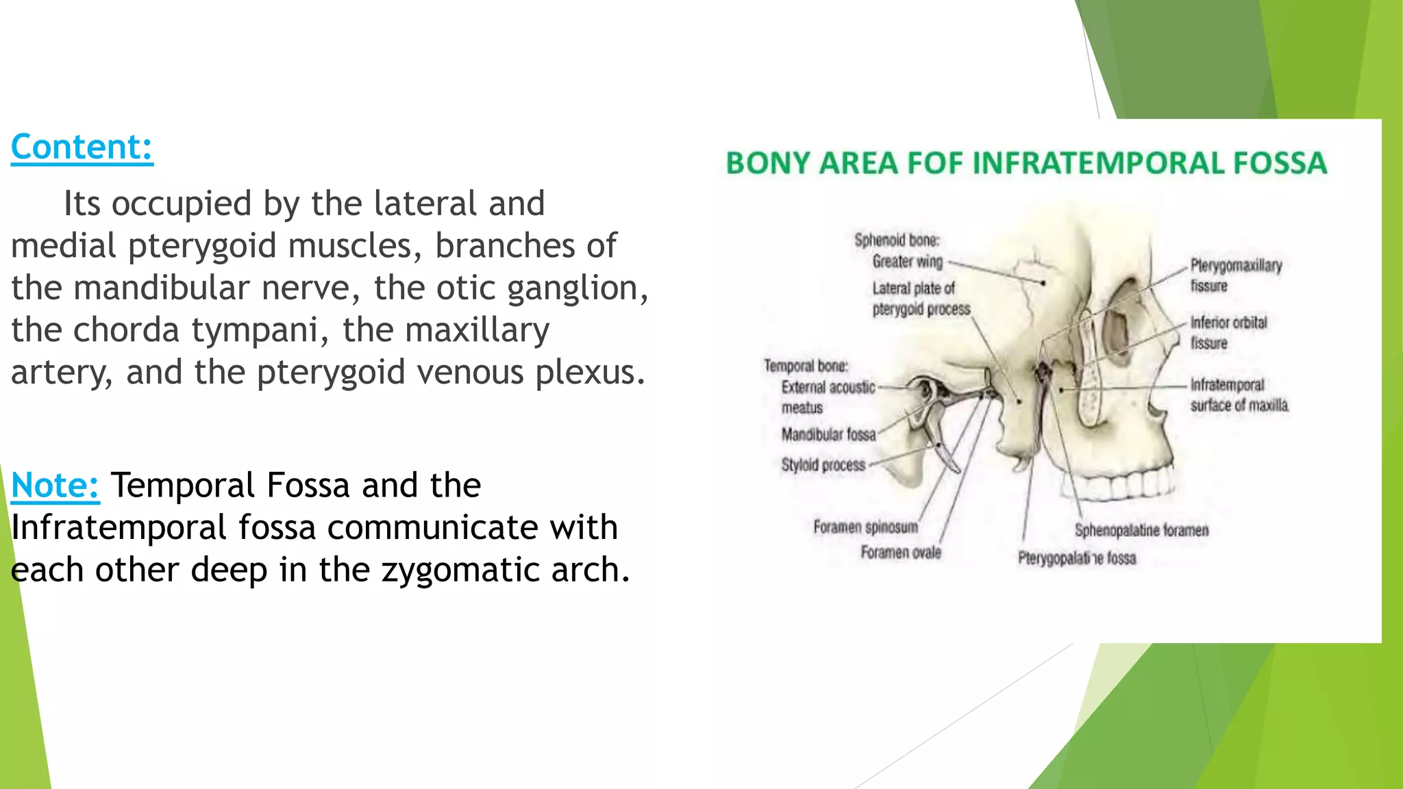
Displays contents within the infratemporal fossa such as muscles and nerve branches.
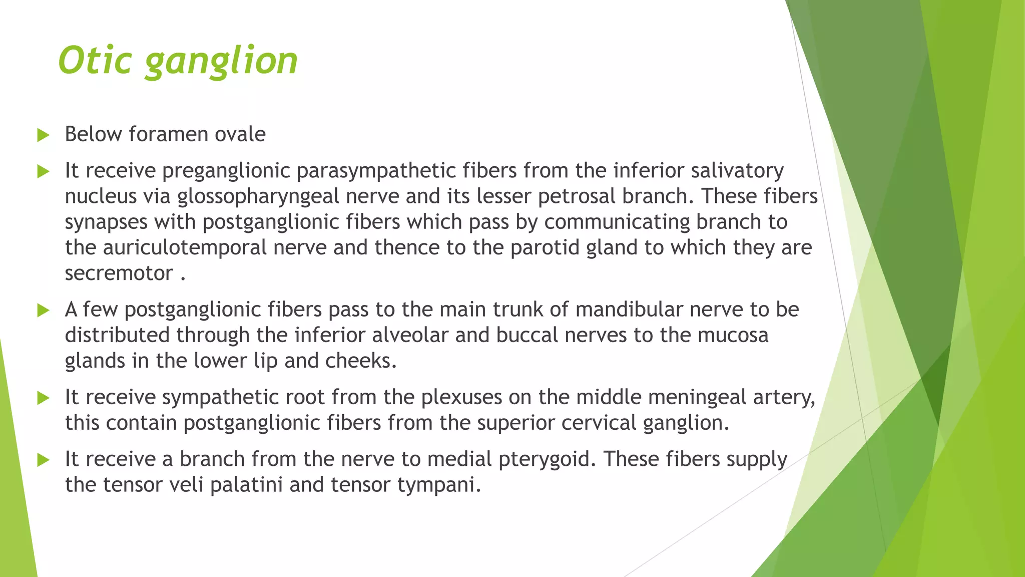
Discusses the otic ganglion's role in parasympathetic innervation pathways.
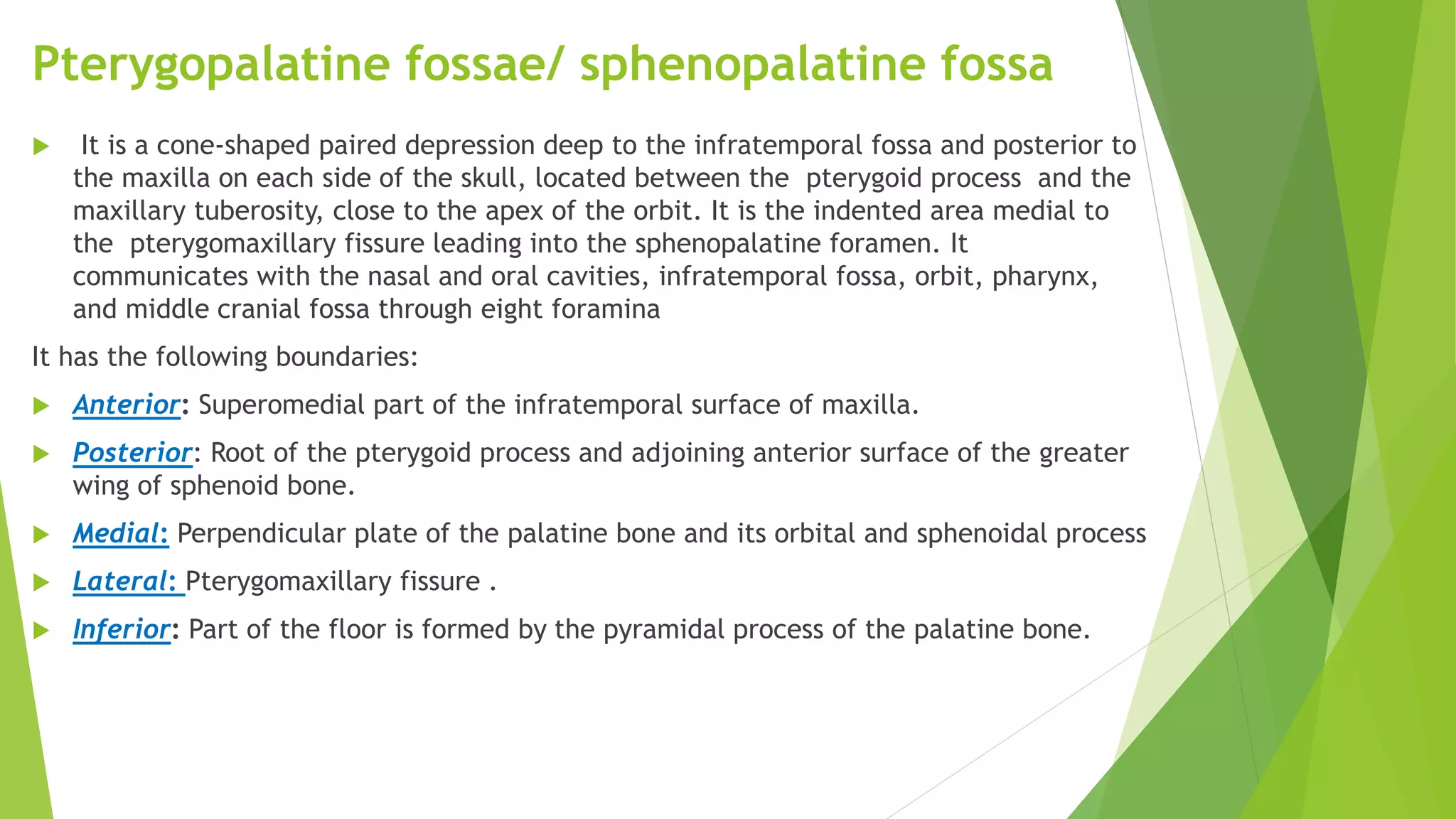
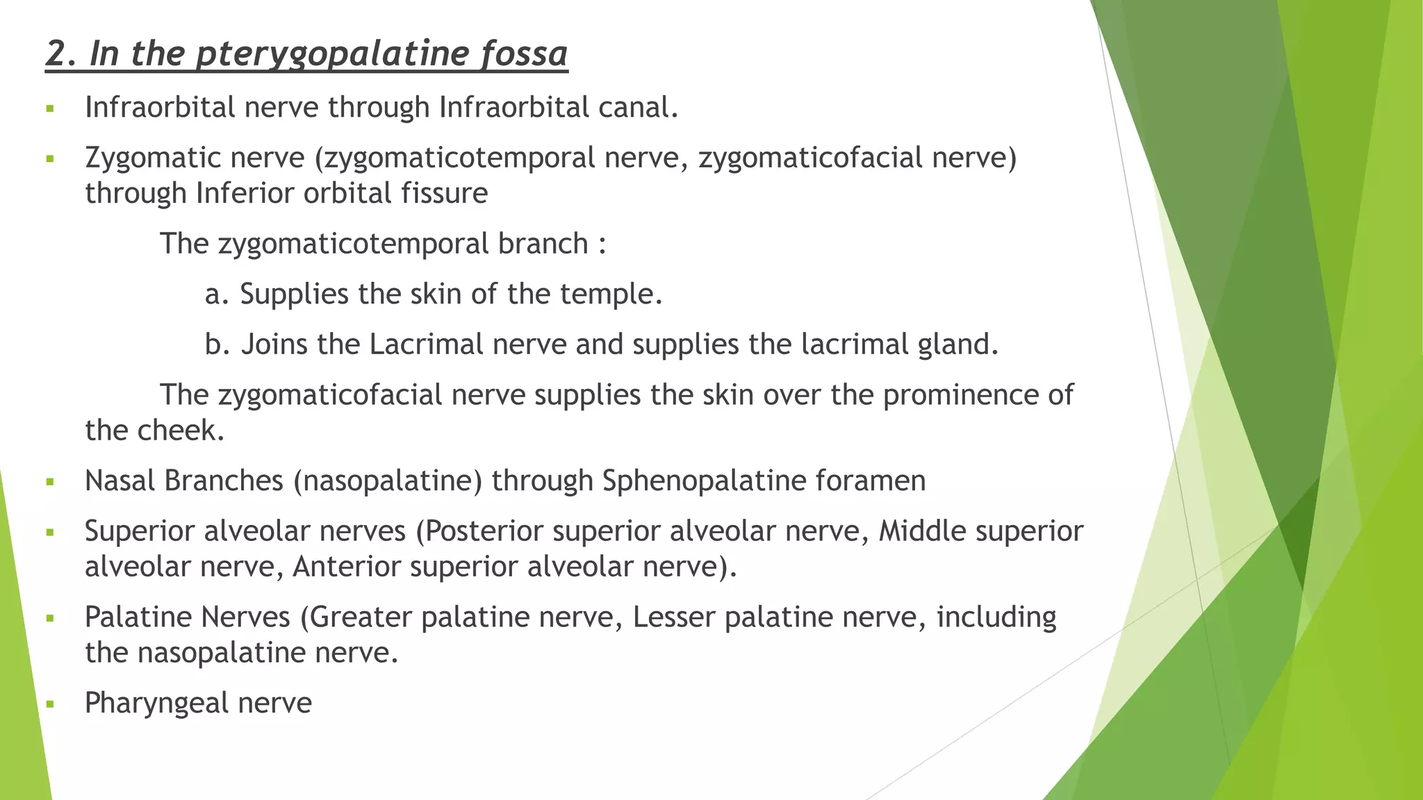
Describing the anatomy of the pterygopalatine fossa and its significance.
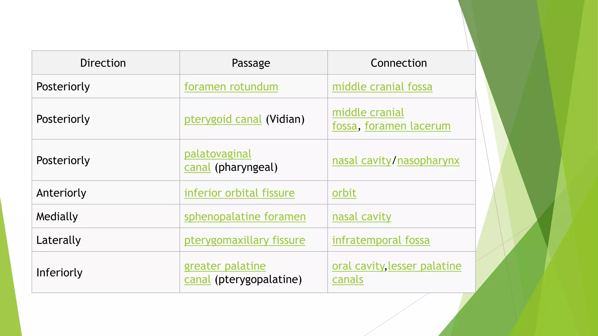
Connections and foramina of the pterygopalatine fossa leading to surrounding regions.
Functions and connections of the pterygopalatine ganglion in the nervous system.
Details on the distribution of postganglionic fibers from the pterygopalatine ganglion.
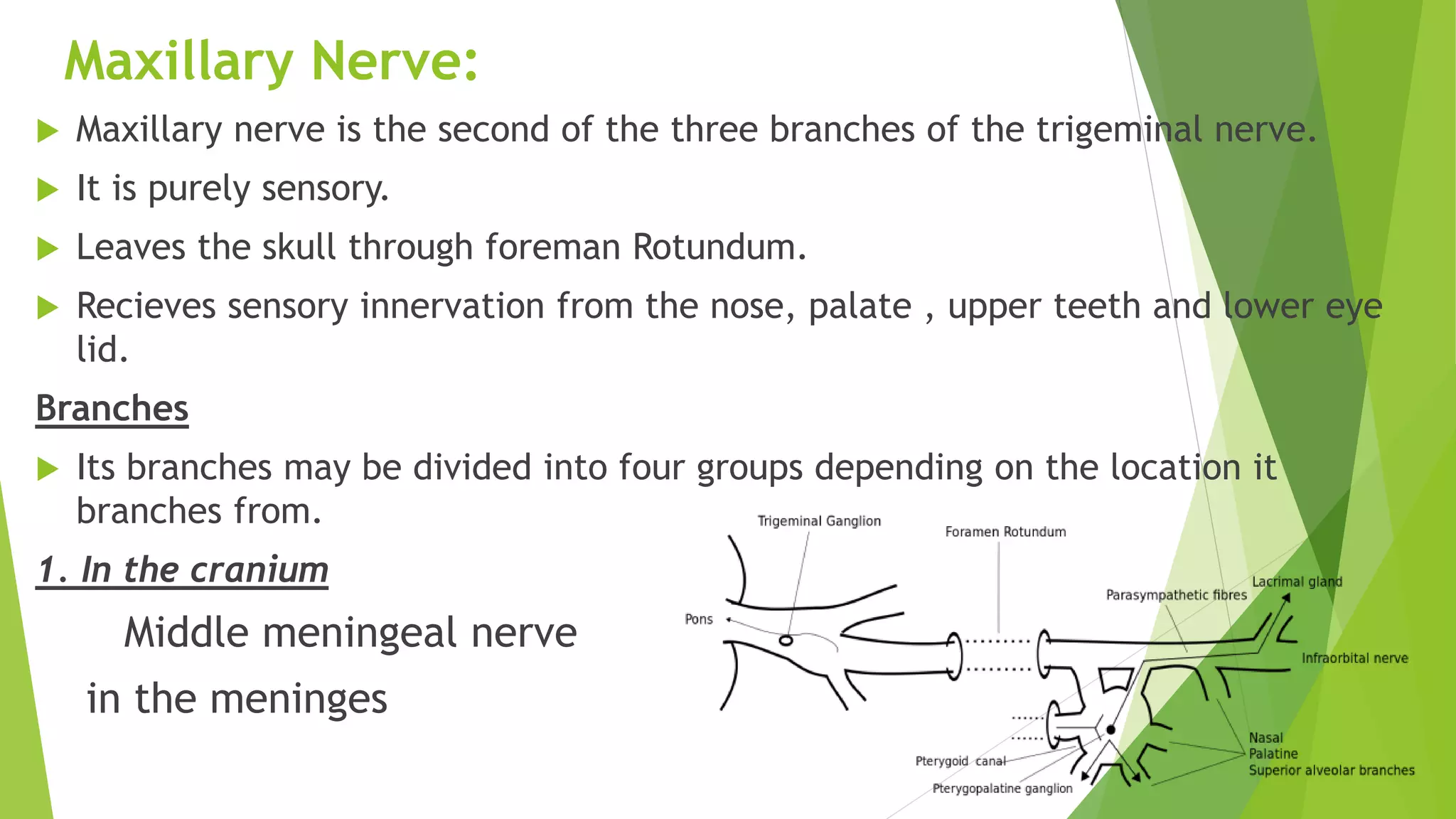
Introduction to the maxillary nerve, identifying its location and branches.
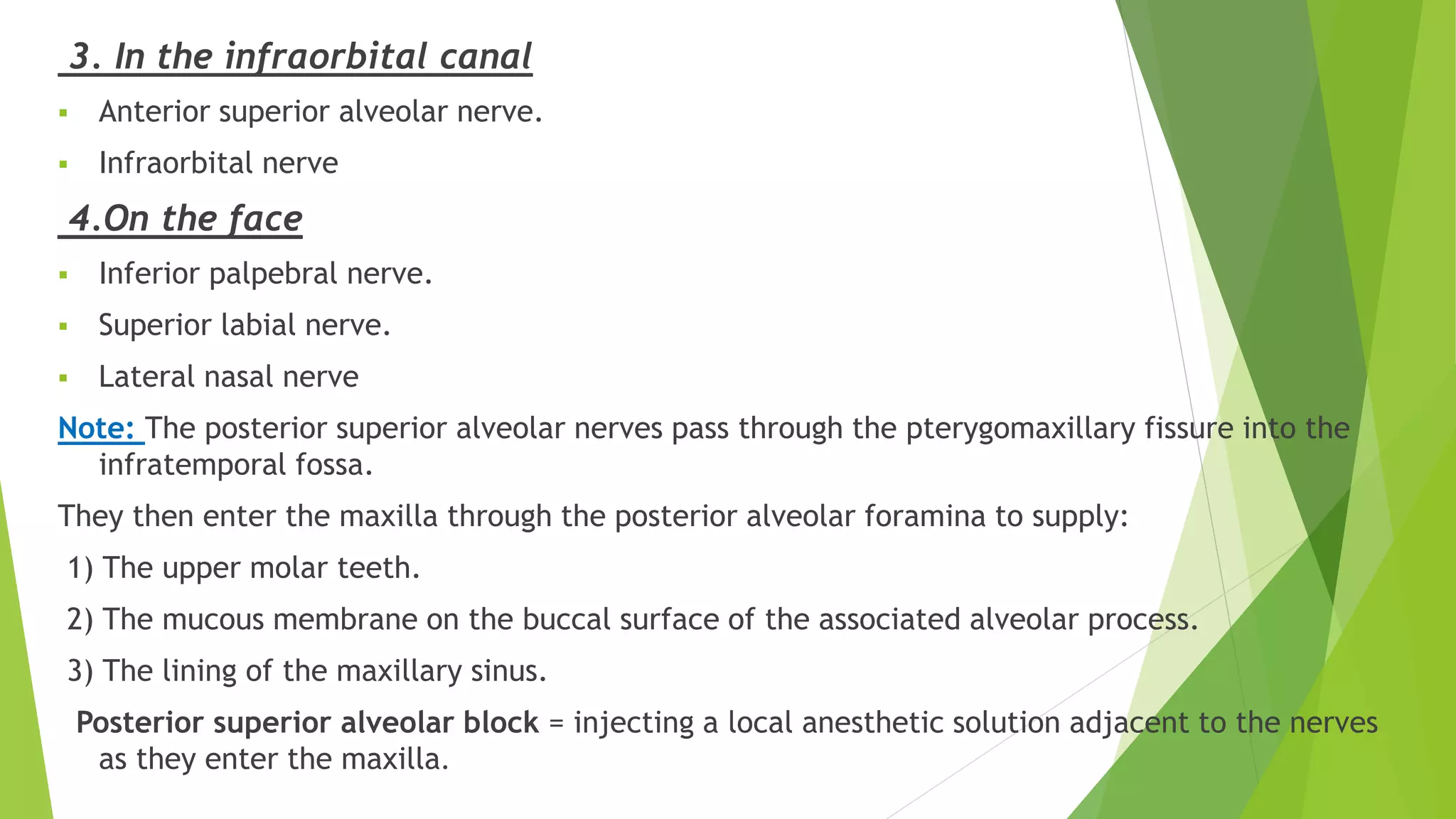
Overview of maxillary nerve branches including their sensory functions.
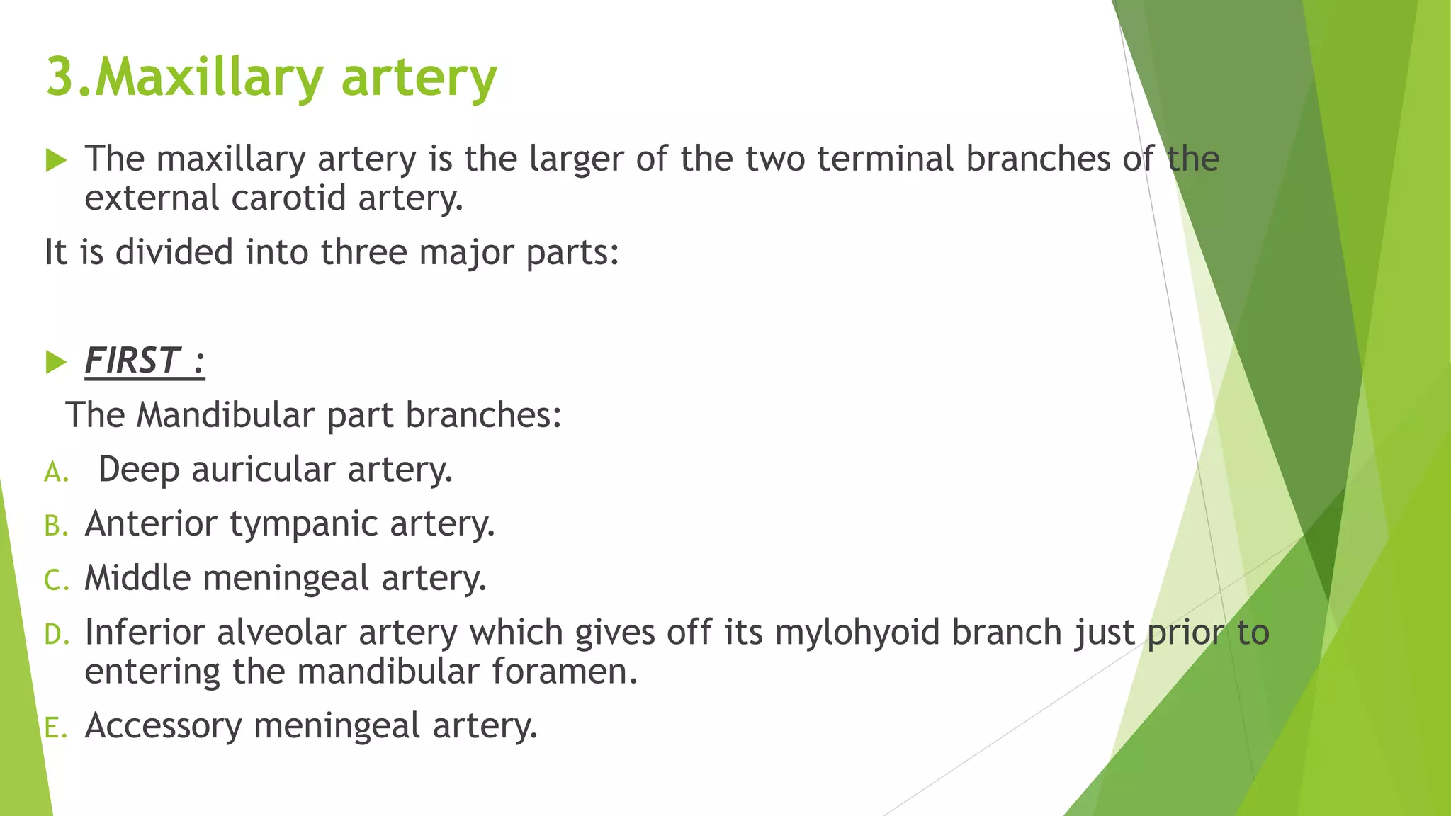
Detailed breakdown of the maxillary artery, its parts, and branches.
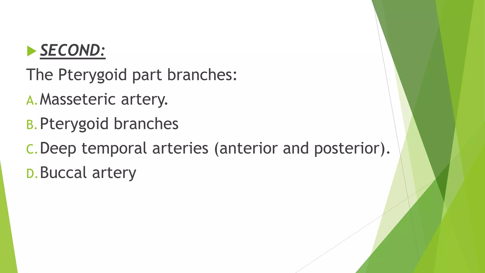
Discusses the pterygoid part branches of the maxillary artery including their functions.
Last set of branches from maxillary artery, summarizing their anatomical significance.
Wrapping up the presentation with acknowledgments.


