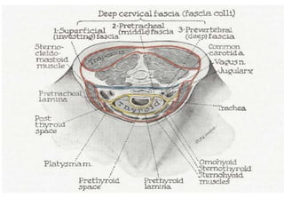
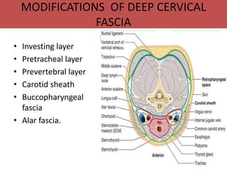
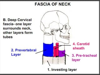
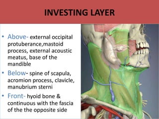
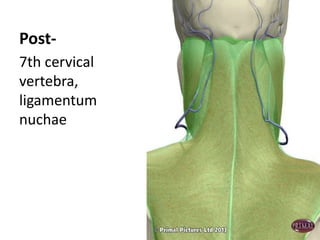
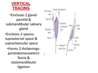
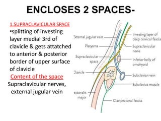
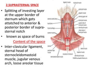
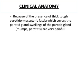
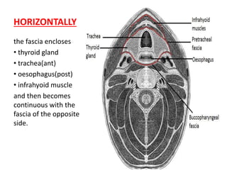
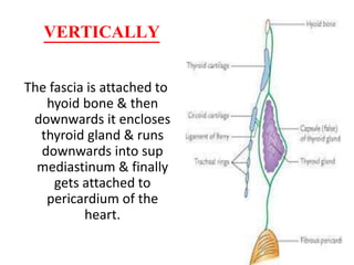
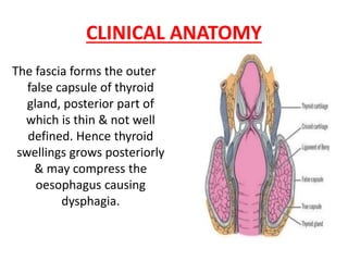
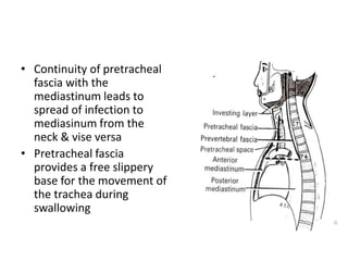
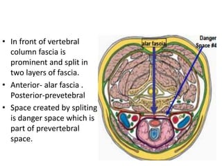
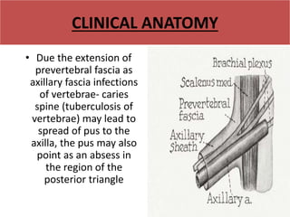
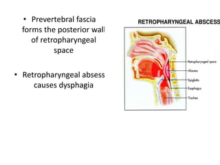
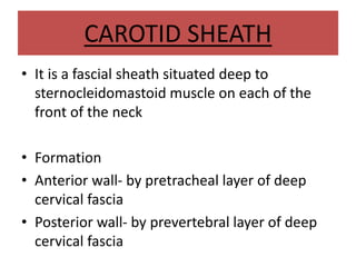
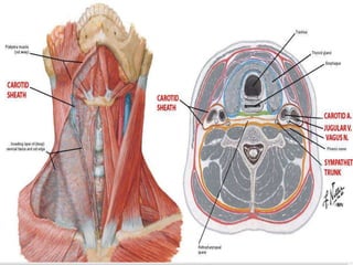
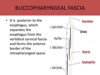
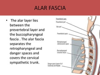
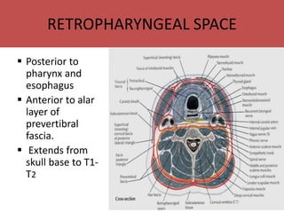
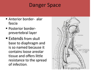
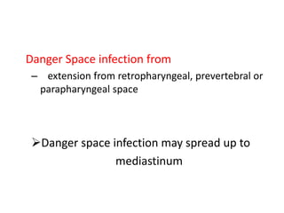
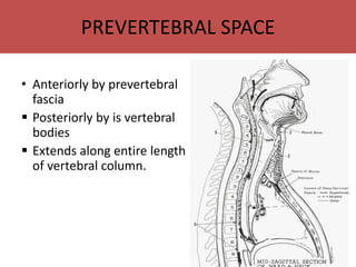
CERVICAL
FASCIA
Dr. Rana Pratap Singh
JRII Surgery
M.L.B. MEDICAL COLLEGE
JHANSI
CERVICAL FASCIA
 Fascia means fibrous connective tissue which
binds together various structures of the body
 2 types- superficial fascia & deep fascia
Superficial fascia is subcutaneous- present just
below the skin.
Deep fascia is present around muscles blood
vessels & organs of the neck
SUPERFICIAL CERVICAL FASCIA
• The superficial cervical fascia is typically a
thin lamina covering the platysma muscle.
• It is hardly demonstrable as a separate
layer but may contain considerable amounts
of adipose tissue especially in females.
DEEP CERVICAL FASCIA
 Deep fascia around the neck is known as deep
cervical fascia or fascia colli as it forms a collar
around the neck
Lies deep to platysma muscle in the interval b/w
muscles, vessels & organs of the cervical region.
Gives various extensions or laminae around
various structures of the neck
MODIFICATIONS OF DEEP CERVICAL
FASCIA
• Investing layer
• Pretracheal layer
• Prevertebral layer
• Carotid sheath
• Buccopharyngeal
fascia
• Alar fascia.
INVESTING LAYER
• Above- external occipital
protuberance,mastoid
process, external acoustic
meatus, base of the
mandible
• Below- spine of scapula,
acromion process, clavicle,
manubrium sterni
• Front- hyoid bone &
continuous with the fascia
of the the opposite side
Post-
7th cervical
vertebra,
ligamentum
nuchae
HORIZONTAL TRACING-
• Encloses 2 muscles-
trapezius &
sternocleidomastoid
• Forms roof of 2
triangle- anterior &
posterior
• Gives 2 lamina for
pretracheal &
prevertebral fascia
• Forms 2 fascial slings for
omohyoid and
diagastric;
VERTICAL
TRACING
•Encloses 2 gland-
parotid &
submandibular salivary
gland
•Encloses 2 spaces-
suprasternal space &
supraclavicular space
•Forms 2 thickenings-
parotidomassaeteric
fascia &
stylomandibular
ligamen
ENCLOSES 2 SPACES-
• Supraclavicular space
1.SUPRACLAVICULAR SPACE
•splitting of investing
layer medial 3rd of
clavicle & gets attatched
to anterior & posterior
border of upper surface
of clavicle
Content of the space
Supraclavicular nerves,
external jugular vein
2.SUPRASTERNAL SPACE
• Splitting of investing layer
at the upper border of
sternum which gets
attatched to anterior &
posterior border of supra-
sternal notch
• known as space of burns
Content of the space
• Inter-clavicular ligament,
sternal head of
sternocleidomastoid
muscle, jugular venous
arch, loose areolar tissue
CLINICAL ANATOMY
• Because of the presence of thick tough
parotido-masseteric fascia which covers the
parotid gland swellings of the parotid gland
(mumps, parotitis) are very painfull
PRETRACHEAL FASCIA
• IT Is one of the lamina of deep cervical fascia that
arises deep to sternocleidomastoid muscle
HORIZONTALLY
the fascia encloses
• thyroid gland
• trachea(ant)
• oesophagus(post)
• infrahyoid muscle
and then becomes
continuous with the
fascia of the opposite
side.
VERTICALLY
The fascia is attached to
hyoid bone & then
downwards it encloses
thyroid gland & runs
downwards into sup
mediastinum & finally
gets attached to
pericardium of the
heart.
CLINICAL ANATOMY
The fascia forms the outer
false capsule of thyroid
gland, posterior part of
which is thin & not well
defined. Hence thyroid
swellings grows posteriorly
& may compress the
oesophagus causing
dysphagia.
Thyroid gland moves with
deglutition as the posterior
aspect of the gland is
attatched to the cricoid
cartilage by a thickening of
pretracheal fascia known as
ligament of berry or
suspensory ligament of
thyroid gland.
 During thyroid surgeries the
ligament of berry has
To be cut to mobilise the
thyroid gland.
• Continuity of pretracheal
fascia with the
mediastinum leads to
spread of infection to
mediasinum from the
neck & vise versa
• Pretracheal fascia
provides a free slippery
base for the movement of
the trachea during
swallowing
PREVERTEBRAL FASCIA
• It is one of the lamina of deep cervical fascia that
arises deep to sternocleidomastoid muscle
• Lies in front of cervical vertebrae & muscles in
front of it
• In front of vertebral
column fascia is
prominent and split in
two layers of fascia.
• Anterior- alar fascia .
Posterior-prevetebral
• Space created by spliting
is danger space which is
part of prevertebral
space.
Attachments
Superior- Skull base.
Inferior attachments- T3.
Posterior attachments--
Spinous processes of cervical
and thoracic vertebrae.
Lateral attachments
Transverse processes of cervical and
thoracic vertebrae.
Horizontally
• It forms the floor of
the posterior
triangle & finally
extends upto axilla
as axillary sheath
enclosing the axillary
vessels and nerves
Vertically
• it extends from the skull
base upto the 3rd
thoracic vertebra
CLINICAL ANATOMY
• Due the extension of
prevertebral fascia as
axillary fascia infections
of vertebrae- caries
spine (tuberculosis of
vertebrae) may lead to
spread of pus to the
axilla, the pus may also
point as an absess in
the region of the
posterior triangle
• Prevertebral fascia
forms the posterior wall
of retropharyngeal
space
• Retropharyngeal absess
causes dysphagia
CAROTID SHEATH
• It is a fascial sheath situated deep to
sternocleidomastoid muscle on each of the
front of the neck
• Formation
• Anterior wall- by pretracheal layer of deep
cervical fascia
• Posterior wall- by prevertebral layer of deep
cervical fascia
CONTENT
Internal jugular vein
laterally, coImmon carotid
artery ( in the lower part)
& internal carotid artery (
in the upper part)
medially, vagus nerve in
b/w them in a posterior
plane
Relations-
Anteriorly- ansa cervicalis
Posteriorly – sympathetic
trunk
BUCCOPHARYNGEAL FASCIA
• It is posterior to the
esophagus, which
separates the
esophagus from the
vertebral cervical fascia
and forms the anterior
border of the
retropharyngeal space.
ALAR FASCIA
• The alar layer lies
between the
prevertebral layer and
the buccopharyngeal
fascia . The alar fascia
separates the
retropharyngeal and
danger spaces and
covers the cervical
sympathetic trunk.
RETROPHARYNGEAL SPACE
 Posterior to
pharynx and
esophagus
 Anterior to alar
layer of
prevertibral
fascia.
 Extends from
skull base to T1-
T2
• Pediatrics
– Cause—suppurative
process in lymph nodes
• Nose, adenoids,
nasopharynx, sinuses
• Adults
– Cause—trauma,
instrumentation,
extension from
adjoining deep neck
space
Danger Space
 Anterior border- alar
fascia
 Posterior border-
prevertebral layer
 Extends from skull
base to diaphragm and
is so named because it
contains loose areolar
tissue and offers little
resistance to the spread
of infection.
Danger Space infection from
– extension from retropharyngeal, prevertebral or
parapharyngeal space
Danger space infection may spread up to
mediastinum
PREVERTEBRAL SPACE
• Anteriorly by prevertebral
fascia
 Posteriorly by is vertebral
bodies
 Extends along entire length
of vertebral column.
Prevertebral space infection from
• Infection of the vertebral bodies
• Penetrating injuries.
• Tuberculosis of the spine may breach the
space and form a Pott’s abscess.
Thanks

cervical fascia

  • 1.
    CERVICAL FASCIA Dr. Rana PratapSingh JRII Surgery M.L.B. MEDICAL COLLEGE JHANSI
  • 2.
    CERVICAL FASCIA  Fasciameans fibrous connective tissue which binds together various structures of the body  2 types- superficial fascia & deep fascia Superficial fascia is subcutaneous- present just below the skin. Deep fascia is present around muscles blood vessels & organs of the neck
  • 3.
    SUPERFICIAL CERVICAL FASCIA •The superficial cervical fascia is typically a thin lamina covering the platysma muscle. • It is hardly demonstrable as a separate layer but may contain considerable amounts of adipose tissue especially in females.
  • 4.
    DEEP CERVICAL FASCIA Deep fascia around the neck is known as deep cervical fascia or fascia colli as it forms a collar around the neck Lies deep to platysma muscle in the interval b/w muscles, vessels & organs of the cervical region. Gives various extensions or laminae around various structures of the neck
  • 6.
    MODIFICATIONS OF DEEPCERVICAL FASCIA • Investing layer • Pretracheal layer • Prevertebral layer • Carotid sheath • Buccopharyngeal fascia • Alar fascia.
  • 8.
    INVESTING LAYER • Above-external occipital protuberance,mastoid process, external acoustic meatus, base of the mandible • Below- spine of scapula, acromion process, clavicle, manubrium sterni • Front- hyoid bone & continuous with the fascia of the the opposite side
  • 9.
  • 10.
    HORIZONTAL TRACING- • Encloses2 muscles- trapezius & sternocleidomastoid • Forms roof of 2 triangle- anterior & posterior • Gives 2 lamina for pretracheal & prevertebral fascia
  • 11.
    • Forms 2fascial slings for omohyoid and diagastric;
  • 12.
    VERTICAL TRACING •Encloses 2 gland- parotid& submandibular salivary gland •Encloses 2 spaces- suprasternal space & supraclavicular space •Forms 2 thickenings- parotidomassaeteric fascia & stylomandibular ligamen
  • 13.
    ENCLOSES 2 SPACES- •Supraclavicular space 1.SUPRACLAVICULAR SPACE •splitting of investing layer medial 3rd of clavicle & gets attatched to anterior & posterior border of upper surface of clavicle Content of the space Supraclavicular nerves, external jugular vein
  • 14.
    2.SUPRASTERNAL SPACE • Splittingof investing layer at the upper border of sternum which gets attatched to anterior & posterior border of supra- sternal notch • known as space of burns Content of the space • Inter-clavicular ligament, sternal head of sternocleidomastoid muscle, jugular venous arch, loose areolar tissue
  • 15.
    CLINICAL ANATOMY • Becauseof the presence of thick tough parotido-masseteric fascia which covers the parotid gland swellings of the parotid gland (mumps, parotitis) are very painfull
  • 16.
    PRETRACHEAL FASCIA • ITIs one of the lamina of deep cervical fascia that arises deep to sternocleidomastoid muscle
  • 17.
    HORIZONTALLY the fascia encloses •thyroid gland • trachea(ant) • oesophagus(post) • infrahyoid muscle and then becomes continuous with the fascia of the opposite side.
  • 18.
    VERTICALLY The fascia isattached to hyoid bone & then downwards it encloses thyroid gland & runs downwards into sup mediastinum & finally gets attached to pericardium of the heart.
  • 19.
    CLINICAL ANATOMY The fasciaforms the outer false capsule of thyroid gland, posterior part of which is thin & not well defined. Hence thyroid swellings grows posteriorly & may compress the oesophagus causing dysphagia.
  • 20.
    Thyroid gland moveswith deglutition as the posterior aspect of the gland is attatched to the cricoid cartilage by a thickening of pretracheal fascia known as ligament of berry or suspensory ligament of thyroid gland.  During thyroid surgeries the ligament of berry has To be cut to mobilise the thyroid gland.
  • 21.
    • Continuity ofpretracheal fascia with the mediastinum leads to spread of infection to mediasinum from the neck & vise versa • Pretracheal fascia provides a free slippery base for the movement of the trachea during swallowing
  • 22.
    PREVERTEBRAL FASCIA • Itis one of the lamina of deep cervical fascia that arises deep to sternocleidomastoid muscle • Lies in front of cervical vertebrae & muscles in front of it
  • 23.
    • In frontof vertebral column fascia is prominent and split in two layers of fascia. • Anterior- alar fascia . Posterior-prevetebral • Space created by spliting is danger space which is part of prevertebral space.
  • 24.
    Attachments Superior- Skull base. Inferiorattachments- T3. Posterior attachments-- Spinous processes of cervical and thoracic vertebrae. Lateral attachments Transverse processes of cervical and thoracic vertebrae.
  • 25.
    Horizontally • It formsthe floor of the posterior triangle & finally extends upto axilla as axillary sheath enclosing the axillary vessels and nerves
  • 26.
    Vertically • it extendsfrom the skull base upto the 3rd thoracic vertebra
  • 27.
    CLINICAL ANATOMY • Duethe extension of prevertebral fascia as axillary fascia infections of vertebrae- caries spine (tuberculosis of vertebrae) may lead to spread of pus to the axilla, the pus may also point as an absess in the region of the posterior triangle
  • 28.
    • Prevertebral fascia formsthe posterior wall of retropharyngeal space • Retropharyngeal absess causes dysphagia
  • 29.
    CAROTID SHEATH • Itis a fascial sheath situated deep to sternocleidomastoid muscle on each of the front of the neck • Formation • Anterior wall- by pretracheal layer of deep cervical fascia • Posterior wall- by prevertebral layer of deep cervical fascia
  • 30.
    CONTENT Internal jugular vein laterally,coImmon carotid artery ( in the lower part) & internal carotid artery ( in the upper part) medially, vagus nerve in b/w them in a posterior plane Relations- Anteriorly- ansa cervicalis Posteriorly – sympathetic trunk
  • 32.
    BUCCOPHARYNGEAL FASCIA • Itis posterior to the esophagus, which separates the esophagus from the vertebral cervical fascia and forms the anterior border of the retropharyngeal space.
  • 33.
    ALAR FASCIA • Thealar layer lies between the prevertebral layer and the buccopharyngeal fascia . The alar fascia separates the retropharyngeal and danger spaces and covers the cervical sympathetic trunk.
  • 34.
    RETROPHARYNGEAL SPACE  Posteriorto pharynx and esophagus  Anterior to alar layer of prevertibral fascia.  Extends from skull base to T1- T2
  • 35.
    • Pediatrics – Cause—suppurative processin lymph nodes • Nose, adenoids, nasopharynx, sinuses • Adults – Cause—trauma, instrumentation, extension from adjoining deep neck space
  • 36.
    Danger Space  Anteriorborder- alar fascia  Posterior border- prevertebral layer  Extends from skull base to diaphragm and is so named because it contains loose areolar tissue and offers little resistance to the spread of infection.
  • 37.
    Danger Space infectionfrom – extension from retropharyngeal, prevertebral or parapharyngeal space Danger space infection may spread up to mediastinum
  • 38.
    PREVERTEBRAL SPACE • Anteriorlyby prevertebral fascia  Posteriorly by is vertebral bodies  Extends along entire length of vertebral column.
  • 39.
    Prevertebral space infectionfrom • Infection of the vertebral bodies • Penetrating injuries. • Tuberculosis of the spine may breach the space and form a Pott’s abscess.
  • 40.