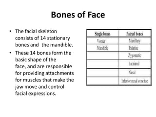

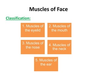


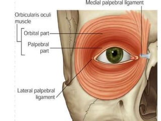
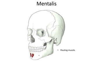
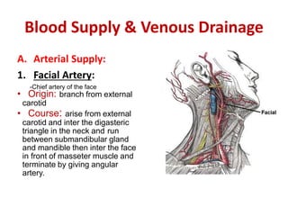
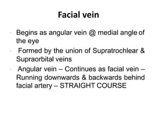
![B. Orbicularis Oris
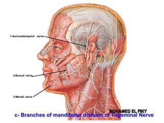
ORBITAL
PART
[OUTER]
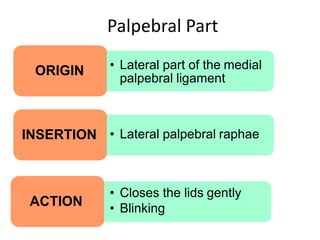
PALPEBRAL
PART
[INNER]
LACRIMAL
PART
[SMALL]
PARTS OF ORBICULARIS OCULI](https://image.slidesharecdn.com/anatomyofface-210210130332/85/Anatomy-of-face-18-320.jpg)


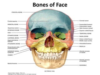
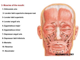
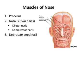
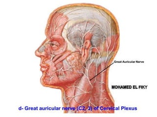
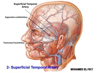
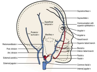
![Orbicularis oris
Two parts:
1. Intrinsic part [Deep stratum]
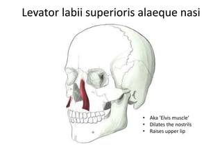
Origin: Superior incisivus from maxilla
& Inferior incisivus from
mandible
Insertion: Angle of mouth
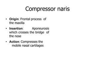
Action: Closes & purses the mouth
2. Extrinsic part [Two strata]
Origin:
• Thickest middle stratum – Buccinator
• Thick superficial stratum – Elevators &
depressors of lips & angles
Insertion: Lips & the angle of the mouth
Action: Closes & purses the mouth](https://image.slidesharecdn.com/anatomyofface-210210130332/85/Anatomy-of-face-26-320.jpg)

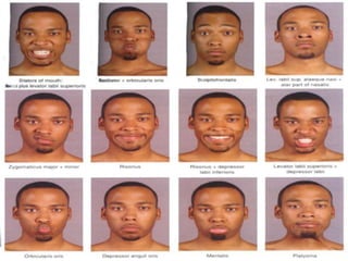
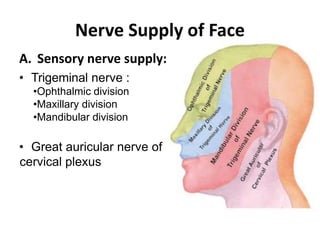

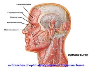
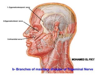


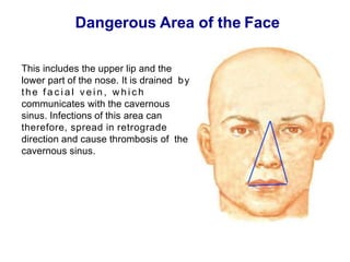


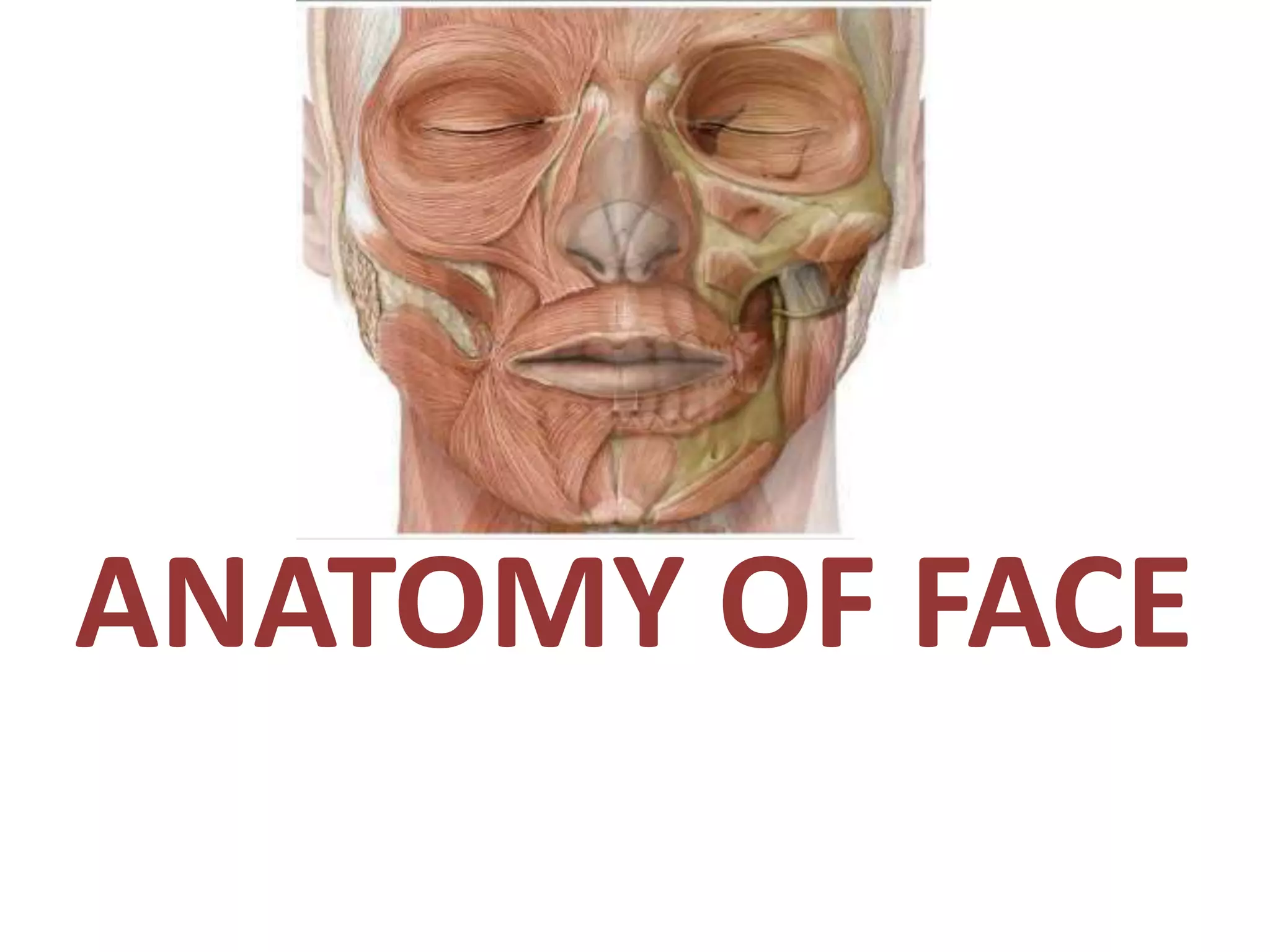
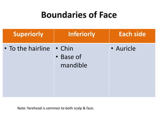
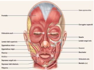
This document provides an overview of the anatomy of the face, including its boundaries, layers, bones, muscles, nerves, blood supply, and lymphatic drainage. The facial skeleton consists of 14 bones that form the basic shape of the face and provide attachments for facial muscles. These muscles are divided into groups that control expressions and movements of the eyes, mouth, nose, ears, and neck. The face has a rich blood supply from the facial artery and veins. Sensation is provided by the trigeminal nerve and motor function by the facial nerve, which innervates most facial muscles.


![CTEV [ clubfoot] DR ARUN LAL ,DR MOHAMED ASHRAF travancore medical college k...](https://cdn.slidesharecdn.com/ss_thumbnails/ctevclubfootdrarunlaldrmohamedashraftravancoremedicalcollegekollamkeralaindia-260208063247-18fc466c-thumbnail.jpg?width=640&height=640&fit=bounds)
![PERI-PROSTHETIC FRACTURE NAIL-PLATE CONSTRUCT [NPC].pptx](https://cdn.slidesharecdn.com/ss_thumbnails/drarunkumardrmohamedashrafperiprostheticfrasturenail-plateconstructnpc-260209164459-7e9d15a1-thumbnail.jpg?width=640&height=640&fit=bounds)


