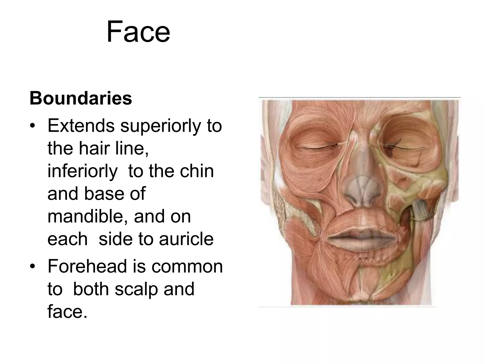
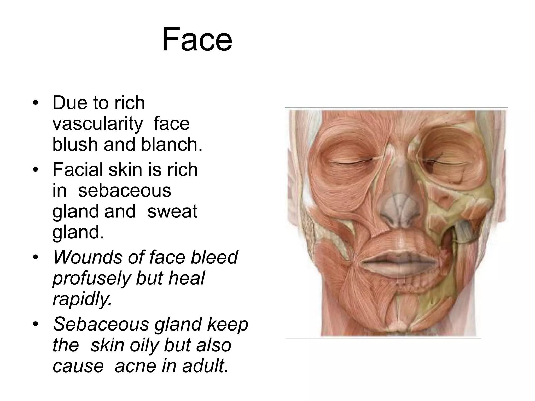
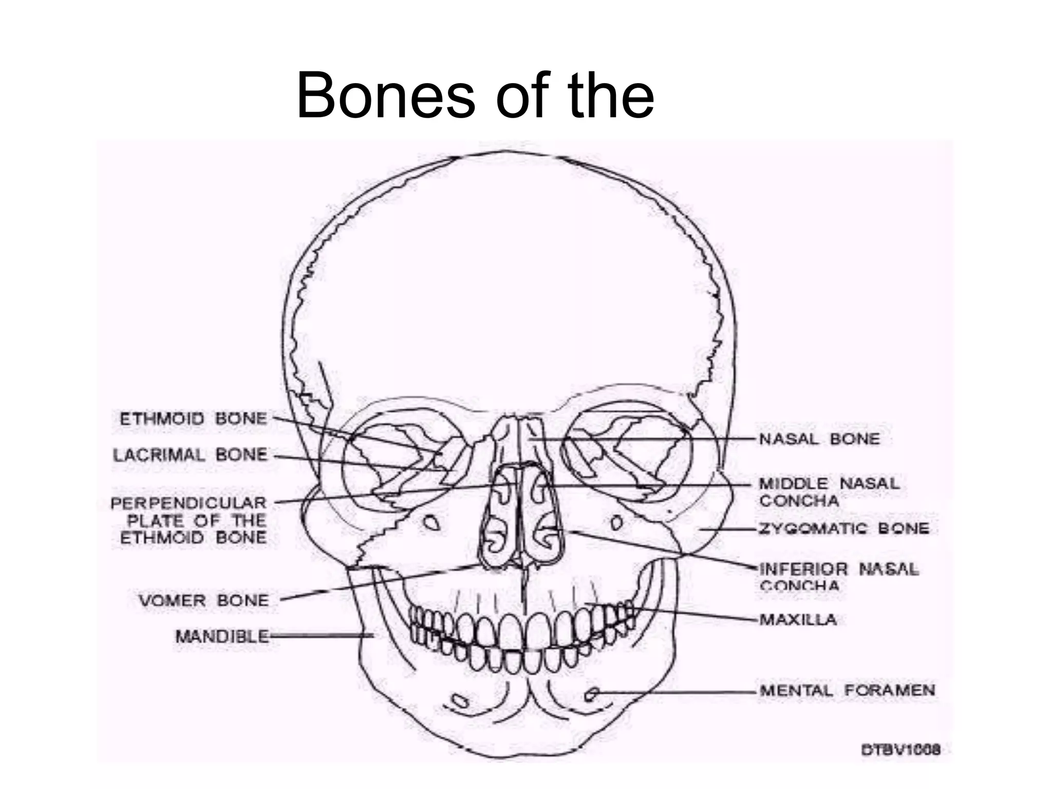
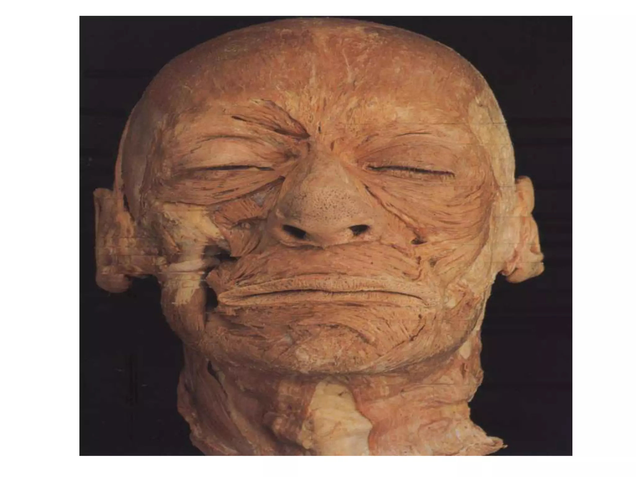
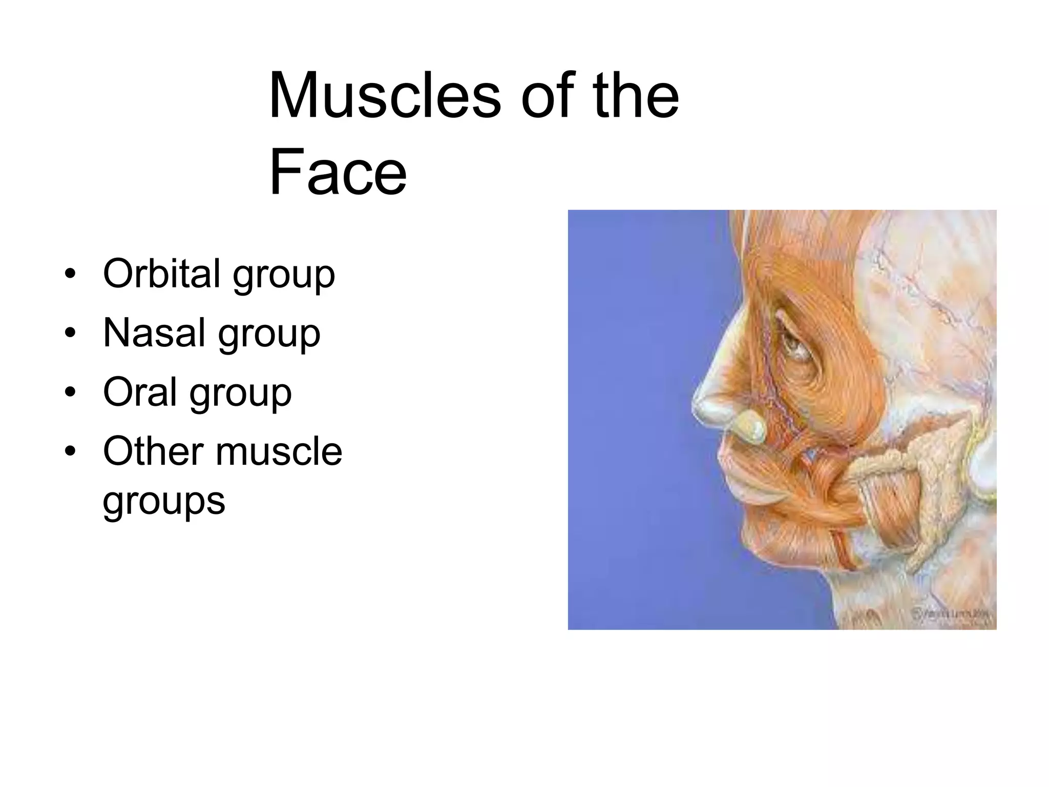
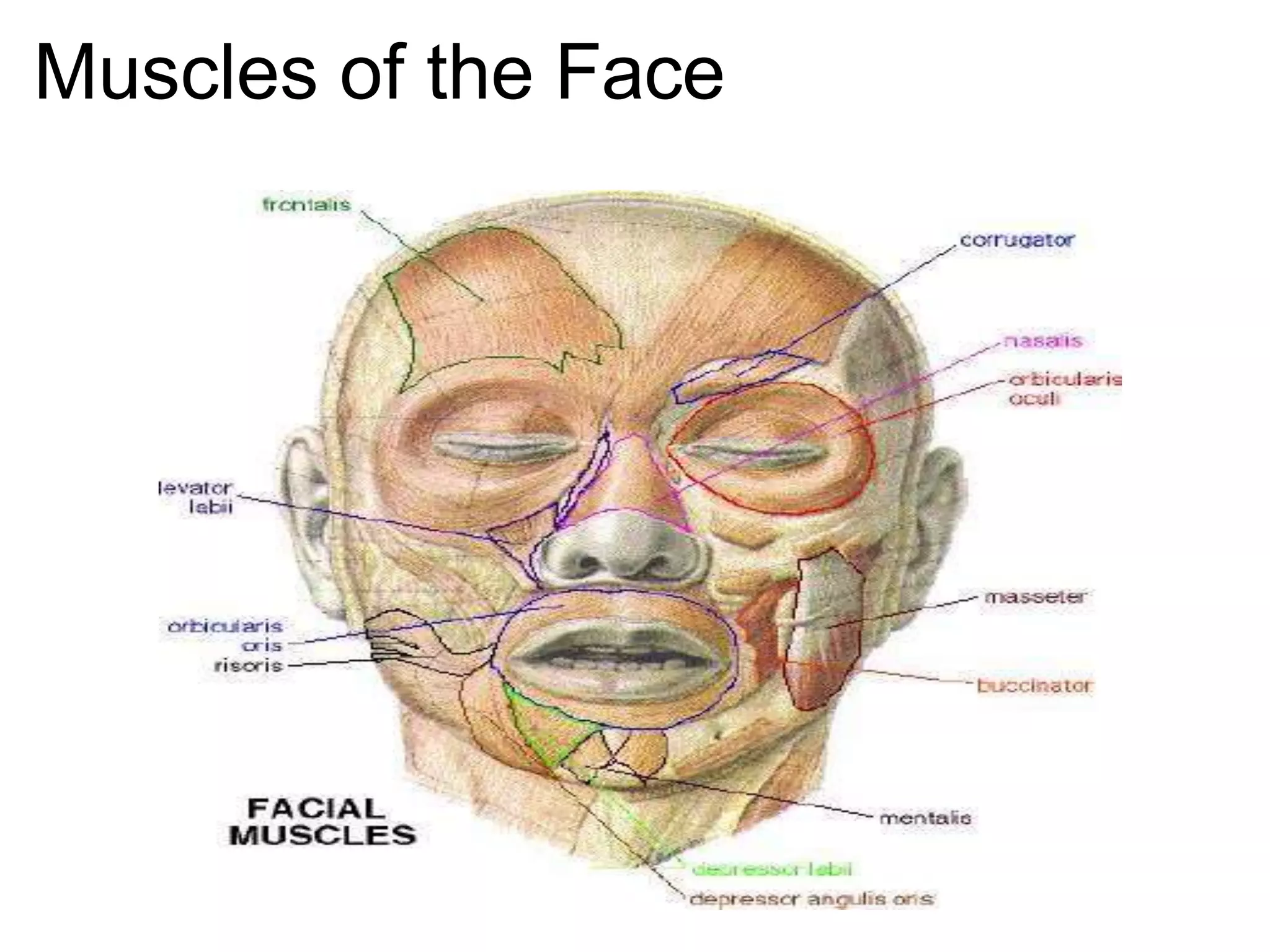
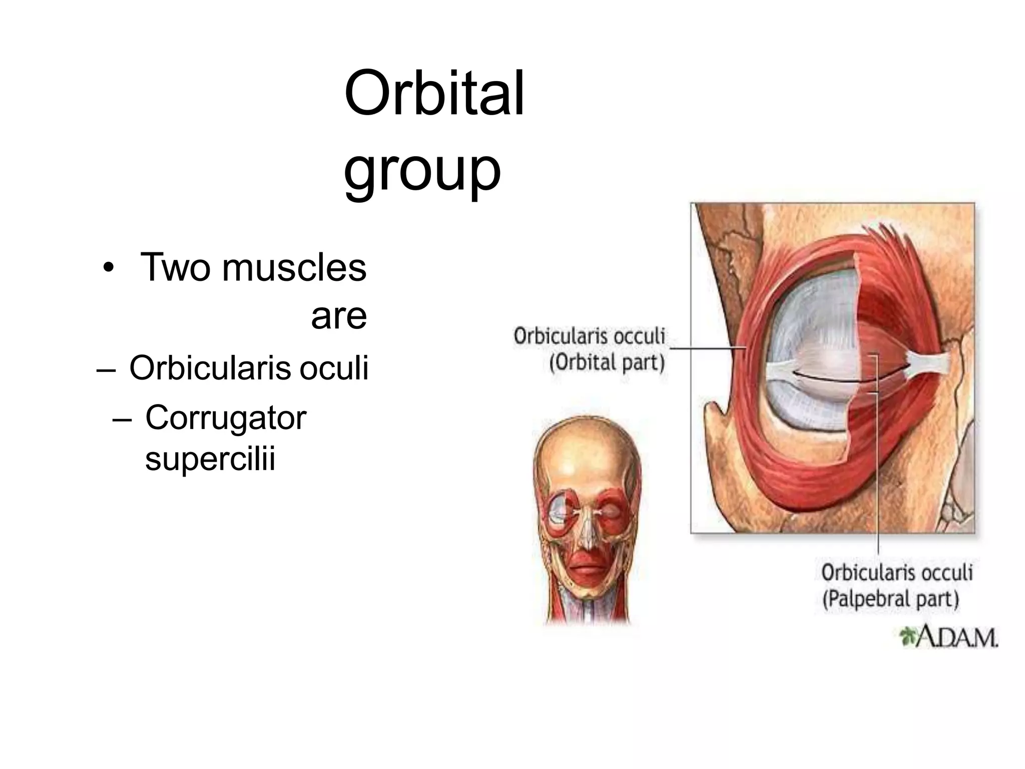
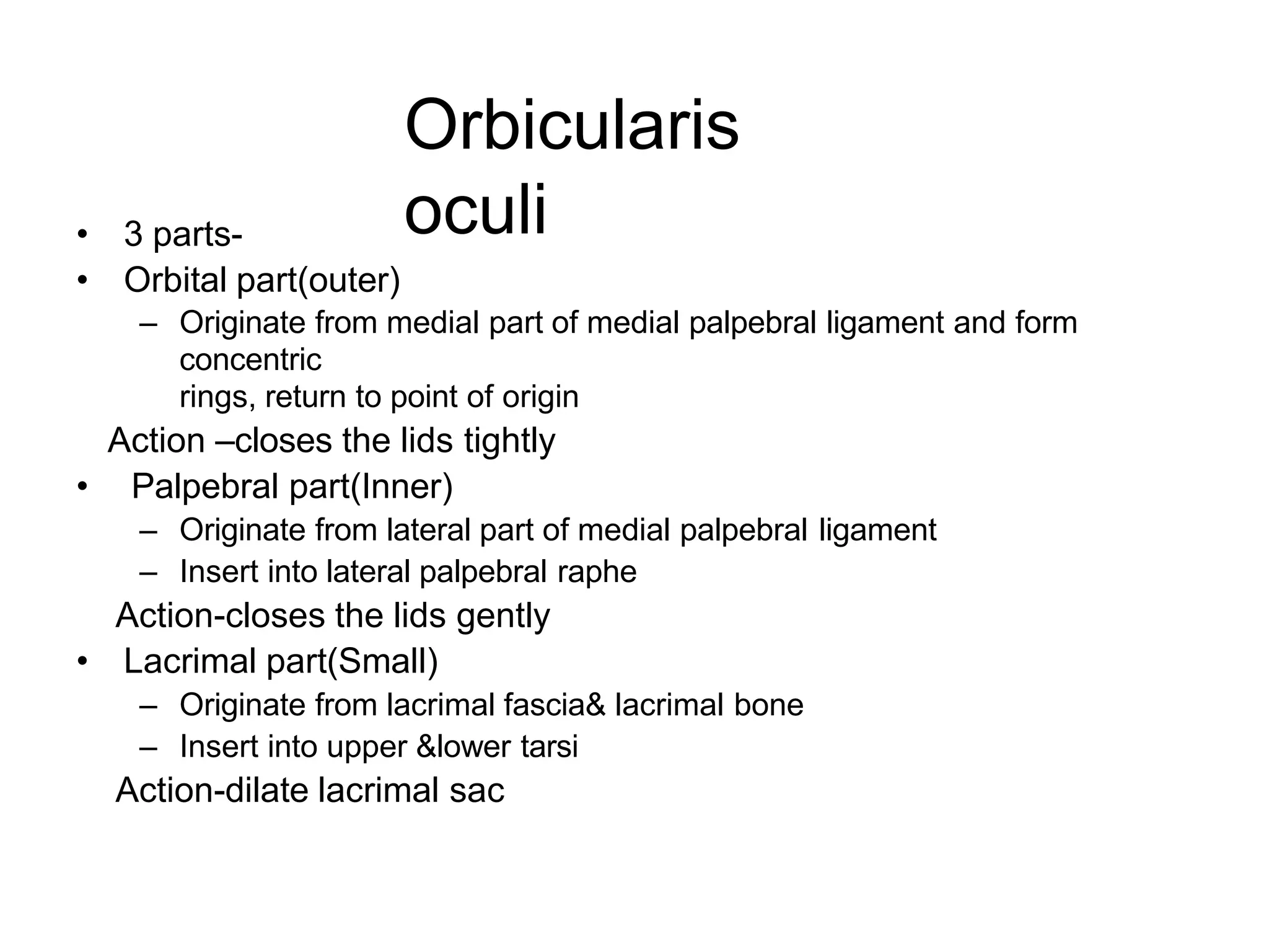
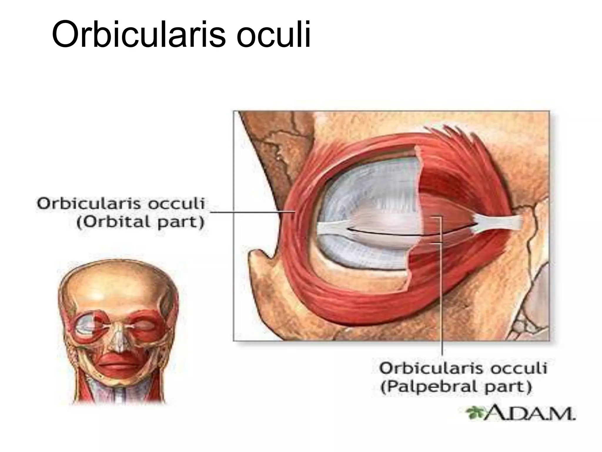
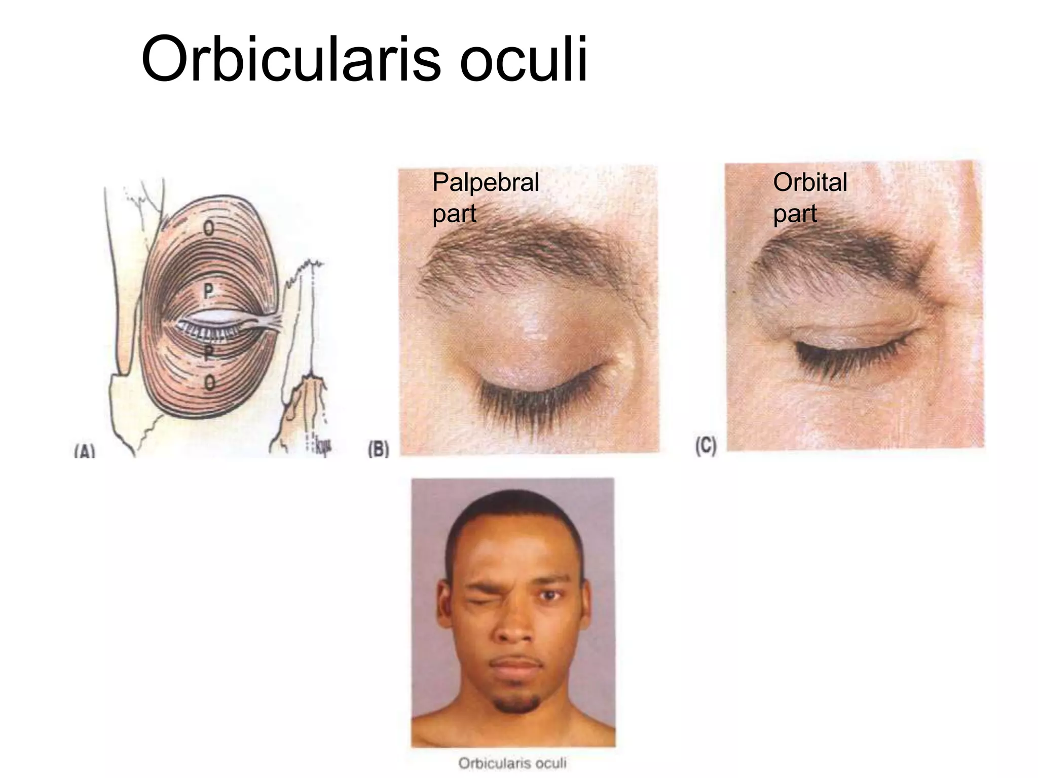
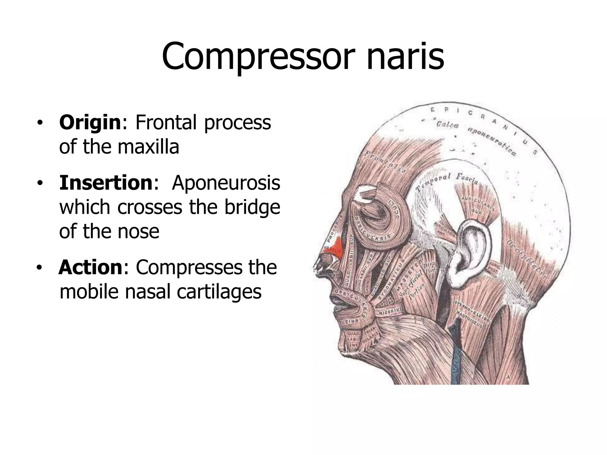
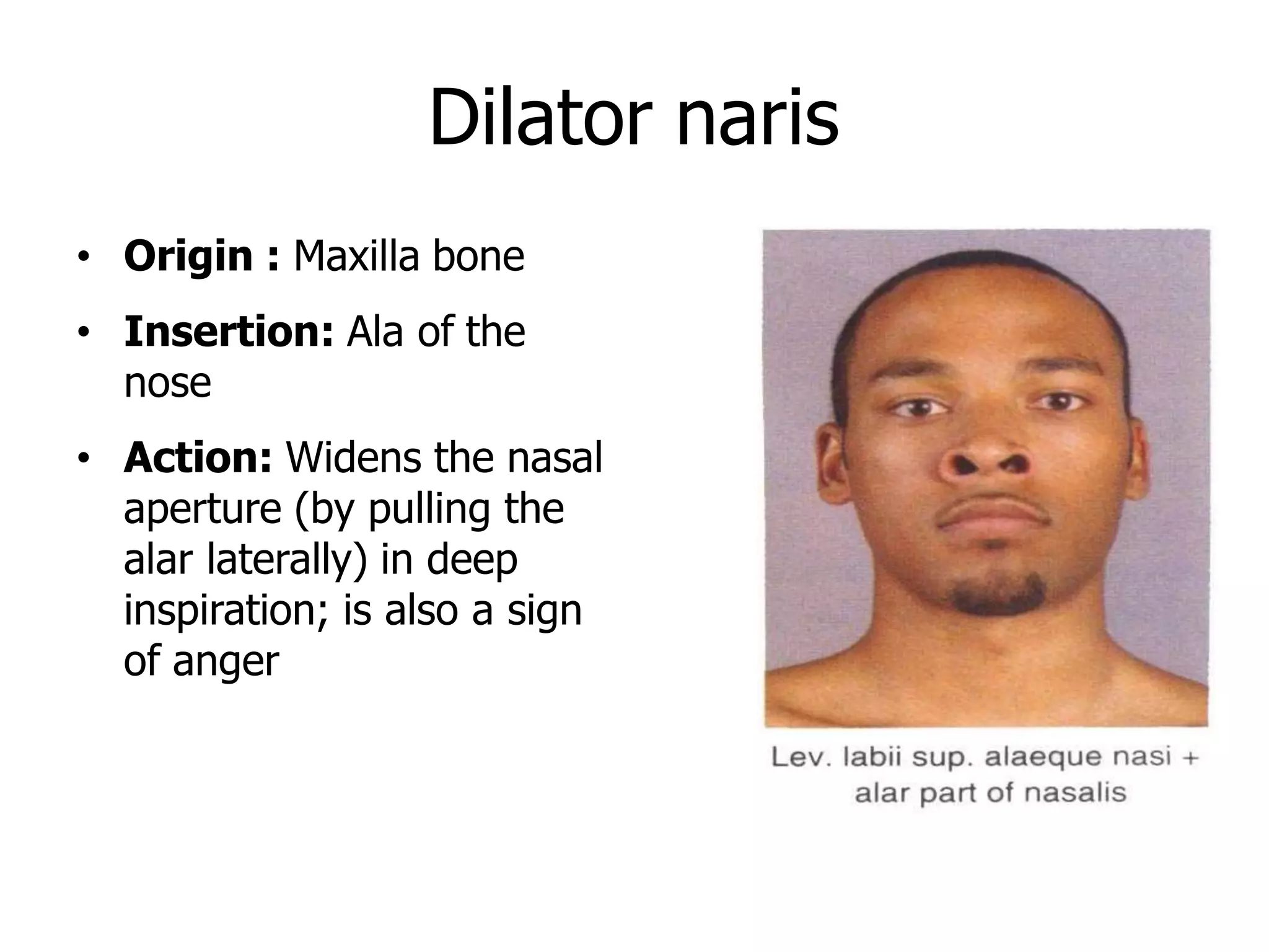
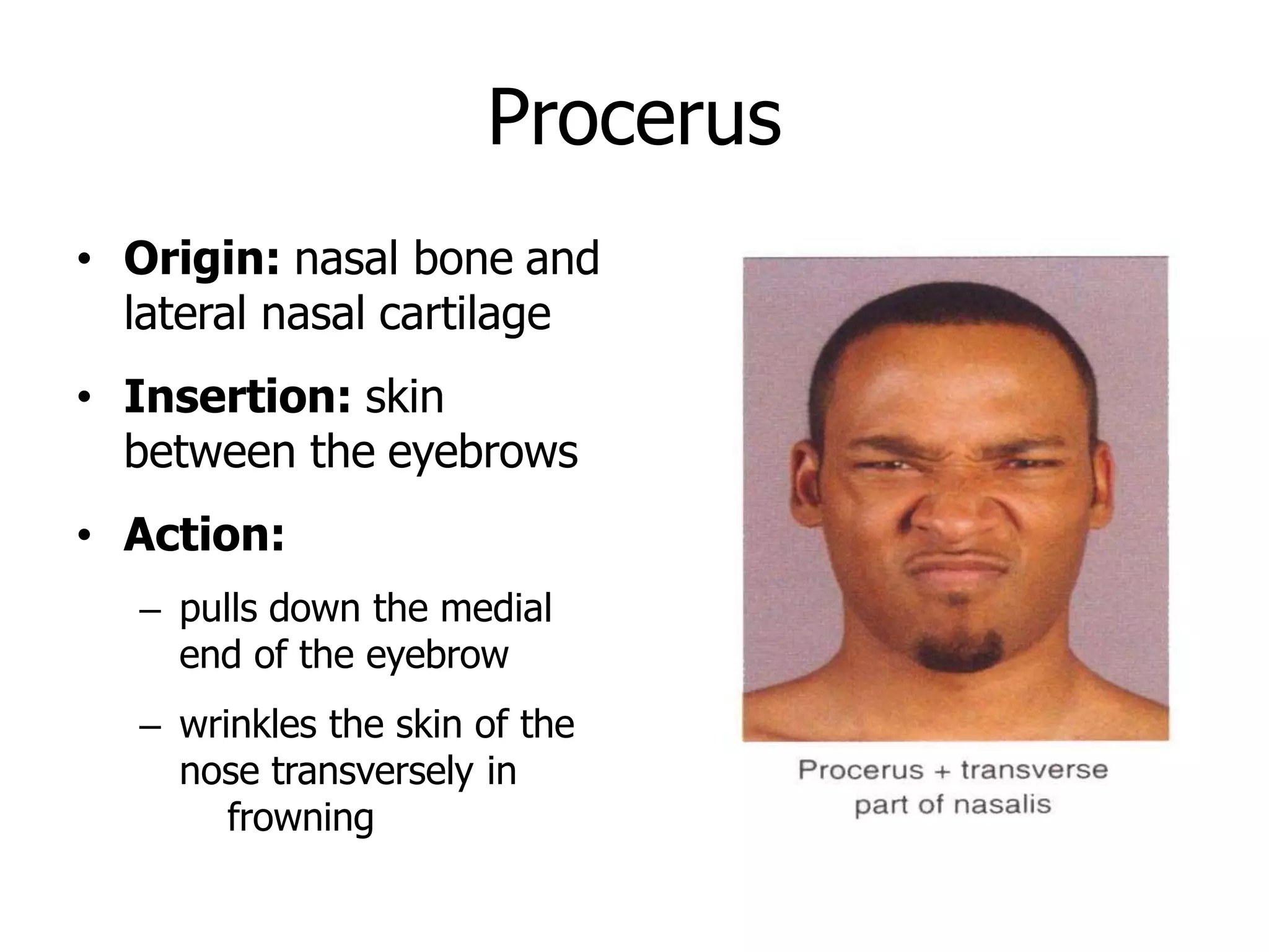
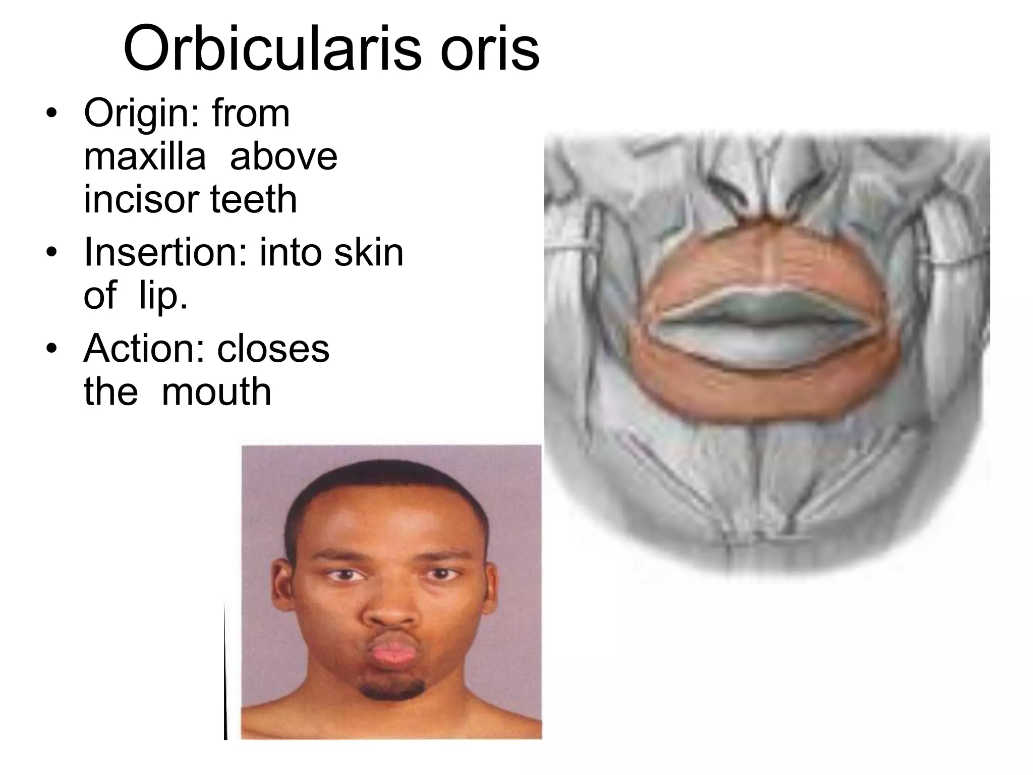
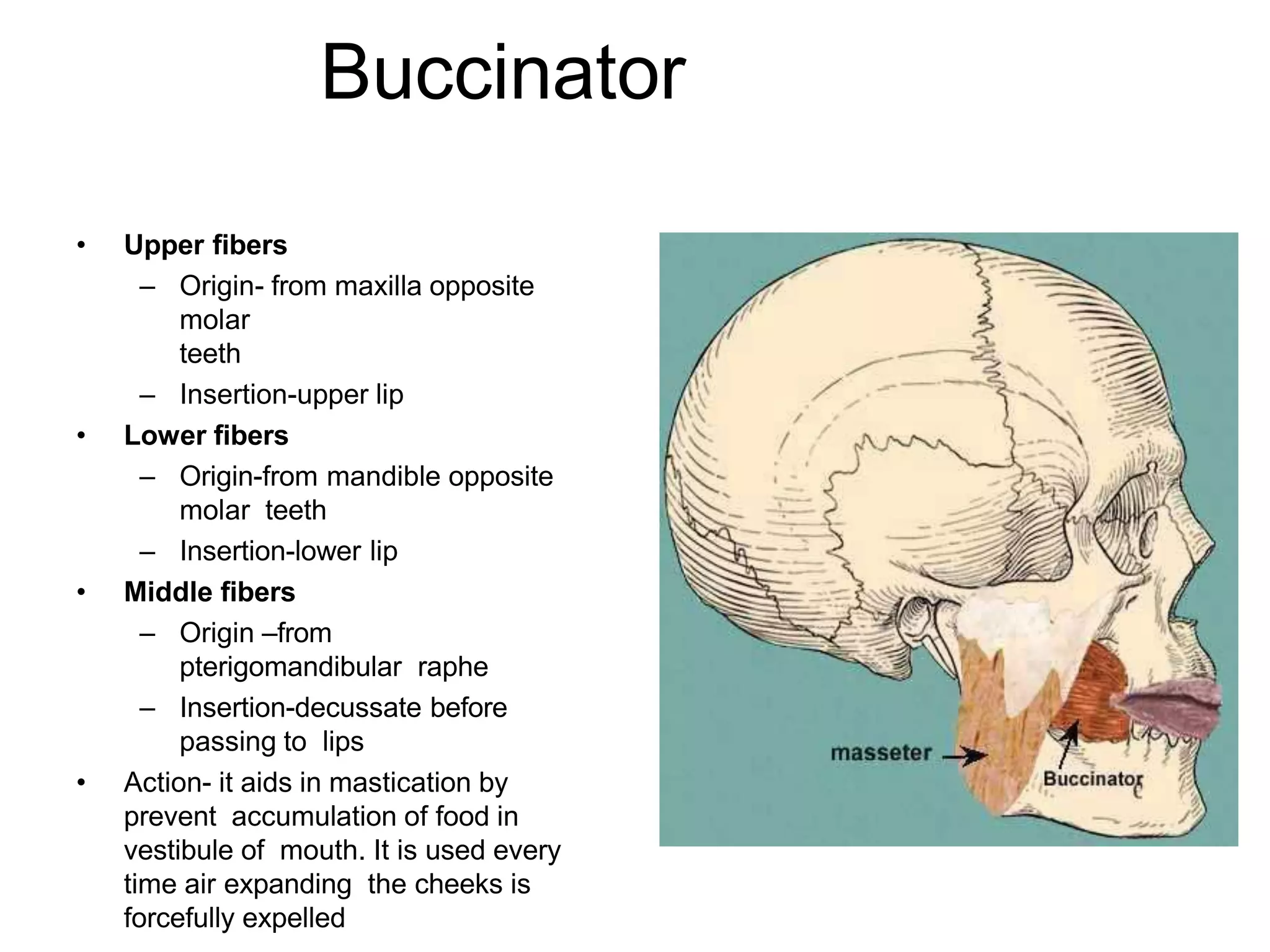
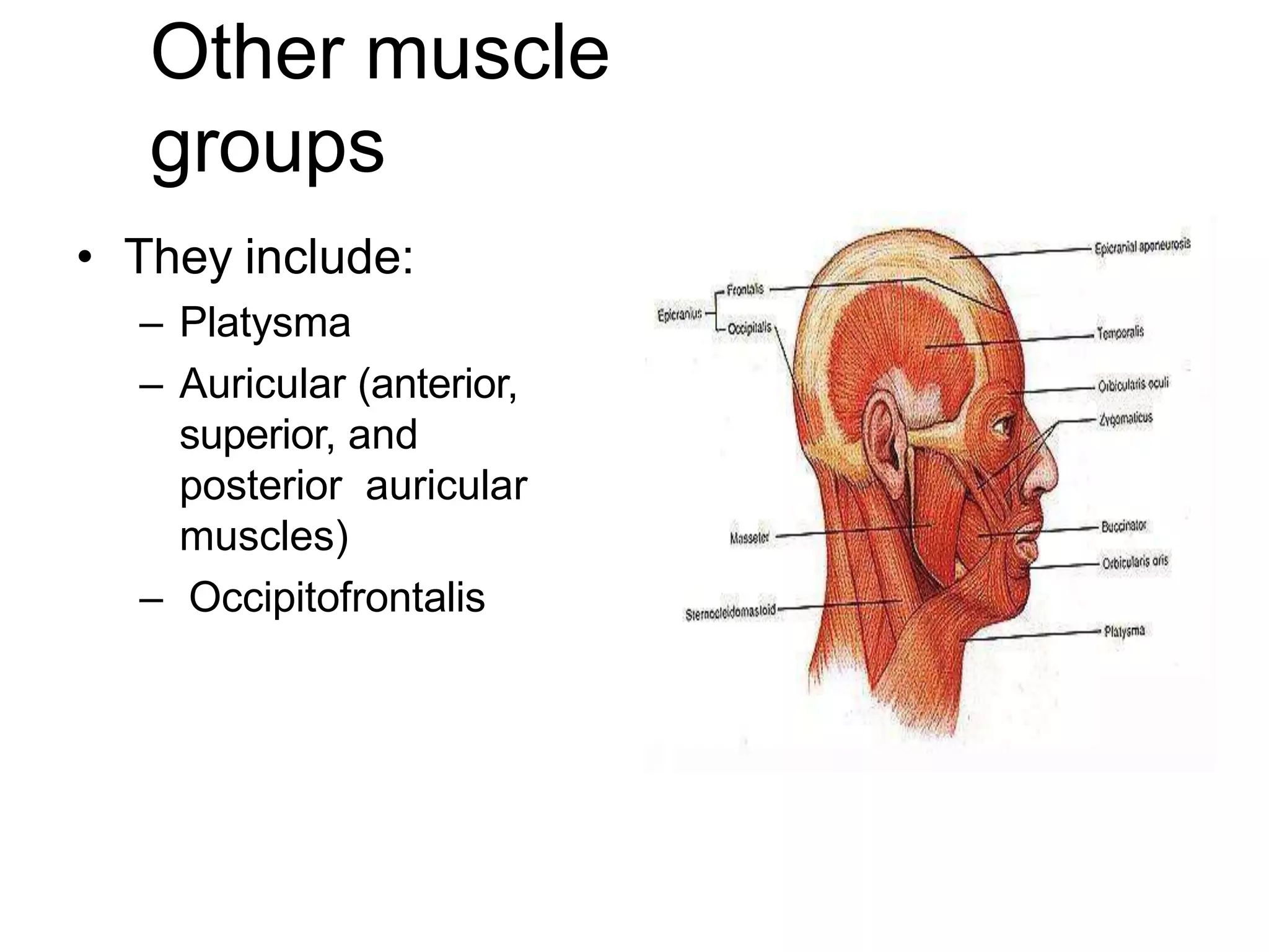
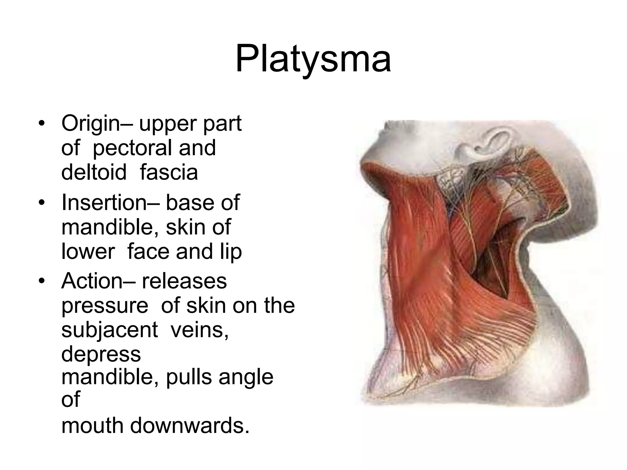
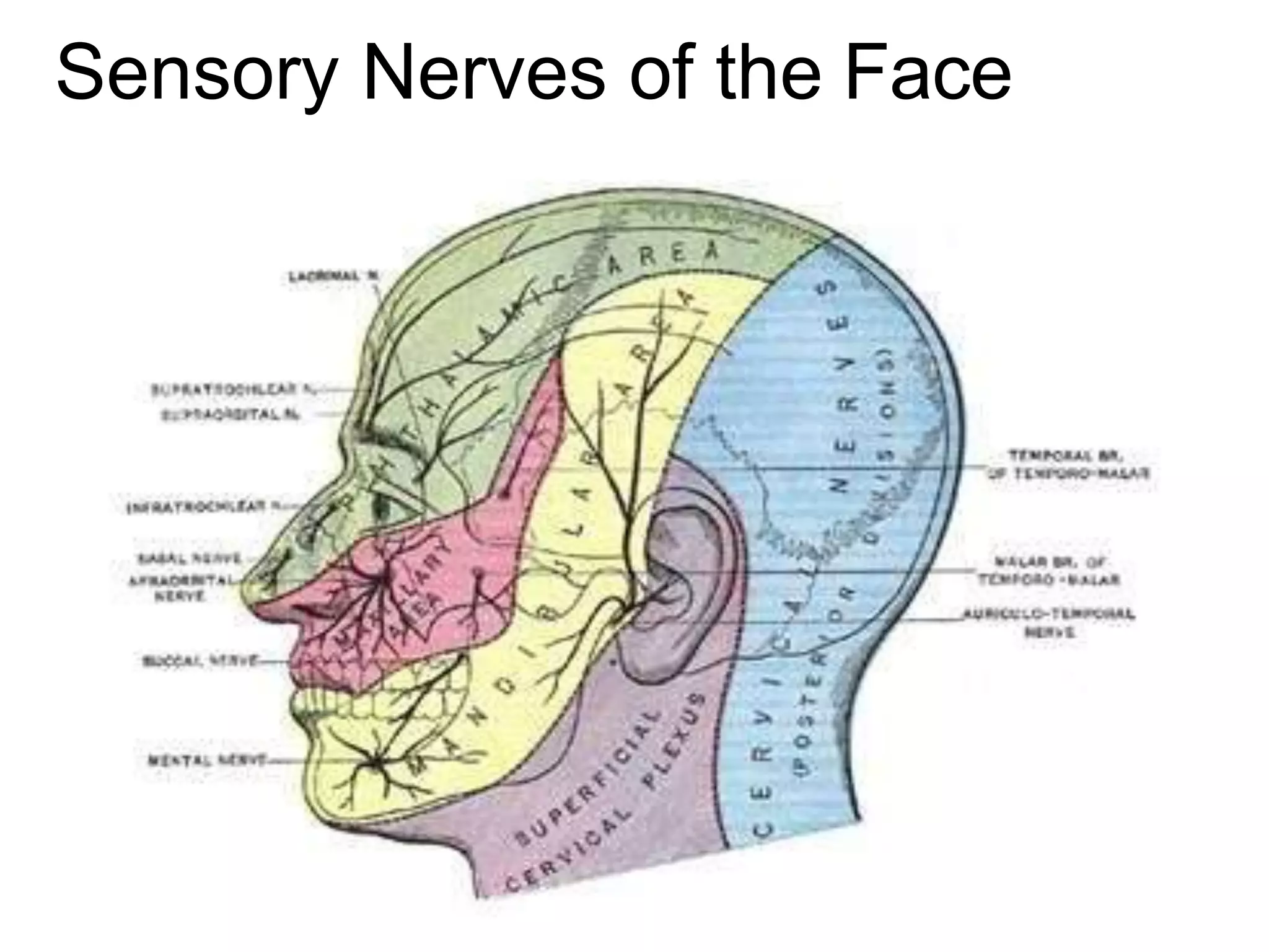
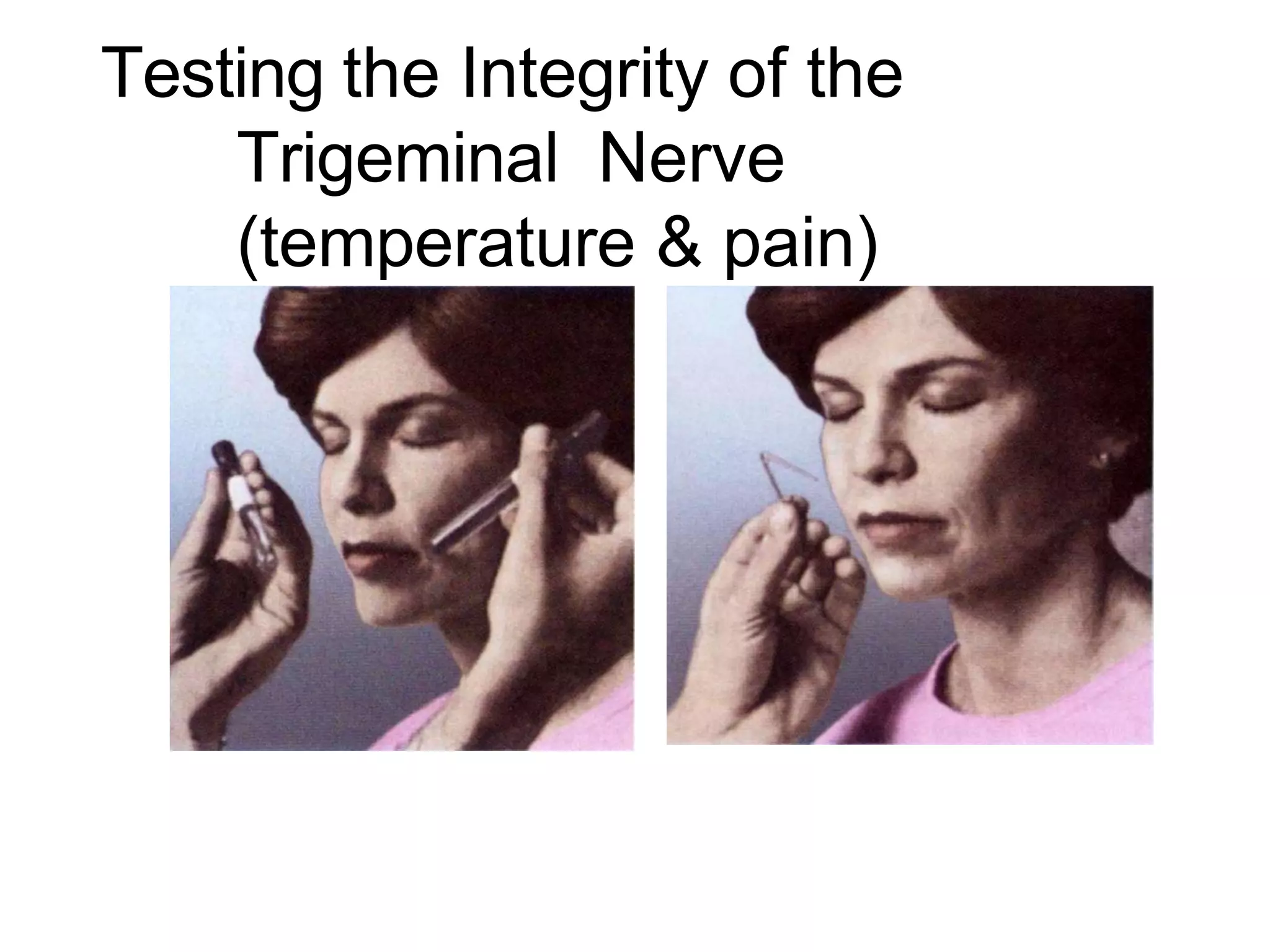
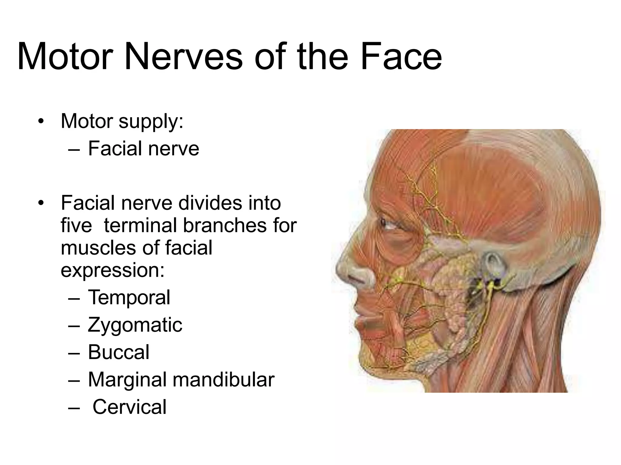
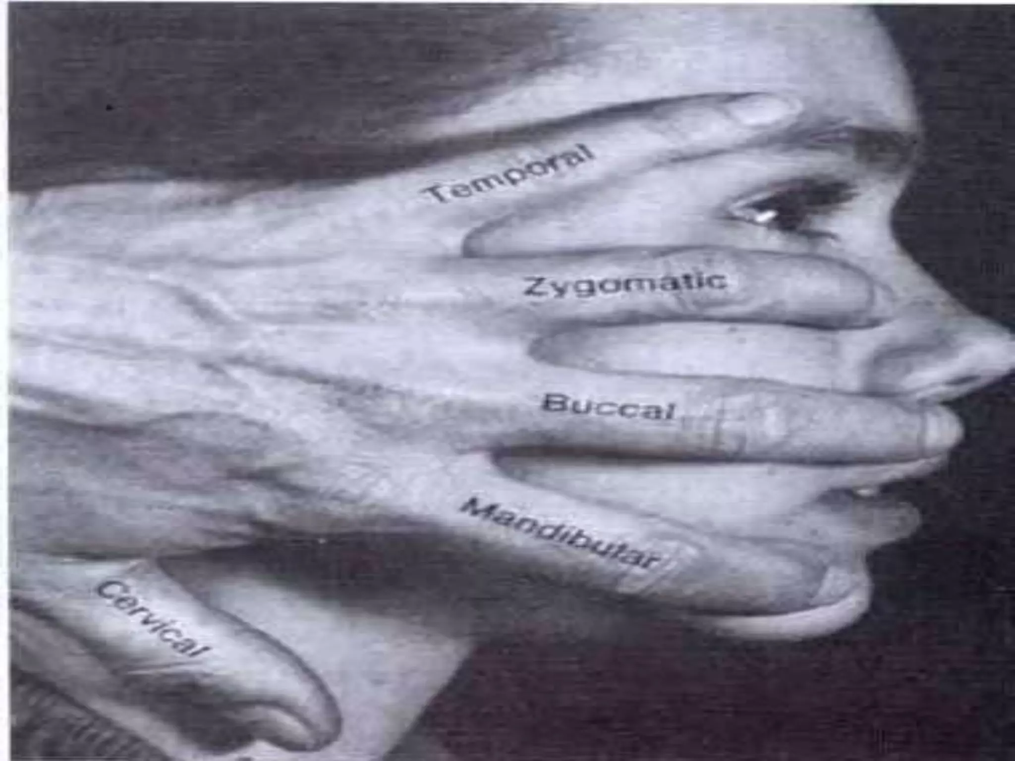
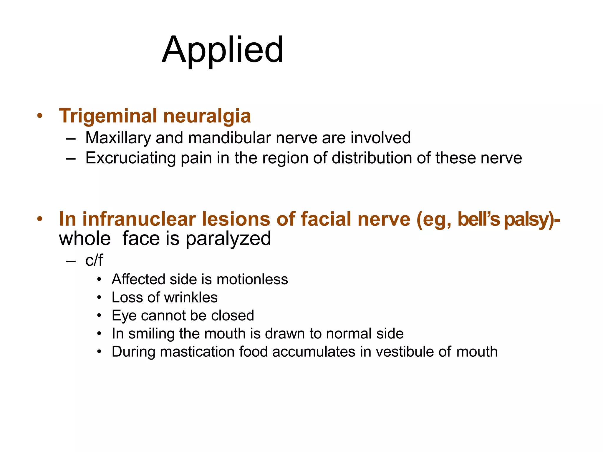
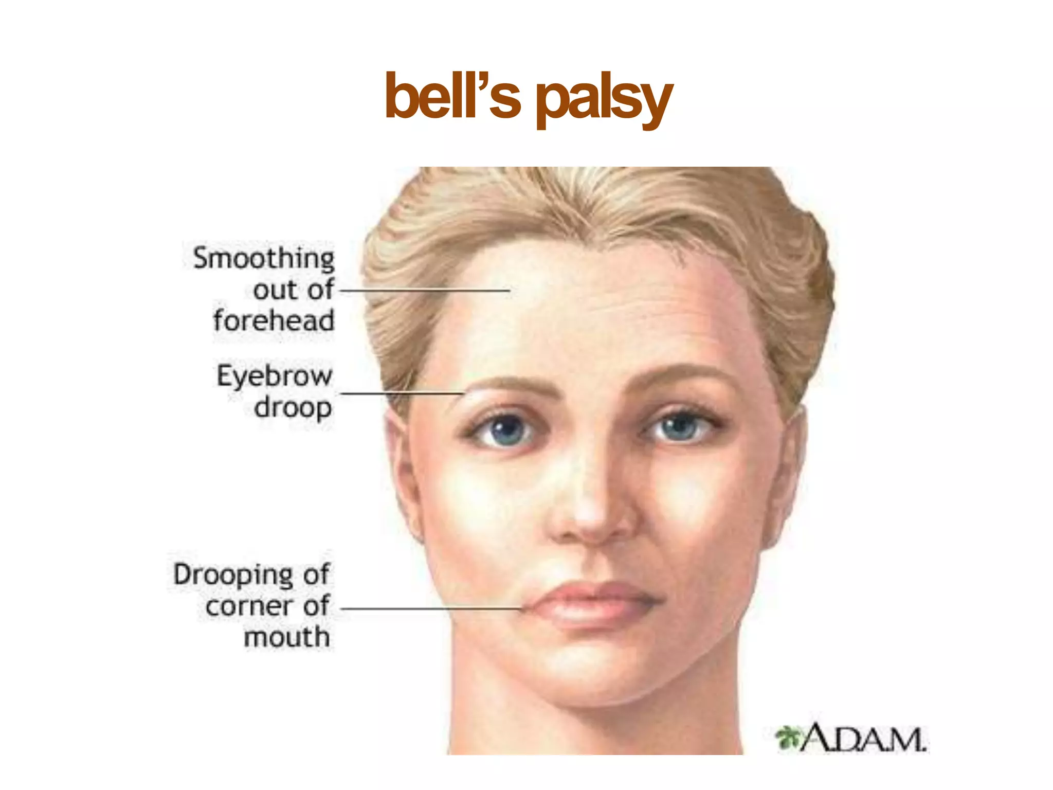
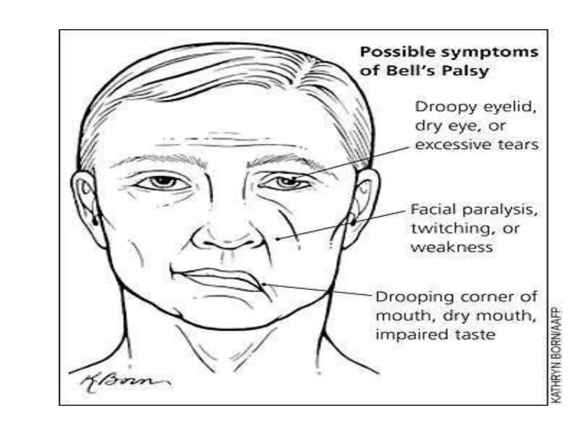
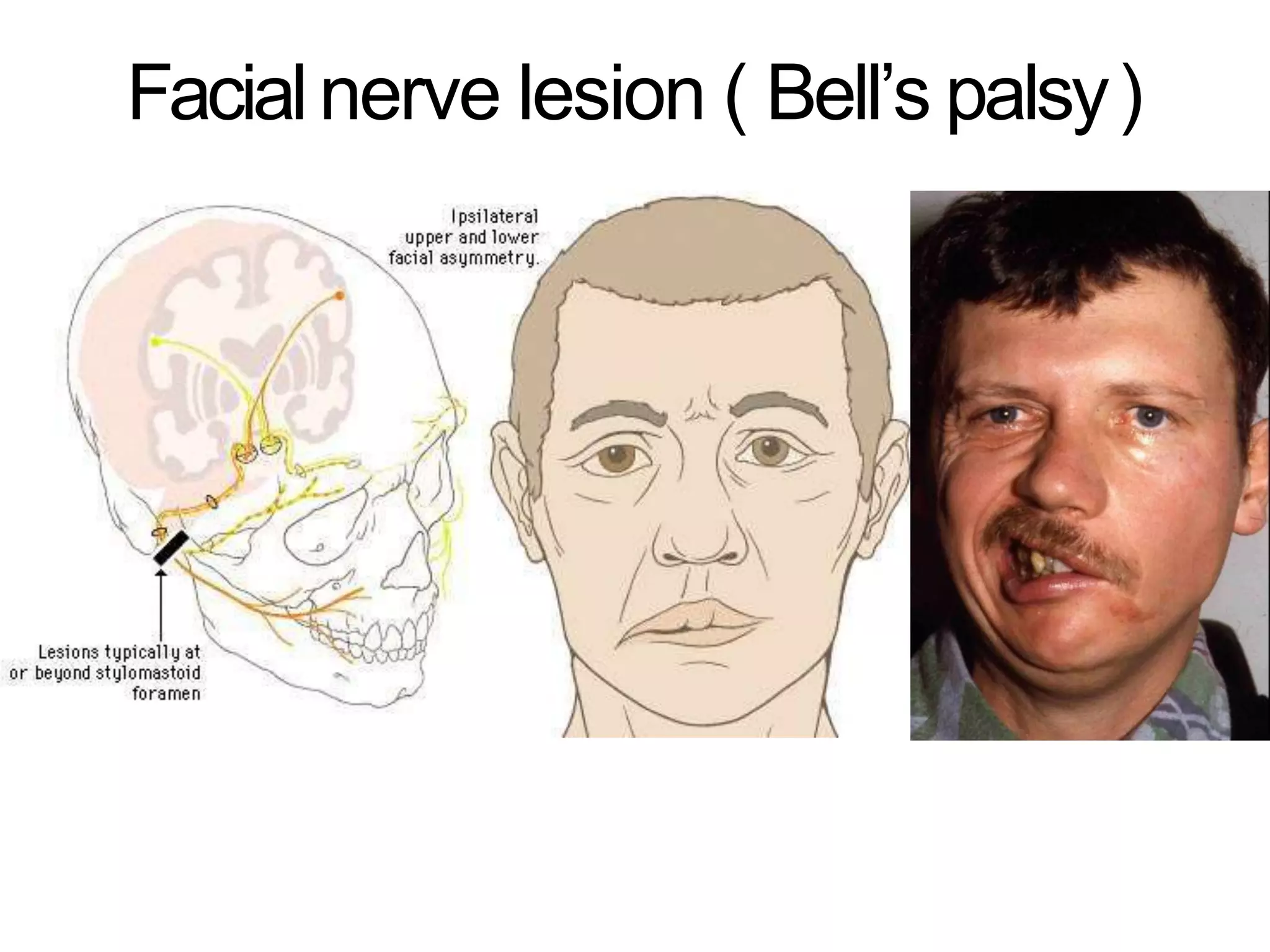
This document discusses the anatomy of the face. It covers the boundaries of the face and key facts about facial skin and wound healing. It then describes the muscles of facial expression, including the orbital, nasal, oral and other minor muscle groups. These muscles are innervated by the facial nerve. The document outlines the sensory and motor innervation of the face by branches of the trigeminal and facial nerves respectively. Finally, it provides examples of clinical conditions that involve the facial nerves, such as Bell's palsy where paralysis of the facial nerve causes an inability to close the eye or smile on the affected side.
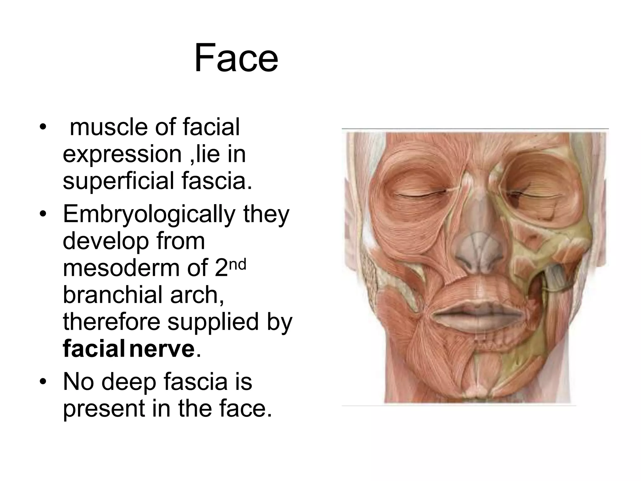
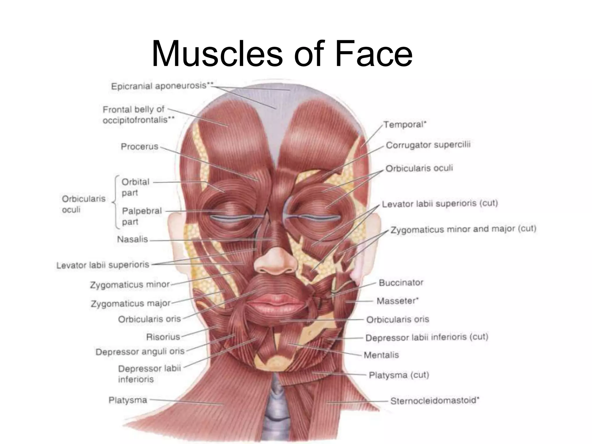
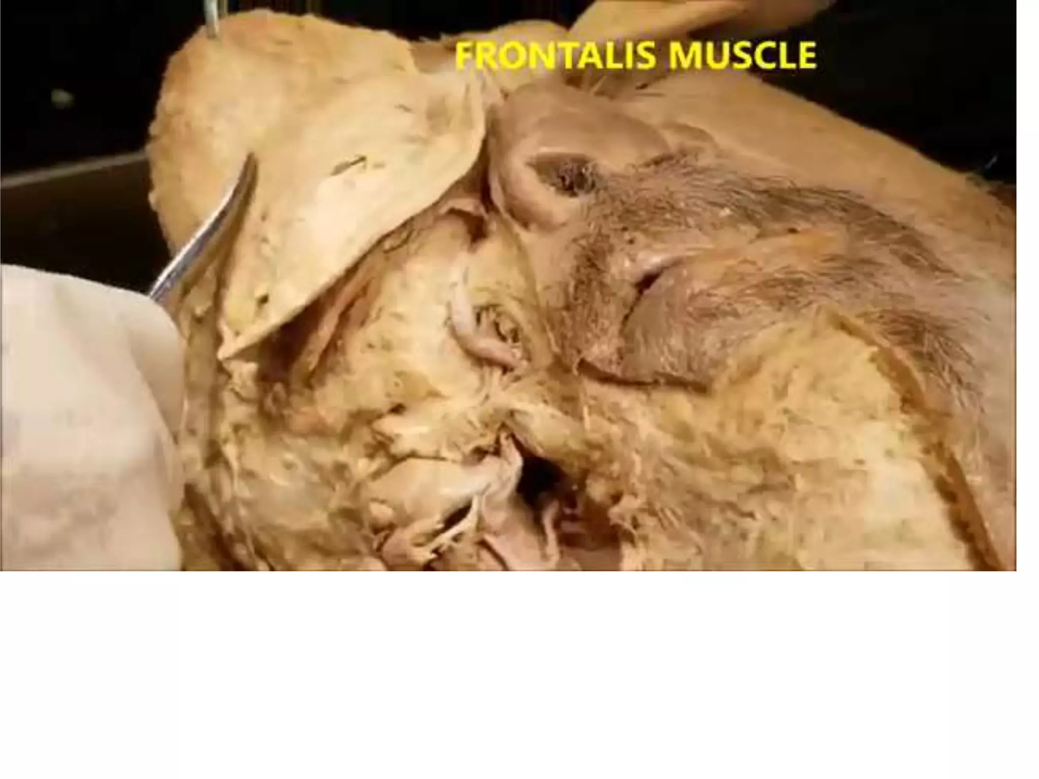
2. LEARNING
OBJECTIVES
• Muscles of facial expressions
• Arterial supply of face
• Venous supply of face
• Sensory and motor supply of face
• Clinical scenarios
3. Face
Boundaries
• Extends superiorly to
the hair line,
inferiorly to the chin
and base of
mandible, and on
each side to auricle
• Forehead is common
to both scalp and
face.
4. Face
• Due to rich
vascularity face
blush and blanch.
• Facial skin is rich
in sebaceous
gland and sweat
gland.
• Wounds of face bleed
profusely but heal
rapidly.
• Sebaceous gland keep
the skin oily but also
cause acne in adult.
5. Face
• muscle of facial
expression ,lie in
superficial fascia.
• Embryologically they
develop from
mesoderm of 2nd
branchial arch,
therefore supplied by
facialnerve.
• No deep fascia is
present in the face.
8. Muscles of the Face
• The muscles of the face develop from the
2nd pharyngeal arch and are innervated by
branches of the facial nerve [VII].
• They are in the superficial fascia, with
origins from either bone or fascia, and
insertions into the skin.
• these muscles control expressions of the
face.
• They act as sphincters and dilators of the
orifices of the face (i.e. the orbits, nose, and
mouth).
9.
10. Muscles of the
Face
• Orbital group
• Nasal group
• Oral group
• Other muscle
groups
13. Orbicularis
oculi• 3 parts-
• Orbital part(outer)
– Originate from medial part of medial palpebral ligament and form
concentric
rings, return to point of origin
Action –closes the lids tightly
• Palpebral part(Inner)
– Originate from lateral part of medial palpebral ligament
– Insert into lateral palpebral raphe
Action-closes the lids gently
• Lacrimal part(Small)
– Originate from lacrimal fascia& lacrimal bone
– Insert into upper &lower tarsi
Action-dilate lacrimal sac
17. Corrugator supercillii
• Origin : superciliary arch
• Insertion: skin of the
eyebrow
• Action: produces vertical
wrinkles of the forehead
in frowning as an
expression of annoyance
18. Nasal group
• Three muscles
are associated
with the nasal
group:
– Nasalis
– Procerus
– Depressor septi
nasi
20. Compressor naris
• Origin: Frontal process
of the maxilla
• Insertion: Aponeurosis
which crosses the bridge
of the nose
• Action: Compresses the
mobile nasal cartilages
21. Dilator naris
• Origin : Maxilla bone
• Insertion: Ala of the
nose
• Action: Widens the nasal
aperture (by pulling the
alar laterally) in deep
inspiration; is also a sign
of anger
22. Procerus
• Origin: nasal bone and
lateral nasal cartilage
• Insertion: skin
between the eyebrows
• Action:
– pulls down the medial
end of the eyebrow
– wrinkles the skin of the
nose transversely in
frowning
23. Oral group
• The muscles in the oral group move the lips and
cheek:
– Orbicularis oris
– Buccinator
– Lower group of oral muscles
• depressor anguli oris
• depressor labii inferioris
• Mentalis
– Upper group of oral muscles
• risorius
• zygomaticus major and zygomaticus minor
• levator labii superioris
• levator labii superioris alaeque nasi
• levator anguli oris
24. Orbicularis oris
• Origin: from
maxilla above
incisor teeth
• Insertion: into skin
of lip.
• Action: closes
the mouth
25. Buccinator
• Upper fibers
– Origin- from maxilla opposite
molar
teeth
– Insertion-upper lip
• Lower fibers
– Origin-from mandible opposite
molar teeth
– Insertion-lower lip
• Middle fibers
– Origin –from
pterigomandibular raphe
– Insertion-decussate before
passing to lips
• Action- it aids in mastication by
prevent accumulation of food in
vestibule of mouth. It is used every
time air expanding the cheeks is
forcefully expelled
26. Other muscle
groups
• They include:
– Platysma
– Auricular (anterior,
superior, and
posterior auricular
muscles)
– Occipitofrontalis
27. Platysma
• Origin– upper part
of pectoral and
deltoid fascia
• Insertion– base of
mandible, skin of
lower face and lip
• Action– releases
pressure of skin on the
subjacent veins,
depress
mandible, pulls angle
of
mouth downwards.
30. Sensory Nerves of the
Face
• The skin of the face is
supplied by the
trigeminal nerve (V),
except for the small area
over the angle of the
mandible and the
parotid gland which is
supplied by the great
auricular nerve (C2 and
3).
• The trigeminal nerve
(V) divides into three
major divisions-the
ophthalmic (V1),
maxillary (V2), and
34. Motor Nerves of the Face
• Motor supply:
– Facial nerve
• Facial nerve divides into
five terminal branches for
muscles of facial
expression:
– Temporal
– Zygomatic
– Buccal
– Marginal mandibular
– Cervical
35.
36. Applied
• Trigeminal neuralgia
– Maxillary and mandibular nerve are involved
– Excruciating pain in the region of distribution of these nerve
• In infranuclear lesions of facial nerve (eg, bell’spalsy)-
whole face is paralyzed
– c/f
• Affected side is motionless
• Loss of wrinkles
• Eye cannot be closed
• In smiling the mouth is drawn to normal side
• During mastication food accumulates in vestibule of mouth