SlideShare a Scribd company logo
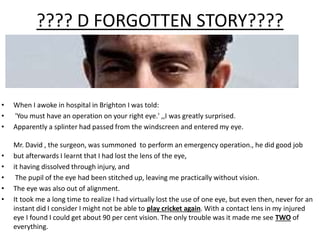
???? D FORGOTTEN STORY????
• When I awoke in hospital in Brighton I was told:
• 'You must have an operation on your right eye.' ,,I was greatly surprised.
• Apparently a splinter had passed from the windscreen and entered my eye.
Mr. David , the surgeon, was summoned to perform an emergency operation., he did good job
• but afterwards I learnt that I had lost the lens of the eye,
• it having dissolved through injury, and
• The pupil of the eye had been stitched up, leaving me practically without vision.
• The eye was also out of alignment.
• It took me a long time to realize I had virtually lost the use of one eye, but even then, never for an
instant did I consider I might not be able to play cricket again. With a contact lens in my injured
eye I found I could get about 90 per cent vision. The only trouble was it made me see TWO of
everything.
• What might have happened that day??
• Which all structures got damaged in orbit??
• Who is HE??? Is he really important!!!
??????*any guesses*?????
A TIGER WITH A SINGLE EYE!!!
ANATOMY OF THE ORBIT
Presenter:- Pushkar Dhir
Moderator:- Dr.Neha Khanduja
INTRODUCTION
• Quadrilateral pyramid shaped Bony cavities situated
one on either side of root of nose.
• Major functions –
1. Provide the socket for the rotatory movements of
the eye
2. Protect the eyeballs.
• Medial wall- parallel
& approximately 2.5cm away
• Lateral wall-90 degree.
• Medial with lateral- 45 degree.
• Orbital axis- 22.5 degree,
Divergent, Forwards and
laterally.
22.5
Optical Axis
Orbital Axis
• Entrance height-3.5cm
• Entrance width 4-4.5cm
DIMENSIONS
*Depthoforbitvariesfrom
4.4-5cm
BONY ORBIT
Composed of 7 bones-
1. Ethmoid
2. Frontal
3. Lacrimal
4. Maxilla
5. Palatine
6. Sphenoid
7. Zygomatic
.
Orbital RIM
• Superior orbital rim -> frontal bone.
• Inferior orbital rim -> maxillary
bone medially
and zygomatic
bone laterally.
• Medial orbital rim -> Frontal process of
maxilla
• Lateral orbital rim -> Zygoma
• In Medial 1/3rd
of Superior orbital rim Supraorbital notch.
(supraorbital nerve & artery passes
to forehead)
Orbital Roof
• 1.Orbital plate of Frontal bone & 2.Lesser wing of sphenoid.
• Relations- anterior cranial fossa (frontal lobe & meninges),frontal sinus, frontal nerve,
trochlear nerve and supraorbital artery.
Landmarks-
1.Fossa for lacrimal gland. 2. Fovea or trochlear fossa.
3.Supraorbital notch. 4. Optic Foramen(+nt in lesser wing)
Applied anat :-Defect in roof- pulsatile proptosis due to transmission of CSF pulsation to
orbit.
•FOSSA FOR THE LACRIMAL GLAND-
•LOCATION: behind the zygomatic
process of the frontal
bone
TROCHLEAR FOSSA (FOVEA)
LOCATION: 4 mm from the
orbital margin
CONTENTS: insertion of
tendinous pulley of
Superior Oblique
(In sme cases there is a
spicule of bone (Spina
trochlearis)
Nasolacrimal
sac & duct
Cribra orbitalia:
-apertures apparent on the medial side of anterior portion of the lacrimal fossa
-for veins from diploë to the orbit ; Best marked in the fetus and infant.
At JUNCTION OF THE ROOF AND MEDIAL WALL, the suture line lies in proximity to
CRIBRIFORM PLATE of ethmoid RUPTURE of dura mater CSF enter orbit/nose.
Lacrimal bone is easily
penetrated during ENDO
DCR
*If maxillary
component is
predominenet
then its
difficult to do
EDNO DCR
1
2 3
4
Fronto-ethmoidal suture line:- dissection done above this
line will lead to cranial cavity
Ant.&
Post.
lacrimal
crest
Weber`s
Suture
Branches of
infraorbital artery
pass via this groove
to supply the nasal
mucosa.
Bleeding may occur
4m these vessels
during DCR surgeries
• Front - Backwards- Frontal process of Maxilla, Lacrimal bone , Orbital plate
of Ethmoid (Largest), Lesser wing of sphenoid.
Applied anatomy-
1. Thinnest portion of wall - Lamina papyracea (it is component of ethmoid
bone). Fractured in blow out fractures.
(Thick posteriorly at sphenoid and anteriorly at lacrimal crest.)
2. Infections and neoplasms of ethmoid sinus-orbital cellulitis and proptosis.
3. Weber`s Suture :- Also known as sutura longitudinalis imperfecta
Runs parallel to anterior lacrimal crest.
Ant. & Post.ethmoidal sinus is located 24mm &36 mm from anterior lacrimal crest respectively.
Ant. & Post. Ehtmoidal arteries pass through it.
CLINICAL SIGNIFICANCE OF MEDIAL WALL
• Medial wall extremely fragile (presence of ethmoidal air cells and nasal cavity)
• Accidental lateral displacement of medial wall- traumatic hypertelorism
• Medial wall provides alternate access route to the orbit through the sinus
• Ethmoid
- Thinnest bone of the orbit
- Vascular connections with ethmoid sinus through foramina
- Inflammation in the ethmoid sinus spreads readily to the orbit
• Tumours of the nasal cavity can breach the lamina papyracea to involve the orbit
• Lacrimal bone can be easily penetrated during endoscopic DCR
• During surgery, hemorrhage is most troublesome due to injury to ethmoidal
vessels.
ORBITAL FLOOR
•Formed by-
•Orbital plate of maxilla (major)
•Orbital surface of Zygomatic bone (anterolateral)
•Orbital plate of Palatine bone
Shortest orbital wall Traingular
• Landmarks-
 Inferior orbital fissure, end backward in pterygopalatine fossa.
 Infraorbital groove (post) and canal (ant)
• Applied anatomy-
1. As it is roof of maxillary sinus- 0.5-1mm thick. Maxillary carcinoma invading up in
orbit may cause proptosis.
2. Blow out fracture- infraorbital nerves & vessels
usually get involved.
C/F:- Diplopia
Restricted movements(upgaze)
Paresthesia
Enophthalmos
ORBIT LATERAL WALL
• Thickest and strongest.
Greater wing of sphenoid
and zygomatic bone forms it
• Applied anatomy- Anterior
half of globe is vulnerable to
lateral trauma.
• Landmarks-
 Whitnall’s tubercle-
zygomatic bone.
 4-5 mm behind lateral orbital
rim & 1 cm below fronto-
zygomatic suture line.
 Structure attaching:-
1. Lateral canthal tendon
2. Lateral rectus check
ligament.
3. Suspensory ligament of
lower eyelid (lockwoods
ligament)
4. Orbital septum
5. Lacrimal gland fascia
*C/S:-
Tubercle is
spared in
maxillary
resection in
CA, as it gives
attachment to
lockwood etc,
can lead to
diplopia if
resected.
2.The Spina recti lateralis :—
• Its a small bony projection situated on the inferior margin of the SOF at the
junction of its wide and narrow portions.
• Gives rise to lateral rectus muscle.
3.Zygomatic Groove
• Extent:-From the anterior end of the inferior orbital fissure to a foramen in the
zygomatic bone.
• Contents:
- Zygomatic nerve
- Zygomatic vessel.
• Lateral wall protects ONLY THE POSTERIOR HALF of the eyeball, hence palpation of
retrobulbar tumours is easier.
• Frontal process of zygoma & zygomatic process of frontal bone protect the globe from lateral
trauma- known as facial buttress area.
• Just behind the facial buttress area, is the zygomaticosphenoid suture, which is the preferred
site for lateral orbitotomy.
C/S of Lateral Wall
ORBITAL FISSURE & FORAMINA
*Also k/a
Spenoidal fissure
*Structure Passing :-
(Superior LFT + NAO)
*C/S :- Tolosa Hunt Syndrome
(Inflammation of the superior
orbital fissure and apex 
ophthalmoplegia &venous
outflow obstruction.
*Superior Orbital Syndrome
(Rochon-duvigneaud Syndrome)-
# of SOF  CN involves
Diplopia,ophthalmoplegia,exopht
halmos,ptosis
*Manner of involvement of nerves helps to
predict the site and extent of the lesion.
Divisions of III’rd nerve ± VI’th nerve
Annulus of Zinn (Purely intraconal lesion)
III’rd, IV’th and VI’th nerve
Entire length of the fissure involved
IOF/Sphenomaxillary Fissure
1.Venous drainage from the inferior part of the orbit
to
the pterygoid plexus
2.neural branches from the pterygopalatine ganglion
3.zygomatic nerve
4. infraorbital nerve
Connective Tissue System of Orbit
1. Periorbita/Orbital Periosteum
*Loosely adherent to the bones
*Sensory innervation by branches of V’th
nerve
Applied Anat:- Tumors and pus
collected in the subperiosteal space
cause thickening of subperiosteum
and can cause proptosis and elevated
IOP.
• Eg- dermoid cyst, epidermoid cyst,
mucocele, subperiosteal abcess,
myeloma, osteomatous tumors,
haematoma and fibrous dysplasia.
• Plain X- rays most useful in diagnosis.
2. Orbital septal system
Includes the connective tissue septa
which are suspended from the periorbita
to form a complex radial and
circumferential interconnecting slings.
3. TENON’S CAPSULE( FASCIA BULBI OR BULBAR SHEATH)
• Dense, elastic and vascular connective tissue that surrounds the globe (except over the cornea).
• Begins anteriorly at the perilimbal sclera, extends around the globe to the optic nerve, and fuses
with the dural sheath and the sclera.
• Separated from the sclera by periscleral lymph space, which is in continuation with subdural and
subarachnoid spaces
• Applied anatomy-
1. After enucleation, implants are placed within tenon’s capsule or posterior to it within muscle cone.
2. Inflammatory pseudotumor cause florid tenonitis to cause proptosis.
ORBITAL SPACES
CENTRAL SPACE
Within the muscle cone
SUBTENONS SPACE
Space b/w sclera &
sub tenon capsule
Contents-
1. ON and its meninges.
2. Sup and inf- oculomotor nerve.
3. Abducent
4. Nasociliary
5. Ciliary ganglion
6. Ophthalmic artery
7. SOV
8. Central orbital fat
Applied Anat:-
1. Tumors- axial proptosis. Eg-
cavernous haemangioma,
solitary neurofibroma,
neurilemmoma, schwannoma,
ON glioma
*Pus collected in this space is
drained by incision of Tenon’s
capsule through the conjunctiva
-*Site for drug instillation
• Thanks for listening to anatomy of
D orbit!!!!!!...nt dis
one genius..
Comp: oh…m sry
Grrr…r u
sleepy…??? Put
the right slide
Comp: ok
The
ORBIT

More Related Content

What's hot

Anatomy of orbit
Anatomy of orbitAnatomy of orbit
Anatomy of orbit
veeru1984
 
Anatomy of the eyelids
Anatomy of the eyelids Anatomy of the eyelids
Anatomy of the eyelids
Vaibhav Kanduri
 
Anatomy and physiology of extraocular muscles
Anatomy and physiology of extraocular musclesAnatomy and physiology of extraocular muscles
Anatomy and physiology of extraocular muscles
priyanka bharti
 
Anatomy of the eyelids.
Anatomy of the eyelids.Anatomy of the eyelids.
Anatomy of the eyelids.
samirlal
 
Orbit Anatomy and Surgical Spaces
Orbit Anatomy and Surgical Spaces Orbit Anatomy and Surgical Spaces
Orbit Anatomy and Surgical Spaces
Vikas Khatri
 
Orbital blowout fracture
Orbital blowout fracture Orbital blowout fracture
Orbital blowout fracture
Cairo university
 
Orbital anatomy
Orbital anatomyOrbital anatomy
Orbital anatomy
Dr Kawshik Nag
 
Retrobulbar haemorrhage
Retrobulbar haemorrhageRetrobulbar haemorrhage
Retrobulbar haemorrhage
DrRudra Chakraborty
 
Orbital spaces
Orbital spacesOrbital spaces
Orbital spaces
ankita mahapatra
 
Anatomy of Orbit
Anatomy of Orbit Anatomy of Orbit
Anatomy of Orbit
Ashwani Kumar
 
Anatomy of pupillary pathways
Anatomy of pupillary pathwaysAnatomy of pupillary pathways
Anatomy of pupillary pathwaysHasika Ravula
 
Anatomy of optic nerve
Anatomy of optic nerveAnatomy of optic nerve
Anatomy of optic nerve
Sudheer Kumar
 
Keratoprosthesis
KeratoprosthesisKeratoprosthesis
Keratoprosthesis
peterroy90
 
Proptosis investigation & management.rdh
Proptosis investigation & management.rdhProptosis investigation & management.rdh
Proptosis investigation & management.rdh
radhe4827
 
Anatomy of orbit
Anatomy of orbitAnatomy of orbit
Anatomy of orbit
ChrisPius
 
Dacryocystorhinostomy
DacryocystorhinostomyDacryocystorhinostomy
Dacryocystorhinostomy
atin bindal
 
Anatomy of orbit ophthalm
Anatomy of orbit ophthalmAnatomy of orbit ophthalm
Anatomy of orbit ophthalm
DrArino John
 

What's hot (20)

Anatomy of orbit
Anatomy of orbitAnatomy of orbit
Anatomy of orbit
 
Anatomy of the eyelids
Anatomy of the eyelids Anatomy of the eyelids
Anatomy of the eyelids
 
Anatomy and physiology of extraocular muscles
Anatomy and physiology of extraocular musclesAnatomy and physiology of extraocular muscles
Anatomy and physiology of extraocular muscles
 
Anatomy of the eyelids.
Anatomy of the eyelids.Anatomy of the eyelids.
Anatomy of the eyelids.
 
Orbit Anatomy and Surgical Spaces
Orbit Anatomy and Surgical Spaces Orbit Anatomy and Surgical Spaces
Orbit Anatomy and Surgical Spaces
 
Orbital blowout fracture
Orbital blowout fracture Orbital blowout fracture
Orbital blowout fracture
 
Orbital anatomy
Orbital anatomyOrbital anatomy
Orbital anatomy
 
Retrobulbar haemorrhage
Retrobulbar haemorrhageRetrobulbar haemorrhage
Retrobulbar haemorrhage
 
Orbital spaces
Orbital spacesOrbital spaces
Orbital spaces
 
Anatomy of Orbit
Anatomy of Orbit Anatomy of Orbit
Anatomy of Orbit
 
Anatomy of pupillary pathways
Anatomy of pupillary pathwaysAnatomy of pupillary pathways
Anatomy of pupillary pathways
 
Anatomy of optic nerve
Anatomy of optic nerveAnatomy of optic nerve
Anatomy of optic nerve
 
Keratoprosthesis
KeratoprosthesisKeratoprosthesis
Keratoprosthesis
 
Orbit
OrbitOrbit
Orbit
 
Proptosis investigation & management.rdh
Proptosis investigation & management.rdhProptosis investigation & management.rdh
Proptosis investigation & management.rdh
 
Anatomy of orbit
Anatomy of orbitAnatomy of orbit
Anatomy of orbit
 
Dacryocystorhinostomy
DacryocystorhinostomyDacryocystorhinostomy
Dacryocystorhinostomy
 
Anatomy of orbit ophthalm
Anatomy of orbit ophthalmAnatomy of orbit ophthalm
Anatomy of orbit ophthalm
 
Retina
RetinaRetina
Retina
 
Abducent nerve
Abducent nerveAbducent nerve
Abducent nerve
 

Similar to Anatomy of orbit by pushkar dhir

By pd anatomy of the orbit
By pd anatomy of the orbitBy pd anatomy of the orbit
By pd anatomy of the orbitPushkar Dhir
 
Anatomy of orbit
Anatomy of orbitAnatomy of orbit
Anatomy of orbit
veeru1984
 
Anatomy of orbit and its clinical applications.
Anatomy of orbit and its clinical applications.Anatomy of orbit and its clinical applications.
Anatomy of orbit and its clinical applications.
RahulGupta39947
 
The Orbit
The OrbitThe Orbit
The Orbit
BirkhaBogati
 
The Orbit
The OrbitThe Orbit
Orbital surgeries
Orbital surgeriesOrbital surgeries
Orbital surgeries
PRAKRITIYAGNAM
 
orbit anatomy.pptx
orbit anatomy.pptxorbit anatomy.pptx
orbit anatomy.pptx
SouvikMukherjee95
 
Orbital anatomy and orbital frcture
Orbital anatomy and orbital frctureOrbital anatomy and orbital frcture
Orbital anatomy and orbital frcture
Indian dental academy
 
ORBIT.pptx
ORBIT.pptxORBIT.pptx
ORBIT.pptx
KAZIMAHBUBURRASHID
 
Antomy of orbit 25 4-19
Antomy of orbit 25 4-19Antomy of orbit 25 4-19
Antomy of orbit 25 4-19
Dr. Devi Shankar
 
orbit malavika NEW.pptx
orbit malavika NEW.pptxorbit malavika NEW.pptx
orbit malavika NEW.pptx
MalavikaAG
 
Anatomy of orbit
Anatomy of orbitAnatomy of orbit
Anatomy of orbit
Arjun Sapkota
 
Orbit anatomy
Orbit anatomyOrbit anatomy
Orbit anatomy
Prajakta Matey
 
anatomy of orbital
anatomy of orbital anatomy of orbital
Orbit final
Orbit finalOrbit final
Orbit final
sauvik2014
 
Orbital apex syndrome
Orbital apex syndromeOrbital apex syndrome
Orbital apex syndrome
Reshma Peter
 
Anatomy of Orbit and its clinical importance
Anatomy of Orbit and its clinical importanceAnatomy of Orbit and its clinical importance
Anatomy of Orbit and its clinical importance
Ashish Gupta
 
Orbital structure
Orbital structure Orbital structure
Orbital structure
Frenky Ramiro
 
Anatomy of the Bony orbit
Anatomy of the Bony orbit Anatomy of the Bony orbit
Anatomy of the Bony orbit
Dr.Nischal Ghimire
 
Anatomy of the orbit
Anatomy of the orbit Anatomy of the orbit
Anatomy of the orbit
SAMEEKSHA AGRAWAL
 

Similar to Anatomy of orbit by pushkar dhir (20)

By pd anatomy of the orbit
By pd anatomy of the orbitBy pd anatomy of the orbit
By pd anatomy of the orbit
 
Anatomy of orbit
Anatomy of orbitAnatomy of orbit
Anatomy of orbit
 
Anatomy of orbit and its clinical applications.
Anatomy of orbit and its clinical applications.Anatomy of orbit and its clinical applications.
Anatomy of orbit and its clinical applications.
 
The Orbit
The OrbitThe Orbit
The Orbit
 
The Orbit
The OrbitThe Orbit
The Orbit
 
Orbital surgeries
Orbital surgeriesOrbital surgeries
Orbital surgeries
 
orbit anatomy.pptx
orbit anatomy.pptxorbit anatomy.pptx
orbit anatomy.pptx
 
Orbital anatomy and orbital frcture
Orbital anatomy and orbital frctureOrbital anatomy and orbital frcture
Orbital anatomy and orbital frcture
 
ORBIT.pptx
ORBIT.pptxORBIT.pptx
ORBIT.pptx
 
Antomy of orbit 25 4-19
Antomy of orbit 25 4-19Antomy of orbit 25 4-19
Antomy of orbit 25 4-19
 
orbit malavika NEW.pptx
orbit malavika NEW.pptxorbit malavika NEW.pptx
orbit malavika NEW.pptx
 
Anatomy of orbit
Anatomy of orbitAnatomy of orbit
Anatomy of orbit
 
Orbit anatomy
Orbit anatomyOrbit anatomy
Orbit anatomy
 
anatomy of orbital
anatomy of orbital anatomy of orbital
anatomy of orbital
 
Orbit final
Orbit finalOrbit final
Orbit final
 
Orbital apex syndrome
Orbital apex syndromeOrbital apex syndrome
Orbital apex syndrome
 
Anatomy of Orbit and its clinical importance
Anatomy of Orbit and its clinical importanceAnatomy of Orbit and its clinical importance
Anatomy of Orbit and its clinical importance
 
Orbital structure
Orbital structure Orbital structure
Orbital structure
 
Anatomy of the Bony orbit
Anatomy of the Bony orbit Anatomy of the Bony orbit
Anatomy of the Bony orbit
 
Anatomy of the orbit
Anatomy of the orbit Anatomy of the orbit
Anatomy of the orbit
 

More from Pushkar Dhir

Siderosis &chalcosis & IOFB
Siderosis &chalcosis & IOFBSiderosis &chalcosis & IOFB
Siderosis &chalcosis & IOFB
Pushkar Dhir
 
Protocol s
Protocol sProtocol s
Protocol s
Pushkar Dhir
 
Cilia
CiliaCilia
Importance of counselling in eye care services100
Importance of counselling in eye care services100Importance of counselling in eye care services100
Importance of counselling in eye care services100
Pushkar Dhir
 
Orthoptic evaluation 1
Orthoptic evaluation 1Orthoptic evaluation 1
Orthoptic evaluation 1
Pushkar Dhir
 
By pd examination of a squint
By pd examination of a squintBy pd examination of a squint
By pd examination of a squint
Pushkar Dhir
 
B scan by Pushkar Dhir
B scan by Pushkar DhirB scan by Pushkar Dhir
B scan by Pushkar Dhir
Pushkar Dhir
 
History & materials of conatct lens by pushkar dhir
History & materials of conatct lens by pushkar dhirHistory & materials of conatct lens by pushkar dhir
History & materials of conatct lens by pushkar dhir
Pushkar Dhir
 
By pd aphakia & pseudophakia
By pd aphakia & pseudophakiaBy pd aphakia & pseudophakia
By pd aphakia & pseudophakiaPushkar Dhir
 
Npcb by pushkar dhir
Npcb by pushkar dhirNpcb by pushkar dhir
Npcb by pushkar dhirPushkar Dhir
 
Extra ocu.movement by pushkar dhir
Extra ocu.movement by pushkar dhirExtra ocu.movement by pushkar dhir
Extra ocu.movement by pushkar dhirPushkar Dhir
 
Visual acuity by pd
Visual acuity by pdVisual acuity by pd
Visual acuity by pdPushkar Dhir
 
Penetrating keratoplasty by pushkar dhir
Penetrating keratoplasty by pushkar dhirPenetrating keratoplasty by pushkar dhir
Penetrating keratoplasty by pushkar dhirPushkar Dhir
 
Examination of pupil by pushkar dhir
Examination of pupil by pushkar dhirExamination of pupil by pushkar dhir
Examination of pupil by pushkar dhirPushkar Dhir
 

More from Pushkar Dhir (15)

Siderosis &chalcosis & IOFB
Siderosis &chalcosis & IOFBSiderosis &chalcosis & IOFB
Siderosis &chalcosis & IOFB
 
Protocol s
Protocol sProtocol s
Protocol s
 
Cilia
CiliaCilia
Cilia
 
Importance of counselling in eye care services100
Importance of counselling in eye care services100Importance of counselling in eye care services100
Importance of counselling in eye care services100
 
Orthoptic evaluation 1
Orthoptic evaluation 1Orthoptic evaluation 1
Orthoptic evaluation 1
 
By pd examination of a squint
By pd examination of a squintBy pd examination of a squint
By pd examination of a squint
 
B scan by Pushkar Dhir
B scan by Pushkar DhirB scan by Pushkar Dhir
B scan by Pushkar Dhir
 
History & materials of conatct lens by pushkar dhir
History & materials of conatct lens by pushkar dhirHistory & materials of conatct lens by pushkar dhir
History & materials of conatct lens by pushkar dhir
 
Retinoscopy
RetinoscopyRetinoscopy
Retinoscopy
 
By pd aphakia & pseudophakia
By pd aphakia & pseudophakiaBy pd aphakia & pseudophakia
By pd aphakia & pseudophakia
 
Npcb by pushkar dhir
Npcb by pushkar dhirNpcb by pushkar dhir
Npcb by pushkar dhir
 
Extra ocu.movement by pushkar dhir
Extra ocu.movement by pushkar dhirExtra ocu.movement by pushkar dhir
Extra ocu.movement by pushkar dhir
 
Visual acuity by pd
Visual acuity by pdVisual acuity by pd
Visual acuity by pd
 
Penetrating keratoplasty by pushkar dhir
Penetrating keratoplasty by pushkar dhirPenetrating keratoplasty by pushkar dhir
Penetrating keratoplasty by pushkar dhir
 
Examination of pupil by pushkar dhir
Examination of pupil by pushkar dhirExamination of pupil by pushkar dhir
Examination of pupil by pushkar dhir
 

Recently uploaded

Instructions for Submissions thorugh G- Classroom.pptx
Instructions for Submissions thorugh G- Classroom.pptxInstructions for Submissions thorugh G- Classroom.pptx
Instructions for Submissions thorugh G- Classroom.pptx
Jheel Barad
 
How to Make a Field invisible in Odoo 17
How to Make a Field invisible in Odoo 17How to Make a Field invisible in Odoo 17
How to Make a Field invisible in Odoo 17
Celine George
 
Chapter 3 - Islamic Banking Products and Services.pptx
Chapter 3 - Islamic Banking Products and Services.pptxChapter 3 - Islamic Banking Products and Services.pptx
Chapter 3 - Islamic Banking Products and Services.pptx
Mohd Adib Abd Muin, Senior Lecturer at Universiti Utara Malaysia
 
Additional Benefits for Employee Website.pdf
Additional Benefits for Employee Website.pdfAdditional Benefits for Employee Website.pdf
Additional Benefits for Employee Website.pdf
joachimlavalley1
 
Digital Tools and AI for Teaching Learning and Research
Digital Tools and AI for Teaching Learning and ResearchDigital Tools and AI for Teaching Learning and Research
Digital Tools and AI for Teaching Learning and Research
Vikramjit Singh
 
Adversarial Attention Modeling for Multi-dimensional Emotion Regression.pdf
Adversarial Attention Modeling for Multi-dimensional Emotion Regression.pdfAdversarial Attention Modeling for Multi-dimensional Emotion Regression.pdf
Adversarial Attention Modeling for Multi-dimensional Emotion Regression.pdf
Po-Chuan Chen
 
Mule 4.6 & Java 17 Upgrade | MuleSoft Mysore Meetup #46
Mule 4.6 & Java 17 Upgrade | MuleSoft Mysore Meetup #46Mule 4.6 & Java 17 Upgrade | MuleSoft Mysore Meetup #46
Mule 4.6 & Java 17 Upgrade | MuleSoft Mysore Meetup #46
MysoreMuleSoftMeetup
 
Polish students' mobility in the Czech Republic
Polish students' mobility in the Czech RepublicPolish students' mobility in the Czech Republic
Polish students' mobility in the Czech Republic
Anna Sz.
 
2024.06.01 Introducing a competency framework for languag learning materials ...
2024.06.01 Introducing a competency framework for languag learning materials ...2024.06.01 Introducing a competency framework for languag learning materials ...
2024.06.01 Introducing a competency framework for languag learning materials ...
Sandy Millin
 
The French Revolution Class 9 Study Material pdf free download
The French Revolution Class 9 Study Material pdf free downloadThe French Revolution Class 9 Study Material pdf free download
The French Revolution Class 9 Study Material pdf free download
Vivekanand Anglo Vedic Academy
 
CACJapan - GROUP Presentation 1- Wk 4.pdf
CACJapan - GROUP Presentation 1- Wk 4.pdfCACJapan - GROUP Presentation 1- Wk 4.pdf
CACJapan - GROUP Presentation 1- Wk 4.pdf
camakaiclarkmusic
 
Unit 8 - Information and Communication Technology (Paper I).pdf
Unit 8 - Information and Communication Technology (Paper I).pdfUnit 8 - Information and Communication Technology (Paper I).pdf
Unit 8 - Information and Communication Technology (Paper I).pdf
Thiyagu K
 
Acetabularia Information For Class 9 .docx
Acetabularia Information For Class 9  .docxAcetabularia Information For Class 9  .docx
Acetabularia Information For Class 9 .docx
vaibhavrinwa19
 
BÀI TẬP BỔ TRỢ TIẾNG ANH GLOBAL SUCCESS LỚP 3 - CẢ NĂM (CÓ FILE NGHE VÀ ĐÁP Á...
BÀI TẬP BỔ TRỢ TIẾNG ANH GLOBAL SUCCESS LỚP 3 - CẢ NĂM (CÓ FILE NGHE VÀ ĐÁP Á...BÀI TẬP BỔ TRỢ TIẾNG ANH GLOBAL SUCCESS LỚP 3 - CẢ NĂM (CÓ FILE NGHE VÀ ĐÁP Á...
BÀI TẬP BỔ TRỢ TIẾNG ANH GLOBAL SUCCESS LỚP 3 - CẢ NĂM (CÓ FILE NGHE VÀ ĐÁP Á...
Nguyen Thanh Tu Collection
 
How libraries can support authors with open access requirements for UKRI fund...
How libraries can support authors with open access requirements for UKRI fund...How libraries can support authors with open access requirements for UKRI fund...
How libraries can support authors with open access requirements for UKRI fund...
Jisc
 
Guidance_and_Counselling.pdf B.Ed. 4th Semester
Guidance_and_Counselling.pdf B.Ed. 4th SemesterGuidance_and_Counselling.pdf B.Ed. 4th Semester
Guidance_and_Counselling.pdf B.Ed. 4th Semester
Atul Kumar Singh
 
The basics of sentences session 5pptx.pptx
The basics of sentences session 5pptx.pptxThe basics of sentences session 5pptx.pptx
The basics of sentences session 5pptx.pptx
heathfieldcps1
 
Palestine last event orientationfvgnh .pptx
Palestine last event orientationfvgnh .pptxPalestine last event orientationfvgnh .pptx
Palestine last event orientationfvgnh .pptx
RaedMohamed3
 
Language Across the Curriculm LAC B.Ed.
Language Across the  Curriculm LAC B.Ed.Language Across the  Curriculm LAC B.Ed.
Language Across the Curriculm LAC B.Ed.
Atul Kumar Singh
 
Honest Reviews of Tim Han LMA Course Program.pptx
Honest Reviews of Tim Han LMA Course Program.pptxHonest Reviews of Tim Han LMA Course Program.pptx
Honest Reviews of Tim Han LMA Course Program.pptx
timhan337
 

Recently uploaded (20)

Instructions for Submissions thorugh G- Classroom.pptx
Instructions for Submissions thorugh G- Classroom.pptxInstructions for Submissions thorugh G- Classroom.pptx
Instructions for Submissions thorugh G- Classroom.pptx
 
How to Make a Field invisible in Odoo 17
How to Make a Field invisible in Odoo 17How to Make a Field invisible in Odoo 17
How to Make a Field invisible in Odoo 17
 
Chapter 3 - Islamic Banking Products and Services.pptx
Chapter 3 - Islamic Banking Products and Services.pptxChapter 3 - Islamic Banking Products and Services.pptx
Chapter 3 - Islamic Banking Products and Services.pptx
 
Additional Benefits for Employee Website.pdf
Additional Benefits for Employee Website.pdfAdditional Benefits for Employee Website.pdf
Additional Benefits for Employee Website.pdf
 
Digital Tools and AI for Teaching Learning and Research
Digital Tools and AI for Teaching Learning and ResearchDigital Tools and AI for Teaching Learning and Research
Digital Tools and AI for Teaching Learning and Research
 
Adversarial Attention Modeling for Multi-dimensional Emotion Regression.pdf
Adversarial Attention Modeling for Multi-dimensional Emotion Regression.pdfAdversarial Attention Modeling for Multi-dimensional Emotion Regression.pdf
Adversarial Attention Modeling for Multi-dimensional Emotion Regression.pdf
 
Mule 4.6 & Java 17 Upgrade | MuleSoft Mysore Meetup #46
Mule 4.6 & Java 17 Upgrade | MuleSoft Mysore Meetup #46Mule 4.6 & Java 17 Upgrade | MuleSoft Mysore Meetup #46
Mule 4.6 & Java 17 Upgrade | MuleSoft Mysore Meetup #46
 
Polish students' mobility in the Czech Republic
Polish students' mobility in the Czech RepublicPolish students' mobility in the Czech Republic
Polish students' mobility in the Czech Republic
 
2024.06.01 Introducing a competency framework for languag learning materials ...
2024.06.01 Introducing a competency framework for languag learning materials ...2024.06.01 Introducing a competency framework for languag learning materials ...
2024.06.01 Introducing a competency framework for languag learning materials ...
 
The French Revolution Class 9 Study Material pdf free download
The French Revolution Class 9 Study Material pdf free downloadThe French Revolution Class 9 Study Material pdf free download
The French Revolution Class 9 Study Material pdf free download
 
CACJapan - GROUP Presentation 1- Wk 4.pdf
CACJapan - GROUP Presentation 1- Wk 4.pdfCACJapan - GROUP Presentation 1- Wk 4.pdf
CACJapan - GROUP Presentation 1- Wk 4.pdf
 
Unit 8 - Information and Communication Technology (Paper I).pdf
Unit 8 - Information and Communication Technology (Paper I).pdfUnit 8 - Information and Communication Technology (Paper I).pdf
Unit 8 - Information and Communication Technology (Paper I).pdf
 
Acetabularia Information For Class 9 .docx
Acetabularia Information For Class 9  .docxAcetabularia Information For Class 9  .docx
Acetabularia Information For Class 9 .docx
 
BÀI TẬP BỔ TRỢ TIẾNG ANH GLOBAL SUCCESS LỚP 3 - CẢ NĂM (CÓ FILE NGHE VÀ ĐÁP Á...
BÀI TẬP BỔ TRỢ TIẾNG ANH GLOBAL SUCCESS LỚP 3 - CẢ NĂM (CÓ FILE NGHE VÀ ĐÁP Á...BÀI TẬP BỔ TRỢ TIẾNG ANH GLOBAL SUCCESS LỚP 3 - CẢ NĂM (CÓ FILE NGHE VÀ ĐÁP Á...
BÀI TẬP BỔ TRỢ TIẾNG ANH GLOBAL SUCCESS LỚP 3 - CẢ NĂM (CÓ FILE NGHE VÀ ĐÁP Á...
 
How libraries can support authors with open access requirements for UKRI fund...
How libraries can support authors with open access requirements for UKRI fund...How libraries can support authors with open access requirements for UKRI fund...
How libraries can support authors with open access requirements for UKRI fund...
 
Guidance_and_Counselling.pdf B.Ed. 4th Semester
Guidance_and_Counselling.pdf B.Ed. 4th SemesterGuidance_and_Counselling.pdf B.Ed. 4th Semester
Guidance_and_Counselling.pdf B.Ed. 4th Semester
 
The basics of sentences session 5pptx.pptx
The basics of sentences session 5pptx.pptxThe basics of sentences session 5pptx.pptx
The basics of sentences session 5pptx.pptx
 
Palestine last event orientationfvgnh .pptx
Palestine last event orientationfvgnh .pptxPalestine last event orientationfvgnh .pptx
Palestine last event orientationfvgnh .pptx
 
Language Across the Curriculm LAC B.Ed.
Language Across the  Curriculm LAC B.Ed.Language Across the  Curriculm LAC B.Ed.
Language Across the Curriculm LAC B.Ed.
 
Honest Reviews of Tim Han LMA Course Program.pptx
Honest Reviews of Tim Han LMA Course Program.pptxHonest Reviews of Tim Han LMA Course Program.pptx
Honest Reviews of Tim Han LMA Course Program.pptx
 

Anatomy of orbit by pushkar dhir

  • 1. ???? D FORGOTTEN STORY???? • When I awoke in hospital in Brighton I was told: • 'You must have an operation on your right eye.' ,,I was greatly surprised. • Apparently a splinter had passed from the windscreen and entered my eye. Mr. David , the surgeon, was summoned to perform an emergency operation., he did good job • but afterwards I learnt that I had lost the lens of the eye, • it having dissolved through injury, and • The pupil of the eye had been stitched up, leaving me practically without vision. • The eye was also out of alignment. • It took me a long time to realize I had virtually lost the use of one eye, but even then, never for an instant did I consider I might not be able to play cricket again. With a contact lens in my injured eye I found I could get about 90 per cent vision. The only trouble was it made me see TWO of everything.
  • 2. • What might have happened that day?? • Which all structures got damaged in orbit?? • Who is HE??? Is he really important!!! ??????*any guesses*????? A TIGER WITH A SINGLE EYE!!!
  • 3.
  • 4.
  • 5. ANATOMY OF THE ORBIT Presenter:- Pushkar Dhir Moderator:- Dr.Neha Khanduja
  • 6. INTRODUCTION • Quadrilateral pyramid shaped Bony cavities situated one on either side of root of nose. • Major functions – 1. Provide the socket for the rotatory movements of the eye 2. Protect the eyeballs.
  • 7.
  • 8. • Medial wall- parallel & approximately 2.5cm away • Lateral wall-90 degree. • Medial with lateral- 45 degree. • Orbital axis- 22.5 degree, Divergent, Forwards and laterally. 22.5 Optical Axis Orbital Axis
  • 9. • Entrance height-3.5cm • Entrance width 4-4.5cm DIMENSIONS
  • 11. BONY ORBIT Composed of 7 bones- 1. Ethmoid 2. Frontal 3. Lacrimal 4. Maxilla 5. Palatine 6. Sphenoid 7. Zygomatic .
  • 12. Orbital RIM • Superior orbital rim -> frontal bone. • Inferior orbital rim -> maxillary bone medially and zygomatic bone laterally. • Medial orbital rim -> Frontal process of maxilla • Lateral orbital rim -> Zygoma • In Medial 1/3rd of Superior orbital rim Supraorbital notch. (supraorbital nerve & artery passes to forehead)
  • 13. Orbital Roof • 1.Orbital plate of Frontal bone & 2.Lesser wing of sphenoid. • Relations- anterior cranial fossa (frontal lobe & meninges),frontal sinus, frontal nerve, trochlear nerve and supraorbital artery.
  • 14. Landmarks- 1.Fossa for lacrimal gland. 2. Fovea or trochlear fossa. 3.Supraorbital notch. 4. Optic Foramen(+nt in lesser wing) Applied anat :-Defect in roof- pulsatile proptosis due to transmission of CSF pulsation to orbit. •FOSSA FOR THE LACRIMAL GLAND- •LOCATION: behind the zygomatic process of the frontal bone TROCHLEAR FOSSA (FOVEA) LOCATION: 4 mm from the orbital margin CONTENTS: insertion of tendinous pulley of Superior Oblique (In sme cases there is a spicule of bone (Spina trochlearis) Nasolacrimal sac & duct
  • 15. Cribra orbitalia: -apertures apparent on the medial side of anterior portion of the lacrimal fossa -for veins from diploë to the orbit ; Best marked in the fetus and infant. At JUNCTION OF THE ROOF AND MEDIAL WALL, the suture line lies in proximity to CRIBRIFORM PLATE of ethmoid RUPTURE of dura mater CSF enter orbit/nose.
  • 16.
  • 17. Lacrimal bone is easily penetrated during ENDO DCR *If maxillary component is predominenet then its difficult to do EDNO DCR 1 2 3 4 Fronto-ethmoidal suture line:- dissection done above this line will lead to cranial cavity Ant.& Post. lacrimal crest Weber`s Suture Branches of infraorbital artery pass via this groove to supply the nasal mucosa. Bleeding may occur 4m these vessels during DCR surgeries
  • 18. • Front - Backwards- Frontal process of Maxilla, Lacrimal bone , Orbital plate of Ethmoid (Largest), Lesser wing of sphenoid. Applied anatomy- 1. Thinnest portion of wall - Lamina papyracea (it is component of ethmoid bone). Fractured in blow out fractures. (Thick posteriorly at sphenoid and anteriorly at lacrimal crest.) 2. Infections and neoplasms of ethmoid sinus-orbital cellulitis and proptosis. 3. Weber`s Suture :- Also known as sutura longitudinalis imperfecta Runs parallel to anterior lacrimal crest.
  • 19. Ant. & Post.ethmoidal sinus is located 24mm &36 mm from anterior lacrimal crest respectively. Ant. & Post. Ehtmoidal arteries pass through it.
  • 20. CLINICAL SIGNIFICANCE OF MEDIAL WALL • Medial wall extremely fragile (presence of ethmoidal air cells and nasal cavity) • Accidental lateral displacement of medial wall- traumatic hypertelorism • Medial wall provides alternate access route to the orbit through the sinus • Ethmoid - Thinnest bone of the orbit - Vascular connections with ethmoid sinus through foramina - Inflammation in the ethmoid sinus spreads readily to the orbit • Tumours of the nasal cavity can breach the lamina papyracea to involve the orbit • Lacrimal bone can be easily penetrated during endoscopic DCR • During surgery, hemorrhage is most troublesome due to injury to ethmoidal vessels.
  • 21. ORBITAL FLOOR •Formed by- •Orbital plate of maxilla (major) •Orbital surface of Zygomatic bone (anterolateral) •Orbital plate of Palatine bone Shortest orbital wall Traingular
  • 22. • Landmarks-  Inferior orbital fissure, end backward in pterygopalatine fossa.  Infraorbital groove (post) and canal (ant) • Applied anatomy- 1. As it is roof of maxillary sinus- 0.5-1mm thick. Maxillary carcinoma invading up in orbit may cause proptosis.
  • 23. 2. Blow out fracture- infraorbital nerves & vessels usually get involved. C/F:- Diplopia Restricted movements(upgaze) Paresthesia Enophthalmos
  • 25. • Thickest and strongest. Greater wing of sphenoid and zygomatic bone forms it • Applied anatomy- Anterior half of globe is vulnerable to lateral trauma. • Landmarks-  Whitnall’s tubercle- zygomatic bone.  4-5 mm behind lateral orbital rim & 1 cm below fronto- zygomatic suture line.  Structure attaching:- 1. Lateral canthal tendon 2. Lateral rectus check ligament. 3. Suspensory ligament of lower eyelid (lockwoods ligament) 4. Orbital septum 5. Lacrimal gland fascia *C/S:- Tubercle is spared in maxillary resection in CA, as it gives attachment to lockwood etc, can lead to diplopia if resected.
  • 26. 2.The Spina recti lateralis :— • Its a small bony projection situated on the inferior margin of the SOF at the junction of its wide and narrow portions. • Gives rise to lateral rectus muscle. 3.Zygomatic Groove • Extent:-From the anterior end of the inferior orbital fissure to a foramen in the zygomatic bone. • Contents: - Zygomatic nerve - Zygomatic vessel. • Lateral wall protects ONLY THE POSTERIOR HALF of the eyeball, hence palpation of retrobulbar tumours is easier. • Frontal process of zygoma & zygomatic process of frontal bone protect the globe from lateral trauma- known as facial buttress area. • Just behind the facial buttress area, is the zygomaticosphenoid suture, which is the preferred site for lateral orbitotomy. C/S of Lateral Wall
  • 27. ORBITAL FISSURE & FORAMINA
  • 28. *Also k/a Spenoidal fissure *Structure Passing :- (Superior LFT + NAO) *C/S :- Tolosa Hunt Syndrome (Inflammation of the superior orbital fissure and apex  ophthalmoplegia &venous outflow obstruction. *Superior Orbital Syndrome (Rochon-duvigneaud Syndrome)- # of SOF  CN involves Diplopia,ophthalmoplegia,exopht halmos,ptosis *Manner of involvement of nerves helps to predict the site and extent of the lesion. Divisions of III’rd nerve ± VI’th nerve Annulus of Zinn (Purely intraconal lesion) III’rd, IV’th and VI’th nerve Entire length of the fissure involved IOF/Sphenomaxillary Fissure 1.Venous drainage from the inferior part of the orbit to the pterygoid plexus 2.neural branches from the pterygopalatine ganglion 3.zygomatic nerve 4. infraorbital nerve
  • 29. Connective Tissue System of Orbit 1. Periorbita/Orbital Periosteum *Loosely adherent to the bones *Sensory innervation by branches of V’th nerve Applied Anat:- Tumors and pus collected in the subperiosteal space cause thickening of subperiosteum and can cause proptosis and elevated IOP. • Eg- dermoid cyst, epidermoid cyst, mucocele, subperiosteal abcess, myeloma, osteomatous tumors, haematoma and fibrous dysplasia. • Plain X- rays most useful in diagnosis. 2. Orbital septal system Includes the connective tissue septa which are suspended from the periorbita to form a complex radial and circumferential interconnecting slings.
  • 30. 3. TENON’S CAPSULE( FASCIA BULBI OR BULBAR SHEATH) • Dense, elastic and vascular connective tissue that surrounds the globe (except over the cornea). • Begins anteriorly at the perilimbal sclera, extends around the globe to the optic nerve, and fuses with the dural sheath and the sclera. • Separated from the sclera by periscleral lymph space, which is in continuation with subdural and subarachnoid spaces • Applied anatomy- 1. After enucleation, implants are placed within tenon’s capsule or posterior to it within muscle cone. 2. Inflammatory pseudotumor cause florid tenonitis to cause proptosis.
  • 31. ORBITAL SPACES CENTRAL SPACE Within the muscle cone SUBTENONS SPACE Space b/w sclera & sub tenon capsule Contents- 1. ON and its meninges. 2. Sup and inf- oculomotor nerve. 3. Abducent 4. Nasociliary 5. Ciliary ganglion 6. Ophthalmic artery 7. SOV 8. Central orbital fat Applied Anat:- 1. Tumors- axial proptosis. Eg- cavernous haemangioma, solitary neurofibroma, neurilemmoma, schwannoma, ON glioma *Pus collected in this space is drained by incision of Tenon’s capsule through the conjunctiva -*Site for drug instillation
  • 32. • Thanks for listening to anatomy of D orbit!!!!!!...nt dis one genius.. Comp: oh…m sry Grrr…r u sleepy…??? Put the right slide Comp: ok The ORBIT