2. DEVELOPMENT
Orbit develops around the eyeball
Orbital walls- derived from cranial neural crest cells
which expand to form Frontonasal process &
Maxillary process
Lateral nasal process + Maxillary process = medial,
inferior and lateral orbital walls
Capsule of forebrain forms orbital roof
3. bones differentiate during the 3rd month and later
undergo ossification.
Ossification by membranous type
Frontal, Zygomatic, Maxillary and Palatine bones-
Intramembranous origin
Sphenoid bone- both enchondral and
intramembranous origins
Although eyeball reaches the adult size by
3years of age,orbit undergoes considerable
alterations in size and shape and grows
progressively till puberty.
4.
5. CHANGES IN ORBIT WITH AGE
Shape Height Width
Fetus Oval 14mm 18mm
Newborn Round 27mm 27mm
7 years Quadrilat. 28mm 33mm
Adult Quadrilat. 35mm 40mm
6. Developmental abnormalities
• Craniosfacial dysostois / Crouzon’
syndrome: Proptosis – shallow
orbits, Hypertelorim wide
separation of orbits, V pattern
exotropia
•Oxycephaly-syndactlye / Apert’
syndrome:: Flattened occiput , steep
forehead , supra orbital ridge
Midfacial hypoplasia , parrot beak
nose
9. DIMENSIONS
Volume:30 ml
Rim: horizontally 40 mm and vertically 35
mm
Intra orbital width:25mm
Extra orbital width:100mm
Depth :medially42mm, laterally 50 mm
•Pyramidal bony cavities
•Situated on either side of
root f the nose
10.
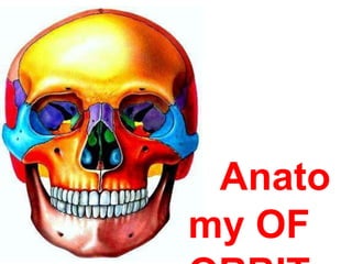
11. Each orbit is made up of 7
bones
Frontal
Ethmoidal
Maxillary
Lacrimal
Zygomatic
Sphenoid
Palatine
12. WALLS OF THE ORBIT
Medial
Lateral
Floor
Roof
15. 1.LACRIMAL GROOVE
Forms the anterior part of medialwall.
Formed by frontal process of maxilla and lacrimal
bone.
Contains the lacrimal sac.
Bounded by anterior and posterior lacrimal crests
Medial to lac groove upper part has ant ethmoidal
sinus and lower part has middle meatus of nose
LAND MARKS
18. APPLIED ANATOMY
Since it is thinnest,ethmoiditis is the commonest
cause of orbital cellulitis,especially inchildren.
Frequently eroded by chronic
inflammatory lesions,neoplasms,cysts.
It is easily fractured during trauma and
during orbitotomy operations.
Hemorrhage can occur due to trauma to
ethmoidal vessels.
19. Accidental lateral displacement of medialwall-
traumatic hypertelorism
Medial wall provides alternate access route to the
orbit through the sinus
Lacrimal bone can be easily penetrated
during endoscopic DCR
22. Triangular in shape.
Slopes upwards and medially
Shortest orbital WALL
Bordered laterally by inferior orbital fissure
and medially by maxilloethmoidal suture
Overlies maxillary sinus
26. Infra orbital fissure :
• Occupies the posterior part of junction
between the lateral wall and floor
• Through this fissure orbit communicates with
infra temporal fossa anteriorly
• And with pterygopalatine fossa Posteriorly
27.
28. APPLIED
ANATOMY
Commonly involved in BLOW
OUT FRACTURES OF THE
ORBIT.infra orbital vessels and
nerves amlost always involved
Easily invaded by tumours
of the maxillary antrum.
31. Superior orbital fissure occupies the posterior part of
the junction between roof & lateral wall.
More anterior wall is transversed by zygomatic
groove and foramena(zygo vesssels and N. pass
through)
Ant part of the wall projection TUBERCLE OF
WHITNALL,gives attachment to lateral check
ligaments of eyeball.
In maxillary resection if tubercle of whitnall damaged
causes diplopia
LAND MARKS
32.
33. APPLIED ANATOMY
• Since lateral wall is almost devoid of
foramina, bleeding is less.
• The Zygomatico-Sphenoid suture
important landmark in creating the flap in
lateral orbitotomy
34. ROOF• Underlies Frontal sinus
and Anterior cranial fossa
• Triangular
• Concave side to side
• Faces downwards, and
slightly forwards
• Formed by-
1. Frontal bone (Orbital
plate)
2. Lesser wing of Sphenoid
35.
36. Relations
• Separates the orbit from anterior cranial fossa
• Frontal air sinus may extend into its
anteromedial part
37. LAND MARKS
1.SUPRAORBITAL NOTCH:
LOCATION:
≈15 mm lateral to the
superomedial angle
TRANSMITS:
- Supraorbital nerve
- Supraorbital vessels
SURFACE ANATOMY:
- At the junction of lateral 2/3rd
and medial 1/3rd
- About two finger breadth from
the medial plane
38. Lacrimal fossa :
• Placed anterolaterally
• Lodges the lacrimal gland
Optic canal :
• Lies posteriorly at the junction of roof and
medial wall
Trochlear fossa :
• Lies anteromedially , provides attachment to
the fibrous pully or trochlea for tendon of the
superior oblique muscle
39.
40.
41. APPLIED ANATOMY
Thin and periorbita peels away easily
Objects piercing upper eyelid penetrate roof and
damage frontal lobe
At the junction of roof and medial wall the suture line
lies in proximity to cribriform plate of ethmoid.Any
trauma rupture of duramater AND CSF escapes
into orbit/nose/both
43. APPLIED ANATOMY
SUPERIOR- Supra orbital notch site for nerve
block
LATERAL -fronto zygomatic suture Prone for
separation following blunt trauma
INFERIOR-At the junction of lateral 2/3rd & medial
1/3rd just within the rim- small depression- origin of
Inferior oblique Prone to fracture and diplopia
44. APEX OF THE ORBIT
OPTIC CANAL and SUP ORBITAL FISSURE
OPTIC CANAL
It transmits the optic nerve (with its meninges)
and ophthalmic artery.
Average length is 6 to 11mm.
It connects the orbit to the middle cranial fossa.
Adult dimensions are achieved by 4-5yrs
Optic nerve glioma or Meningioma may lead
to unilateral enlargement of Opticcanal
47. It is a comma shaped aperture in the orbital
cavity.
It is bounded by greater and lesser wings of
sphenoid.
It is situated lateral to optic canal.
It is divided into upper,middle and lower parts by
common tendinous ring.
48. APPLIED ANATOMY
TOLOSA HUNT SYNDROME-Inflammation of the
superior orbital fissure and apex may result in a
multitude of signs including ophthalmoplegia and
venous outflow obstruction
SUPERIOR ORBITAL SYNDROME-Fracture at
superior orbital fissureInvolvement of cranial
nervesDiplopia, Ophthalmoplegia,
Exophthalmos, Ptosis
50. PERIORBITA
Loosely adherent to the bones
Sensory innervation by branches of V’th nerve
Fixed firmly at
- Orbital margins (Arcus marginale)
- Suture lines
- Various fissures & foramina
- Lacrimal fossa
APPLIED ANATOMY-Surgery in the orbital
roof in the areas of fissures and suture lines
may be complicated by cerebrospinal fluid
leakage .
51.
52. ORBITAL SEPTAL SYSTEM
Includes the connective tissue septa which are
suspended from the periorbita to form a
complex radial and circumferential
interconnecting slings.
These septa surround Extraocular muscles,
Optic nerve, neuro-vascular elements and the
fat lobules.
53. TENON’S CAPSULE
Also known as Fascia bulbi or bulbar sheath.
Dense, elastic and vascular connective tissue
that surrounds the globe (except over the
cornea).
Begins anteriorly at the perilimbal sclera, extends
around the globe to the optic nerve, and fuses with
the dural sheath and the sclera.
Separated from the sclera by periscleral lymph space,
which is in continuation with subdural and
subarachnoid spaces.
54. CONTENTS OF THE ORBIT
Eye ball
Muscles
4 Recti
2 obliques
Levator palpebrae superioris
Muller’s muscle (Musculus orbitalis)
Nerves
Sensory- branches of V’thNerve
Motor- III’rd, IV’th & VI’th Nerve
Autonomic- N. to the Lacrimal gland
Ciliary ganglion
55. Vessels
Arteries-
Internal carotid system- branches of ophthalmic
artery
External carotid system- a branch of internal
maxillary artery
Veins-
Superior ophthalmic vein
Inferior ophthalmic vein
Lymphatics-
none
Lacrimal gland
Lacrimal sac
Orbital fat, reticular tissue & orbital fascia
56. AGE CHANGES IN THE ORBIT
Infantile orbits are more divergent (≈115°) than those
of adults(≈40-45°)
Interorbital distance is smaller in children- may give
false impression of squint
Periorbita much thicker and stronger at birth than
in adults
Roof much larger than floor in infancy
Optic canal has no length at birth- a foramen
- at 1 year of age≈ 4mm
57.
58. LACRIMAL APPARATUS
• It is concerned with the tear formation &
transport.
• Lacrimal passage includes :
• Lacrimal gland and its ducts ( secretory part)
• Conjunctival sac
• Lacrimal puncta
• Lacrimal canaliculi
• Lacrimal sac
• Nasolacrimal duct
Drainage part
62. Osteology
o The lacrimal sac is seen in a
depression in inferomedial orbital rim .
o Maxillary and lacrimal bones.
o Bordered by the anterior lacrimal crest
(maxillary bone) & posterior lacrimal crest
(lacrimal bone).
63.
64. • The nasolacrimal canal originates at base of
lacrimal groove.
• Formed by the maxillary bone laterally and the
lacrimal and inferior turbinate bones medially.
• The width of superior opening is 4–6 mm.
• The duct courses posteriorly and laterally in
the bone for 12 mm to drain into the inferior
meatus of the nasal cavity.
65.
66. Secretory system
• It includes lacrimal gland, accessory glands
• Lacrimal gland is above & anterolateral to
globe.
• Secretes tears into superior fornix.
• Tears moisten & lubricates the : cornea ,
conjunctiva.
67.
68.
69. Lacrimal gland
• Yellowish soft lobulted serous gland.
• It consists of
Large Orbital Part
Smaller Palpebral Part
70. The orbital part
• It has the shape and size of an
almond.
• Lodged in the lacrimal fossa in the
anterolateral part of the roof of the orbit
• Posterior to the orbital septum
71. The palpebral part
• ⅓ size of the orbital part.
• Lodged in the lateral part of upper eyelid.
• Continuous with the orbital part around the
lateral margin of the aponeurosis of the levator
palpebrae superioris.
72. Accessory glands
• Are small, compound, branched, tubular
glands .
• Located in the middle of lid (Wolfring glands) or
superior & inferior fornices (Krause glands).
• Ectopic portions of lacrimal gland tissue .
73.
74. vascular supply
• Artery supply : Lacrimal artery , branch of
ophthalmic artery.
• Venous drainages : Ophthalmic Vein.
• Lymphatic drainage : Joins that of
conjunctiva & drain into the preauricular
lymph nodes.
75. Nerve supply
Parasympathetic :
• The parasympathetic secretomotor fibres are drived
from the lacrimal nucleus of facial nerve .
• They reach the Sphenopalatine ganglion via the
Greater superficial petrosal nerve
• The postganglionic fibres join the Maxillary nerve
then through its Zygomatic nerve and further
through its Zygomticotemporal branch
76. They join the lacrimal nerve by a
communication between it and
zygomatico temporal nerve.
They reach the gland through lacrimal nerve.
Some postganglionic fibres from the
sphenopalatine ganglion reach the gland
through the periosteum of the orbit via its
orbital branches.
77. • Sympathetic :
From carotid plexus
• Sensory :
lacrimal nerve , branch of
ophthalmic division of trigeminal
nerve.
80. Duct system :
• The gland has about 12 short, slender ducts.
• They arise from the lower surface of the
gland.
• Open into the lateral part of the superior
fornix of the conjuctiva.
81. Flow of tears
• The lacrimal secretion flows down and is pushed
medially by the movements of the eyelids.
• The tears moisten the eye and accumulate in the
Lacus Lacrimalis which is a triangular depression
between the medial parts of both eyelids.
• It is drained by the Lacrimal canaliculi to the
lacrimal sac.
• From the sac it descends through the
Nasolacrimal duct to reach the inferior
meatus of the nose.
82. Lacrimal canaliculi :
• They are two slender ducts 10 mm in
length.
• They run in the medial parts of the
margins of both eyelids.
• Each duct begins by an opening called
Lacrimal Punctum on the summit of an
elevtion called Lacrimal Papilla.
• They drain the lacrimal fluid into the
Lacrimal Sac.
83.
84. Lacrimal sac :
• This is a small sac lodged in the lacrimal
groove.
• It is about 12 mm in length having blind
upper and lower ends.
• The lower end is continuous with the
Nasolacrimal duct.
• It is covered by lacrimal fascia, which
separates the lacrimal sac from the medial
palpebral ligament anteriorly and from the
lacrimal part of orbicularis oculi posteriorly.
85.
86.
87. Nasolacrimal duct :
• This is a tube , half an inch in length
runs through the nasolacrimal canal.
• It begins from lower end of the lacrimal sac,
run downwards, backwards and laterally to
open into the anterior part of the inferior
meatus of the nose.
The lower end of the duct is guarded by a
mucous fold called lacrimal fold, which acts as
a valve preventing nasal secretion from
ascending up into the duct.