Downloaded 463 times


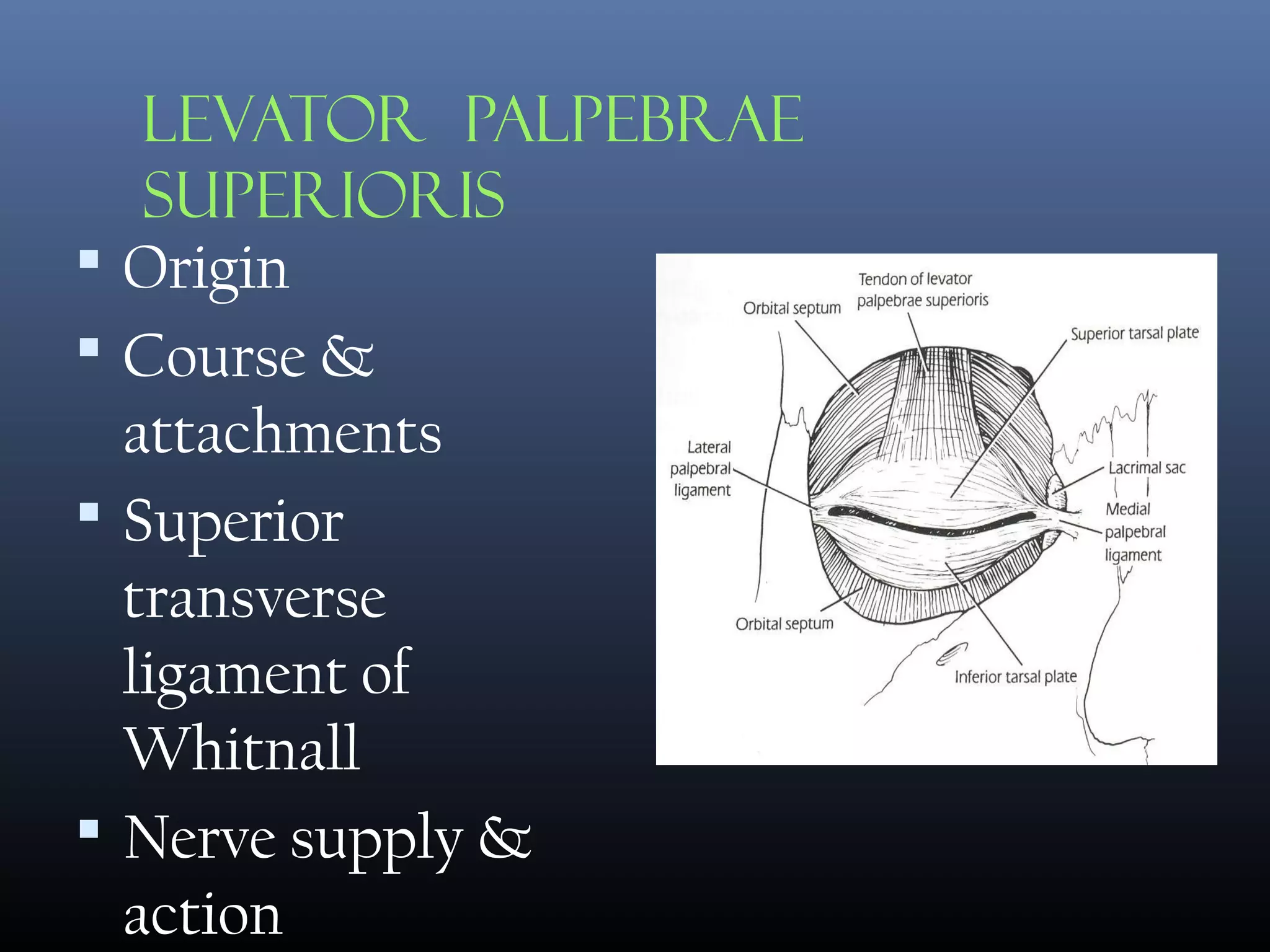


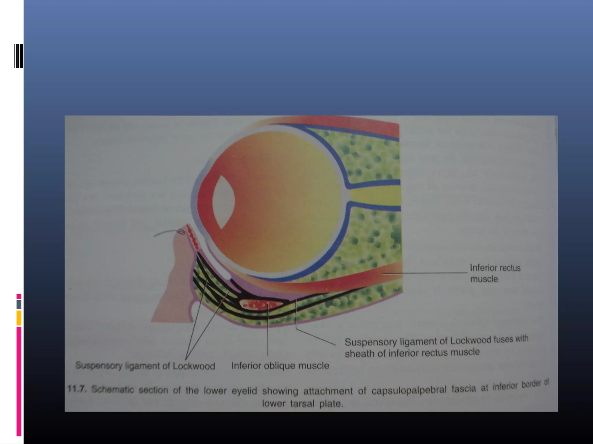


The document provides an extensive overview of embryology, anatomy, and physiology regarding the eyelids, detailing their formation, structure, muscles, and functions. It covers topics such as the development of eyelid folds, the arrangement of various layers, and the mechanisms of eyelid movement including opening and closure facilitated by different muscles. Additionally, it discusses the vascular and nerve supply, along with the roles of various glands associated with the eyelid.


