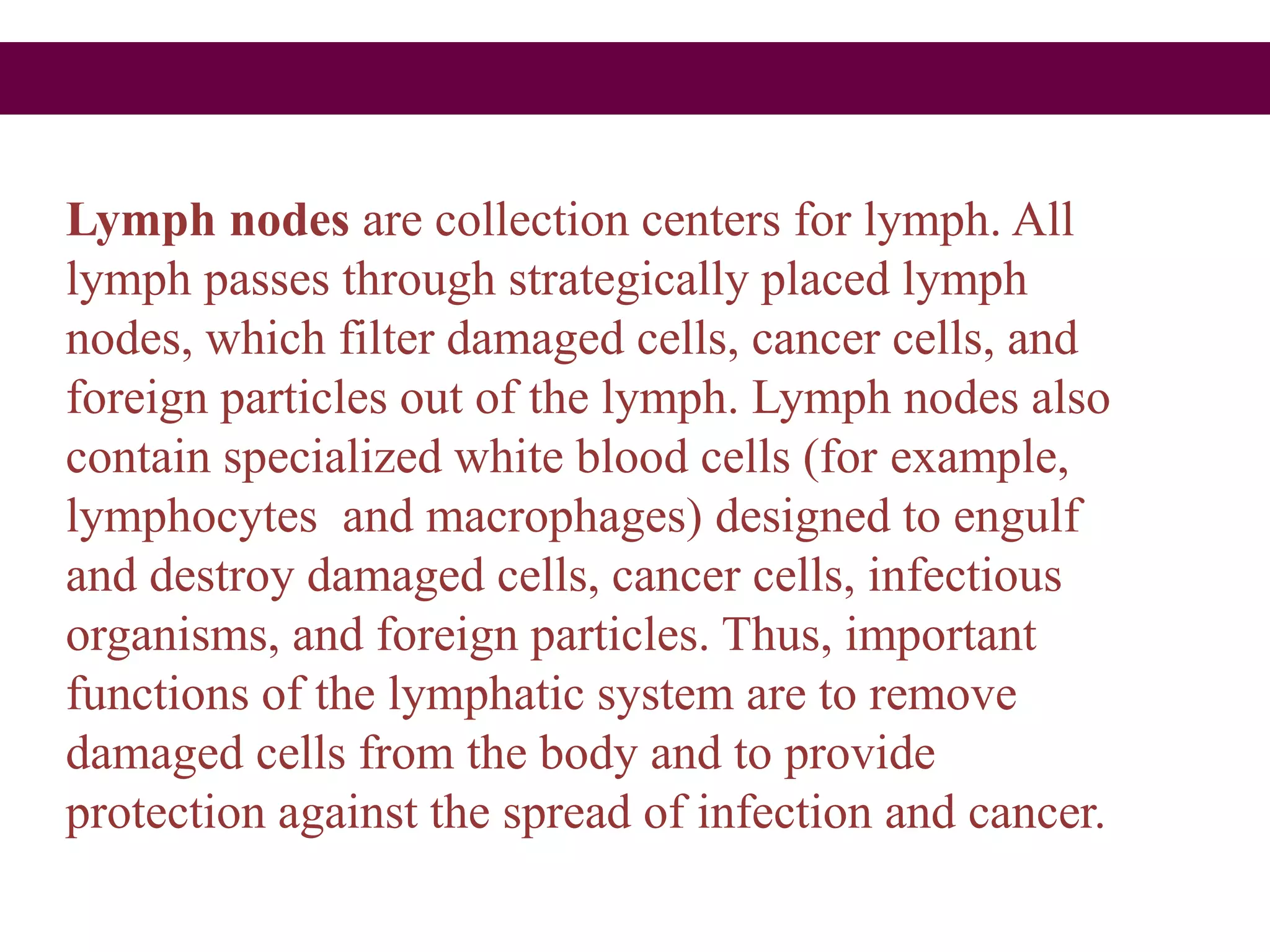
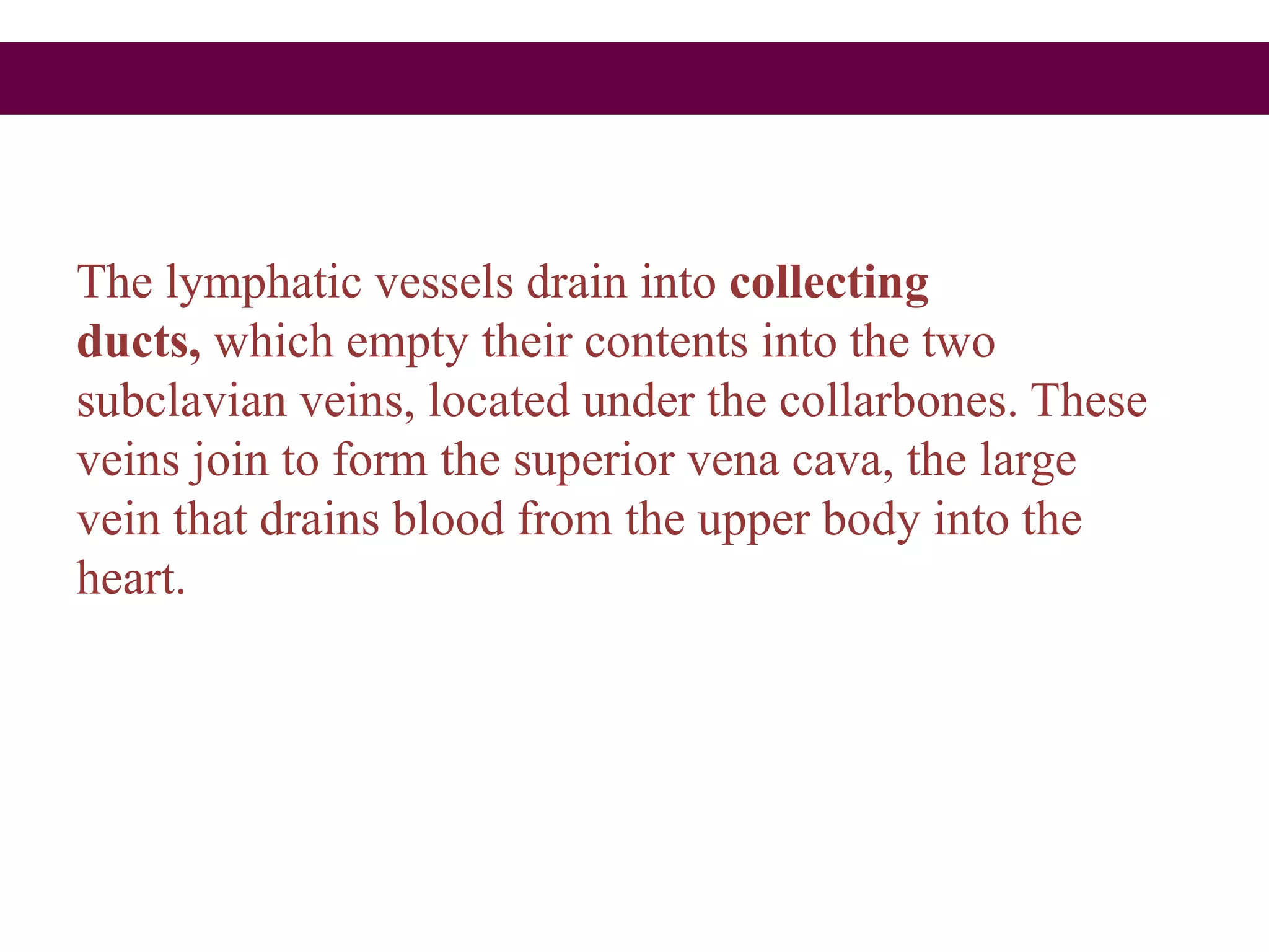
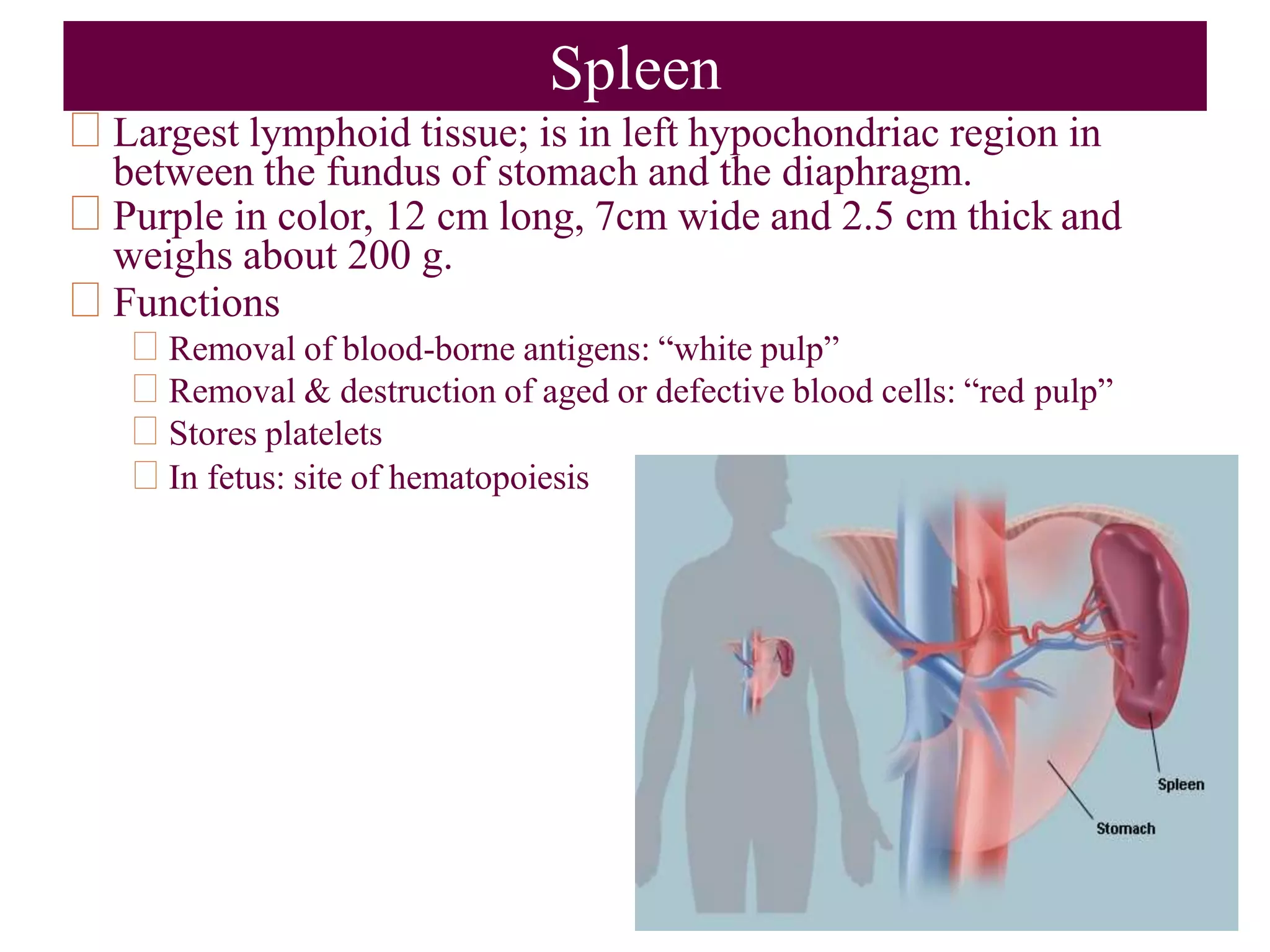
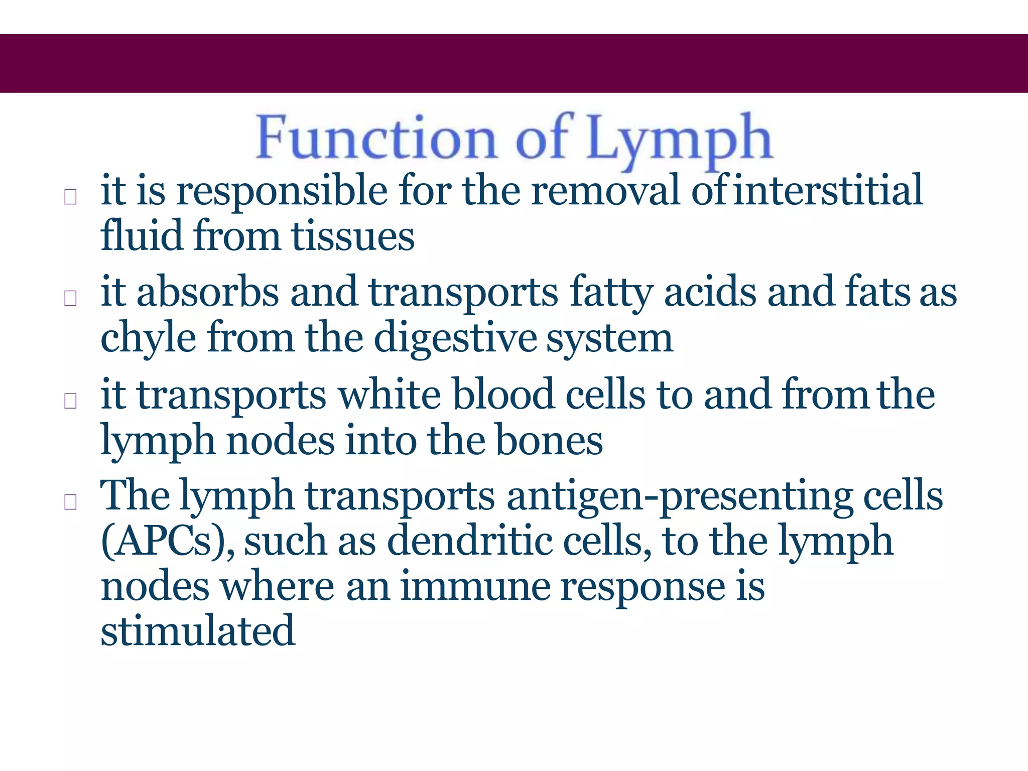
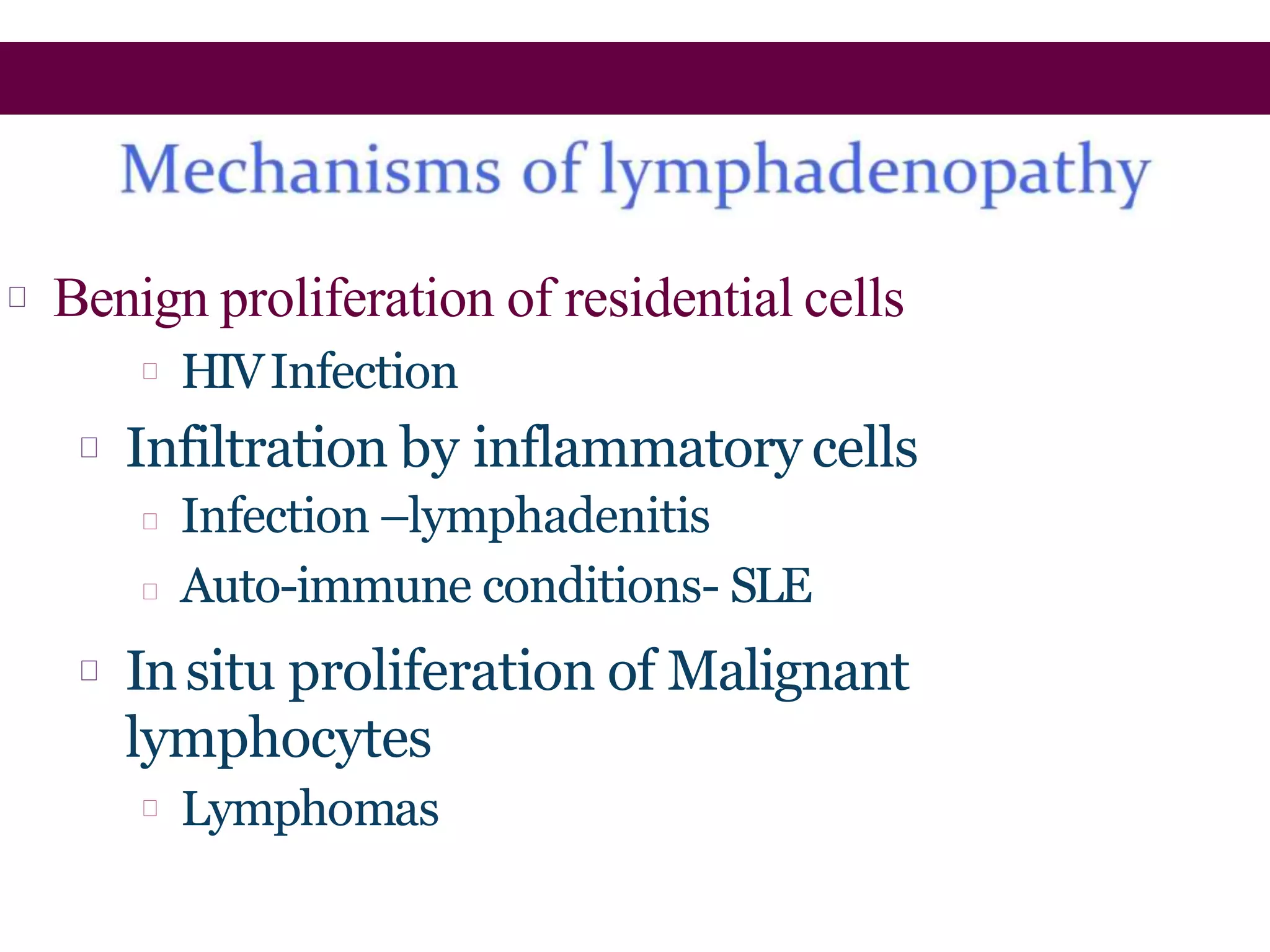
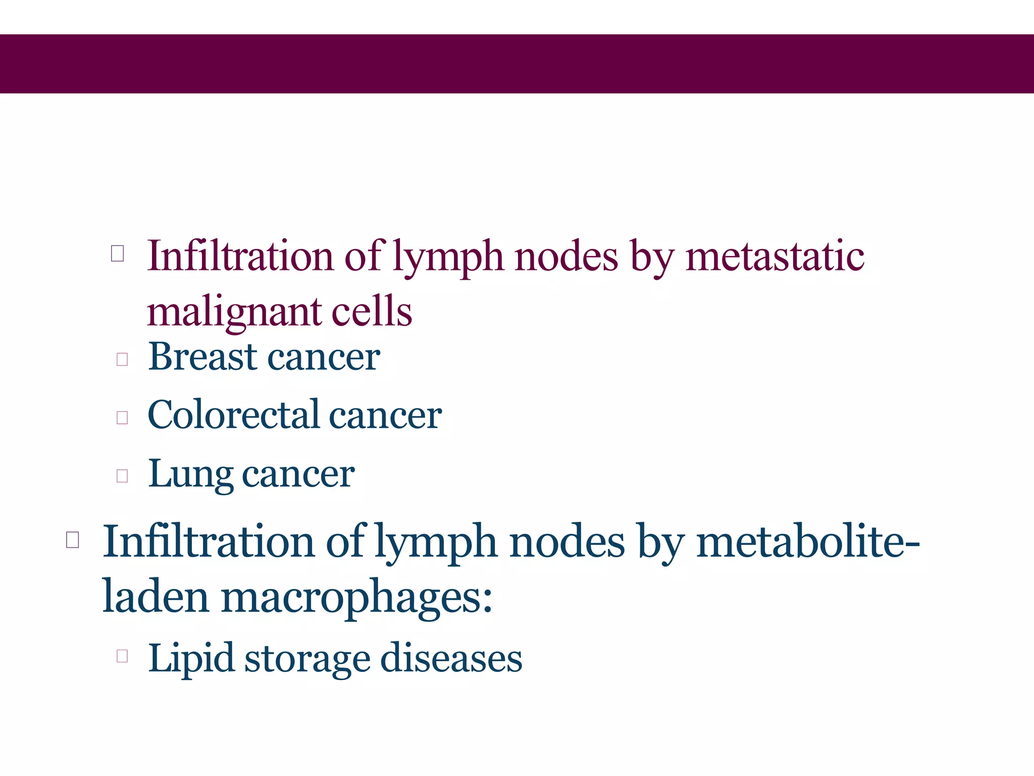
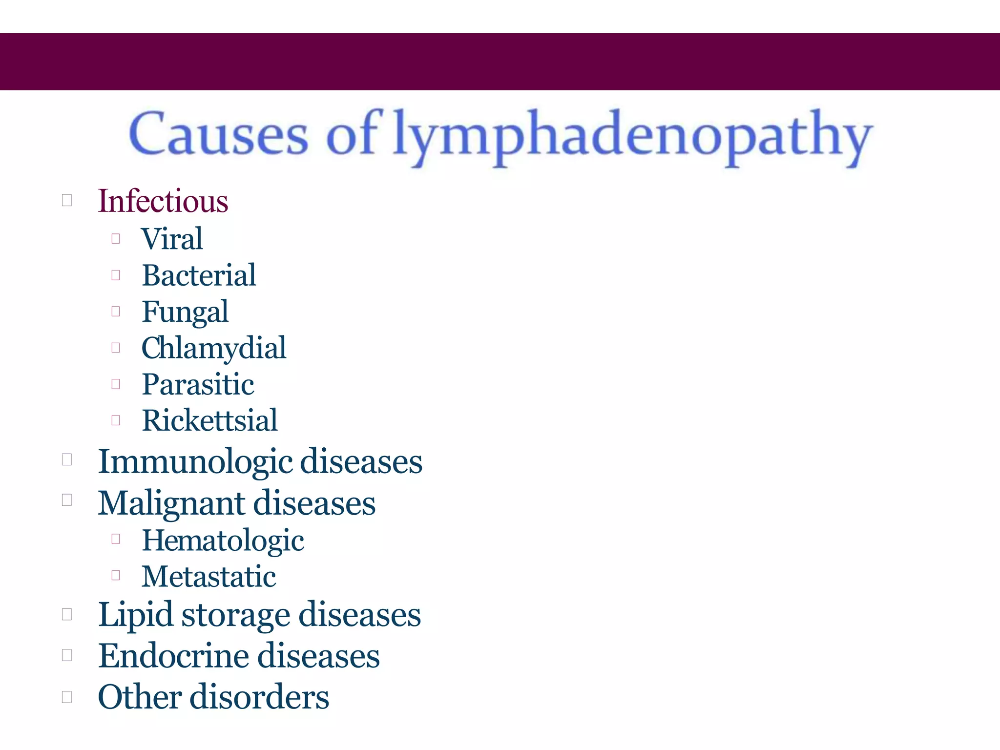
The lymphatic system transports fluid and immune cells throughout the body, removes cellular waste and pathogens, and presents antigens to immune cells. It consists of lymphatic vessels, lymph nodes, the spleen, thymus, tonsils, and bone marrow. Lymph fluid is collected from tissues by lymphatic capillaries and passes through lymph nodes, which filter the lymph and harbor immune cells. The lymph eventually returns to the blood circulation via lymph ducts in the neck. Enlargement of lymph nodes or spleen can indicate infection, inflammation, or cancer that is being assessed through history, exam, labs, and imaging.
Cardiogenic shock is a condition of diminished cardiac output that severely impairs cardiac perfusion. In this condition in which the heart suddenly can't pump enough blood to meet the body's needs.
one of most important topic of vascular surgery , i couldn't find this much in slideshare so , i made a slide and uploaded it . Hope you will enjoy reading :)
anatomy of the lower extremity veins, CVI , ambulatory venous hypertension, varicose veins , clinical examination and performance of various tests of the varicose veins
The lymph node is a small bean-shaped object which factors in the body's immune system. Lymph nodes clean out substances that travel through the lymphatic fluid, and they contain white blood cells that are the body's defense when fighting off ailments. There are many lymph nodes found throughout the body.
At the completion of this unit, learners will be able to: 1. Define lymph & the lymphatic system 2. Identify the organs of lymphatic system 3. Describe the general functions of the lymphatic system 4. Describe how lymph is formed 5. Describe the lymph vessels & how lymph is returned to the blood vessels 6. Describe the structure and functions of the lymph nodes, nodules, spleen and the thymus glands.
THE LYMPHATIC SYSTEM// LYMPH CIRCULATION//LYMPH VESSELS// LYMPH ORGANS Wasim Ak
The lymphatic system is a sub-system of circulatory system and immune system.
It is a type of drainage system of human body which collects all the tissue fluids (constantly leaking out of the bloodstream) and takes back to the major veins through a network of lymph vessels.
The lymphatic system consists of -
Lymph – colourless tissue fluid
Lymphatic organs – Thymus, Bone Marrow, Lymph nodes, Spleen, Tonsils.
Lymph vessels – through which lymph circulation takes place.
It maintains balance between blood and tissues:
Blood volume : 5 – 6L
Interstitial fluid volume: 10 – 11L
Lymph volume: 2 – 3L.
It helps in Immunity.
It fascilitate absorption of fats and hormones:
Breakdown products of fat and fat-soluble vitamins are absorbed into the central lacteals (lymphatic vessels) of the villi.
Normal blood circulation forces fluid out of the bloodstream and that leads in the increase in the interstitial fluid volume.
Due to osmotic pressure this interstitial fluid will be collected by the fine lymphatic capillaries.
Now this fluid is lymph and it has the same mineral distribution as that of blood plasma.
The lymph is transported to lymph nodes and organs where the pathogen will be killed by lymphocytes and lymph will be filtered.
The back flow of lymph is prevented by the valves present in lymph vessels.
Lymph moves from lymphatic vessels to lymphatic trunks, collecting ducts, and ultimately into the Subclavian veins.
micro teaching on communication m.sc nursing.pdfAnurag Sharma
Microteaching is a unique model of practice teaching. It is a viable instrument for the. desired change in the teaching behavior or the behavior potential which, in specified types of real. classroom situations, tends to facilitate the achievement of specified types of objectives.
Ozempic: Preoperative Management of Patients on GLP-1 Receptor Agonists Saeid Safari
Preoperative Management of Patients on GLP-1 Receptor Agonists like Ozempic and Semiglutide
ASA GUIDELINE
NYSORA Guideline
2 Case Reports of Gastric Ultrasound
Factory Supply Best Quality Pmk Oil CAS 28578–16–7 PMK Powder in Stockrebeccabio
Factory Supply Best Quality Pmk Oil CAS 28578–16–7 PMK Powder in Stock
Telegram: bmksupplier
signal: +85264872720
threema: TUD4A6YC
You can contact me on Telegram or Threema
Communicate promptly and reply
Free of customs clearance, Double Clearance 100% pass delivery to USA, Canada, Spain, Germany, Netherland, Poland, Italy, Sweden, UK, Czech Republic, Australia, Mexico, Russia, Ukraine, Kazakhstan.Door to door service
Hot Selling Organic intermediates
Ethanol (CH3CH2OH), or beverage alcohol, is a two-carbon alcohol
that is rapidly distributed in the body and brain. Ethanol alters many
neurochemical systems and has rewarding and addictive properties. It
is the oldest recreational drug and likely contributes to more morbidity,
mortality, and public health costs than all illicit drugs combined. The
5th edition of the Diagnostic and Statistical Manual of Mental Disorders
(DSM-5) integrates alcohol abuse and alcohol dependence into a single
disorder called alcohol use disorder (AUD), with mild, moderate,
and severe subclassifications (American Psychiatric Association, 2013).
In the DSM-5, all types of substance abuse and dependence have been
combined into a single substance use disorder (SUD) on a continuum
from mild to severe. A diagnosis of AUD requires that at least two of
the 11 DSM-5 behaviors be present within a 12-month period (mild
AUD: 2–3 criteria; moderate AUD: 4–5 criteria; severe AUD: 6–11 criteria).
The four main behavioral effects of AUD are impaired control over
drinking, negative social consequences, risky use, and altered physiological
effects (tolerance, withdrawal). This chapter presents an overview
of the prevalence and harmful consequences of AUD in the U.S.,
the systemic nature of the disease, neurocircuitry and stages of AUD,
comorbidities, fetal alcohol spectrum disorders, genetic risk factors, and
pharmacotherapies for AUD.
Title: Sense of Smell
Presenter: Dr. Faiza, Assistant Professor of Physiology
Qualifications:
MBBS (Best Graduate, AIMC Lahore)
FCPS Physiology
ICMT, CHPE, DHPE (STMU)
MPH (GC University, Faisalabad)
MBA (Virtual University of Pakistan)
Learning Objectives:
Describe the primary categories of smells and the concept of odor blindness.
Explain the structure and location of the olfactory membrane and mucosa, including the types and roles of cells involved in olfaction.
Describe the pathway and mechanisms of olfactory signal transmission from the olfactory receptors to the brain.
Illustrate the biochemical cascade triggered by odorant binding to olfactory receptors, including the role of G-proteins and second messengers in generating an action potential.
Identify different types of olfactory disorders such as anosmia, hyposmia, hyperosmia, and dysosmia, including their potential causes.
Key Topics:
Olfactory Genes:
3% of the human genome accounts for olfactory genes.
400 genes for odorant receptors.
Olfactory Membrane:
Located in the superior part of the nasal cavity.
Medially: Folds downward along the superior septum.
Laterally: Folds over the superior turbinate and upper surface of the middle turbinate.
Total surface area: 5-10 square centimeters.
Olfactory Mucosa:
Olfactory Cells: Bipolar nerve cells derived from the CNS (100 million), with 4-25 olfactory cilia per cell.
Sustentacular Cells: Produce mucus and maintain ionic and molecular environment.
Basal Cells: Replace worn-out olfactory cells with an average lifespan of 1-2 months.
Bowman’s Gland: Secretes mucus.
Stimulation of Olfactory Cells:
Odorant dissolves in mucus and attaches to receptors on olfactory cilia.
Involves a cascade effect through G-proteins and second messengers, leading to depolarization and action potential generation in the olfactory nerve.
Quality of a Good Odorant:
Small (3-20 Carbon atoms), volatile, water-soluble, and lipid-soluble.
Facilitated by odorant-binding proteins in mucus.
Membrane Potential and Action Potential:
Resting membrane potential: -55mV.
Action potential frequency in the olfactory nerve increases with odorant strength.
Adaptation Towards the Sense of Smell:
Rapid adaptation within the first second, with further slow adaptation.
Psychological adaptation greater than receptor adaptation, involving feedback inhibition from the central nervous system.
Primary Sensations of Smell:
Camphoraceous, Musky, Floral, Pepperminty, Ethereal, Pungent, Putrid.
Odor Detection Threshold:
Examples: Hydrogen sulfide (0.0005 ppm), Methyl-mercaptan (0.002 ppm).
Some toxic substances are odorless at lethal concentrations.
Characteristics of Smell:
Odor blindness for single substances due to lack of appropriate receptor protein.
Behavioral and emotional influences of smell.
Transmission of Olfactory Signals:
From olfactory cells to glomeruli in the olfactory bulb, involving lateral inhibition.
Primitive, less old, and new olfactory systems with different path
These simplified slides by Dr. Sidra Arshad present an overview of the non-respiratory functions of the respiratory tract.
Learning objectives:
1. Enlist the non-respiratory functions of the respiratory tract
2. Briefly explain how these functions are carried out
3. Discuss the significance of dead space
4. Differentiate between minute ventilation and alveolar ventilation
5. Describe the cough and sneeze reflexes
Study Resources:
1. Chapter 39, Guyton and Hall Textbook of Medical Physiology, 14th edition
2. Chapter 34, Ganong’s Review of Medical Physiology, 26th edition
3. Chapter 17, Human Physiology by Lauralee Sherwood, 9th edition
4. Non-respiratory functions of the lungs https://academic.oup.com/bjaed/article/13/3/98/278874
HOT NEW PRODUCT! BIG SALES FAST SHIPPING NOW FROM CHINA!! EU KU DB BK substit...GL Anaacs
Contact us if you are interested:
Email / Skype : kefaya1771@gmail.com
Threema: PXHY5PDH
New BATCH Ku !!! MUCH IN DEMAND FAST SALE EVERY BATCH HAPPY GOOD EFFECT BIG BATCH !
Contact me on Threema or skype to start big business!!
Hot-sale products:
NEW HOT EUTYLONE WHITE CRYSTAL!!
5cl-adba precursor (semi finished )
5cl-adba raw materials
ADBB precursor (semi finished )
ADBB raw materials
APVP powder
5fadb/4f-adb
Jwh018 / Jwh210
Eutylone crystal
Protonitazene (hydrochloride) CAS: 119276-01-6
Flubrotizolam CAS: 57801-95-3
Metonitazene CAS: 14680-51-4
Payment terms: Western Union,MoneyGram,Bitcoin or USDT.
Deliver Time: Usually 7-15days
Shipping method: FedEx, TNT, DHL,UPS etc.Our deliveries are 100% safe, fast, reliable and discreet.
Samples will be sent for your evaluation!If you are interested in, please contact me, let's talk details.
We specializes in exporting high quality Research chemical, medical intermediate, Pharmaceutical chemicals and so on. Products are exported to USA, Canada, France, Korea, Japan,Russia, Southeast Asia and other countries.
Lung Cancer: Artificial Intelligence, Synergetics, Complex System Analysis, S...Oleg Kshivets
RESULTS: Overall life span (LS) was 2252.1±1742.5 days and cumulative 5-year survival (5YS) reached 73.2%, 10 years – 64.8%, 20 years – 42.5%. 513 LCP lived more than 5 years (LS=3124.6±1525.6 days), 148 LCP – more than 10 years (LS=5054.4±1504.1 days).199 LCP died because of LC (LS=562.7±374.5 days). 5YS of LCP after bi/lobectomies was significantly superior in comparison with LCP after pneumonectomies (78.1% vs.63.7%, P=0.00001 by log-rank test). AT significantly improved 5YS (66.3% vs. 34.8%) (P=0.00000 by log-rank test) only for LCP with N1-2. Cox modeling displayed that 5YS of LCP significantly depended on: phase transition (PT) early-invasive LC in terms of synergetics, PT N0—N12, cell ratio factors (ratio between cancer cells- CC and blood cells subpopulations), G1-3, histology, glucose, AT, blood cell circuit, prothrombin index, heparin tolerance, recalcification time (P=0.000-0.038). Neural networks, genetic algorithm selection and bootstrap simulation revealed relationships between 5YS and PT early-invasive LC (rank=1), PT N0—N12 (rank=2), thrombocytes/CC (3), erythrocytes/CC (4), eosinophils/CC (5), healthy cells/CC (6), lymphocytes/CC (7), segmented neutrophils/CC (8), stick neutrophils/CC (9), monocytes/CC (10); leucocytes/CC (11). Correct prediction of 5YS was 100% by neural networks computing (area under ROC curve=1.0; error=0.0).
CONCLUSIONS: 5YS of LCP after radical procedures significantly depended on: 1) PT early-invasive cancer; 2) PT N0--N12; 3) cell ratio factors; 4) blood cell circuit; 5) biochemical factors; 6) hemostasis system; 7) AT; 8) LC characteristics; 9) LC cell dynamics; 10) surgery type: lobectomy/pneumonectomy; 11) anthropometric data. Optimal diagnosis and treatment strategies for LC are: 1) screening and early detection of LC; 2) availability of experienced thoracic surgeons because of complexity of radical procedures; 3) aggressive en block surgery and adequate lymph node dissection for completeness; 4) precise prediction; 5) adjuvant chemoimmunoradiotherapy for LCP with unfavorable prognosis.
Explore natural remedies for syphilis treatment in Singapore. Discover alternative therapies, herbal remedies, and lifestyle changes that may complement conventional treatments. Learn about holistic approaches to managing syphilis symptoms and supporting overall health.
3. The lymphatic system is a vital part of the immune
system, along with the thymus, bone marrow, spleen,
tonsils, appendix, and Peyer patches in the small
intestine.
Like the venous system, the lymphatic
system transports fluids throughout the body. The
lymphatic system consists of
Thin-walled lymphatic vessels
Lymph nodes
Two collecting ducts
4. Lymphatic vessels, located throughout the body, are
larger than capillaries (the smallest blood vessels, which
connect arteries and veins), and most are smaller than
the smallest veins. Most of the lymphatic vessels have
valves like those in veins to keep the lymph, which can
clot, flowing in the one direction (toward the heart).
Lymphatic vessels drain fluid called lymph from tissues
throughout the body.
5. Lymph begins as fluid that has diffused through the
very thin walls of capillaries into the space between
cells. Most of the fluid is reabsorbed into the
capillaries and the rest is drained into the lymphatic
vessels, which eventually return it to the veins.
Lymph also contains many other substances
including
Proteins, minerals, nutrients, and other substances,
which provide nourishment to tissues
Damaged cells, cancer cells, and foreign particles
(such as bacteria and viruses) that may have entered
the tissue fluids
6. Lymph nodes are collection centers for lymph. All
lymph passes through strategically placed lymph
nodes, which filter damaged cells, cancer cells, and
foreign particles out of the lymph. Lymph nodes also
contain specialized white blood cells (for example,
lymphocytes and macrophages) designed to engulf
and destroy damaged cells, cancer cells, infectious
organisms, and foreign particles. Thus, important
functions of the lymphatic system are to remove
damaged cells from the body and to provide
protection against the spread of infection and cancer.
7. The lymphatic vessels drain into collecting
ducts, which empty their contents into the two
subclavian veins, located under the collarbones. These
veins join to form the superior vena cava, the large
vein that drains blood from the upper body into the
heart.
8. The Lymphatic System
A circulatory system for fluids
Returns fluid to the blood
Removes antigens from the body
Exposes antigens to the immune system
Main structures of the lymphatic system
Lymph
Lymphatic vessels
Lymph nodes
Diffuse Lymphoid tissue, Eg: tonsils
Lymphoid organs, Eg: spleen
&Thymus
Bone marrow
9. Lymph
Lymph is a clear watery fluid, similar in composition to
plasma , with important exception of plasma proteins and
identical in composition to interstitial fluid.
Transports the plasma proteins that seep out of the
capillary beds back to the bloodstream.
It also carries away larger particles, Eg. Bacteria, Cell
debris etc.
Contains lymphocytes which circulates in the lymphatic
system allowing them to patrol the different regions of
the body.
10. The Lymphatic System
Lymphatic vessels
collect tissue fluid
from loose connective
tissue
Carry fluid to
great veins in the
neck
Fluid flows
only toward the
heart
Collect excess tissue
fluid and blood
proteins
11. Orders of Lymphatic Vessels
Lymph capillaries
Smallest lymph vessels
First to receive lymph
Lymphatic collecting vessels
Collect from lymph capillaries
Lymph nodes
Scattered along collecting
vessels
Lymph trunks
Collect lymph from collecting
vessels
Lymph ducts
Empty into veins of the neck
12. Lymphatic Capillaries
Located near blood capillaries
Receive tissue fluid from CT
Increased volume of tissue fluid
Minivalve flaps open and allow fluid to enter
Highly permeability allows entrance of
Tissue fluid
Bacteria, viruses, and cancer cells
Lacteals – specialized lymphatic capillaries
Located in the villi of the small intestines
Receive digested fats
Fatty lymph – chyle
14. Lymphatic Collecting Vessels
Accompany blood vessels
Composed of the same three tunics as blood
vessels
Contain more valves than veins do
Helps direct the flow of blood
Lymph propelled by
Bulging of skeletal muscles
Pulsing of nearby arteries
Tunica media of the lymph vessels
15. Lymph Nodes
Lymph nodes are bean shaped organs along with
lymphatic collecting vessels
Up to 1 inch in size
Cleanse the lymph of pathogens
Human body contains around 500
Lymph nodes are organized in clusters
These nodes are considerably in size: some are as small
as a pin head & the largest are about the size of an
almond
17. Microscopic Anatomy of a Lymph Node
Outer Fibrous capsule – surrounds lymph nodes
Trabeculae – connective tissue strands
The main substance of the node consists of reticular and
lymphatic tissue containing many lymphocytes and
macrophages.
Each node has a concave surface called hilum, where an
artery enters & a vein and efferent vessel leaves.
Lymph vessels
Afferent lymphatic vessels
Efferent lymphatic vessels
18. Microscopic Anatomy of a Lymph Node
Functions:
1.Filtering and phagocytosis
2.Proliferation of lymphocytes.
19. Lymph Trunks
Lymphatic collecting vessels converge
Five major lymph trunks
Lumbar trunks
Receives lymph from lower limbs
Intestinal trunk
Receives chyle from digestive organs
Bronchomediastinal trunks
Collects lymph from thoracic viscera
Subclavian trunks
Receive lymph from upper limbs and thoracic
wall
Jugular trunks
Drain lymph from the head and neck
22. Lymph Ducts
Cisterna chyli
Located at the union of lumbar and intestinal trunks
Thoracic duct
Ascends along vertebral bodies
Empties into venous circulation
Junction of left internal jugular and left subclavian veins
Drains three quarters of the body
Right lymphatic duct
Empties into right internal jugular and subclavian veins
23. Spleen
Largest lymphoid tissue; is in left hypochondriac region in
between the fundus of stomach and the diaphragm.
Purple in color, 12 cm long, 7cm wide and 2.5 cm thick and
weighs about 200 g.
Functions
Removal of blood-borne antigens: “white pulp”
Removal & destruction of aged or defective blood cells: “red pulp”
Stores platelets
In fetus: site of hematopoiesis
25. Lies in the upper part of the medistinum behind the sternum &
extends upwards into the root of the neck.
Weighs about 10-15 g at birth and grows until the individual
reaches puberty. 30-40g by middle age .
Prominent in newborns, almost disappears by old age
Function: T lymphocyte maturation (immunocompetence)
Has no follicles because no B cells
Structure:
Consists of two lobes joined by areolar tissue.
Lobes are enclosed by a fibrous capsule which dips into their
substances, dividing them into lobules that consist of an irregular
branching framework of epithelial cells and lymphocytes.
19
Thymus
29. Aggregated lymphoid
nodules (“Peyer’s Patches”)
About 40 follicles, 1 cm
wide
Distal small
intestine (ileum)
Appendix
Parts of the intestine are so densely packed with
MALT (mucosa-associated lymphoid tissue) that
they are considered lymphoid organs
30. Lymph Node Assessment
Palpate the regional lymph nodes of the head and neck, axillae,
arms, and groin. Use firm, circular movements of the finger pads
and note size, shape, symmetry, consistency, delineation,
mobility, tenderness, sensation, and condition of overlying skin.
Nodes should not be enlarged or painful
Lymphadenopathy refers to the enlargement of lymph nodes
(over 1 cm) with or without tenderness. It may be caused by
inflammation, infection, or malignancy of the nodes or the
regions drained by the nodes.
31. Lymph node enlargement with tenderness suggests
inflammation (lymphadenitis). With bacterial
infection, the nodes may be warm and matted with
localized swelling.
Malignant or metastatic nodes may be hard,
indicating lymphoma; rubbery, indicating Hodgkin’s
disease; or fixed to adjacent structures. Usually they
are not tender.
•Ear infections and scalp and facial lesions, such as
acne, may cause enlargement of the preauricular and
cervical nodes.
32. Anterior cervical nodes are enlarged and infected
with streptococcal pharyngitis and mononucleosis.
Lymphadenitis of the cervical and submandibular
nodes occurs with herpes simplex lesions.
Enlargement of supraclavicular nodes, especially
the left, is highly suggestive of metastatic disease
from abdominal and thoracic cancer.
Axillary lymphadenopathy is associated with breast
cancer.
Lesions of the genitals may produce enlargement of
the inguinal nodes.
33. Persistent generalized lymphadenopathy is
associated with acquired immunodeficiency
syndrome (AIDS) and AIDS-related complex.
Spleen Assessment:
Palpate for the spleen, in the upper
left quadrant of the abdomen. The spleen is normally
not palpable. A palpable spleen in the left upper
abdominal quadrant of an adult may indicate abnormal
enlargement (splenomegaly) and may be associated
with cancer, blood dyscrasias, and viral infection, such
as mononucleosis
34. A dull percussion note in the lowest left ICS at the
anterior axillary line or below the tenth rib at the
midaxillary line suggests splenic enlargement
Percuss for splenic dullness in the
lowest left intercostal space (ICS) at the anterior
axillary line or in the 9th to 10th ICS at the midaxillary
line . Normally, tympany is heard.
35. Lymphatic system
Introduction
Mechanism and causes of lymphadenopathy
Approach to lymphadenopathy: Hx,P/E, Lab
Splenomegaly
Introduction
Causes of splenomegaly
Evaluation of splenomegaly: Hx,P/E, Labstudies,
Imaging...
Evaluation of swellings (Lumps)
36. it is responsible for the removal ofinterstitial
fluid from tissues
it absorbs and transports fatty acids and fats as
chyle from the digestive system
it transports white blood cells to and fromthe
lymph nodes into the bones
The lymph transports antigen-presenting cells
(APCs), such as dendritic cells, to the lymph
nodes where an immune response is
stimulated
37. Diagnostic dilemma
Various causes with spectrum of clinical
course
Localized or Generalized
Normal size of LNs
Larger in adolescents
Inguinal and submandibular
Recurrent trauma and infection
38. Benign proliferation of residential cells
HIVInfection
Infiltration by inflammatory cells
Infection –lymphadenitis
Auto-immune conditions- SLE
Insitu proliferation of Malignant
lymphocytes
Lymphomas
39. Infiltration of lymph nodes by metastatic
malignant cells
Breast cancer
Colorectal cancer
Lung cancer
Infiltration of lymph nodes by metabolite-
laden macrophages:
Lipid storage diseases
41. Focused history
Sx of anemia
Infection
Bleeding
Duration of lymphadenopathy
Acute vs Chronic
Progression of the lymphadenopathy
Waxing & weaning
Slow vs fast
Involvement of adjacent or distant LN
42. Associated symptoms
Pain
Fever, hotness
Sx of obstruction
Localizing symptoms of infections and
malignancy
Draining sinus
Hotness and local pain
Exposures
Radiation
Chemotherapy
Other agents: pets
45. Features characteristic of the lymph node
Location
Size
Consistency
Fixation
Tenderness
Splenomegaly
hepatomegaly
46. • Components
–
–
–
–
–
–
Various laboratory and serologic tests
Imaging
Lymph node biopsy
Bone Marrow study
Other biopsies.
?Empirical treatment
• Depend on various factors
–
–
–
–
Age
Duration
Localized/regional/generalized
Epidemiology and the clinical setting
47. Laboratory tests
CBC& Peripheral Smear
ESR
HIV
RPR/VDR
L ANA
Heterophile Antibody tests
LDH& other tests according to the setting as
well as importance
48. Imaging study for the purpose of
Defining size & distribution more precisely
Distinguishing from other similar structure
Staging
Guiding for FNA
50. • Types of biopsy
–
–
–
Open biopsy
Fine Needle Aspiration
Core Needle Biopsy
• Choice of LN& type of biopsy
–
–
–
–
The most diseased
Supraclavicular/cervical/axillary/inguinal
Ifsingle go for open biopsy as much as possible
accessability
• Possible studies from thespecimen
–
–
–
Pathological
Immunochemistry/immunophenotype
Genetic/molecular studies
52. Spleen is one of the lymphoid organs which is also
called reticuloendothelial system.
Splenomegaly is common clinical condition & it is
never normal
Various causes with diagnostic challenge
Other condition
Massive splenomegaly
Splenic infarction
Ruptured spleen
Splenic abscess
Functional hyposplenism/ asplenia
Hypersplenism
53. Lies in the Peritoneal cavity in the left upper
quadrant.
Adjacent to 9th-11th rib, stomach, colon and
pancreas.
Weight
Male=80-200g
Female= 70-180g
Average =150g (0.2% of Body Weight)
Palpability and size
Not palpable normal ( children, adolescents, thin
adults)
Soft organ unless infiltrated
54. Participates in cellular and humoral immunity
Removes senescent and/or poorly deformable
red cells, bacteria, and other particulates from
the circulation
Under abnormal circumstances the spleen may
become the site of extramedullary
hematopoiesis
Approximately one-third of circulating
platelets are sequestered in the spleen,where
they are in equilibrium with circulating
platelets
55. Splenic abnormalities can include
Increased function (hypersplenism)
Decreased to absent function (hyposplenism,
asplenia)
Abscess, infarction, calcification, cysts
Traumatic or atraumatic rupture
Enlargement (Splenomegaly)
56. Splenic engorgement with sequestration
Chronic inflammation or infection
Lipid deposition
Congenital condition
Splenic infiltration
57. 1.Congestive
Cirrhosis
Heart Failure
Thrombosis of portal, hepatic and splenic veins
2.Malignancy
Lymphomas, usually indolent
Leukemias
Myeloproliferative Disorders
Primary splenic tumours
Metastatic solid tumours
62. Symptoms of splenomegaly
Pain, a sense of fullness, or discomfort in the left
upper quadrant
Pain referred to the left shoulder
Early satiety, due to encroachment onthe
adjacent stomach
Focused history
Underlying conditions
Constitutional symptoms
Travel history
63. Complete physical examination
Cardinal steps in spleen/ abdominal exam
Inspection
Palpation
Bimanual
Ballottement
Middleton’s method ( palpation fromabove)
Percussion
Nixon’s Method
Castell’s Method
Percussion of the Traube’s semilunar space
Auscultation
67. •
If there is an unusual lump anywhere in the
body note the following
–
–
–
–
–
–
–
– Site/appearance Size in
diameter
Shape & nature of surface skin Fixation
Consistency
Tenderness
Pulsation & bruit(auscultation) Transillumination
in a darkened room