Downloaded 10 times


![Lymphatic system[1]](https://image.slidesharecdn.com/v8tdil7slo1obvifzera-signature-460517c25b85fc4e63c8080c3e27df73c8dfae9e0c6544cc7ea6d9e8b5e79cc7-poli-180213064029/85/Lymphatic-system-1-38-320.jpg)
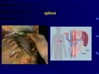
![Lymphatic system[1]](https://image.slidesharecdn.com/v8tdil7slo1obvifzera-signature-460517c25b85fc4e63c8080c3e27df73c8dfae9e0c6544cc7ea6d9e8b5e79cc7-poli-180213064029/85/Lymphatic-system-1-39-320.jpg)
![Lymphatic system[1]](https://image.slidesharecdn.com/v8tdil7slo1obvifzera-signature-460517c25b85fc4e63c8080c3e27df73c8dfae9e0c6544cc7ea6d9e8b5e79cc7-poli-180213064029/85/Lymphatic-system-1-40-320.jpg)
![Lymphatic system[1]](https://image.slidesharecdn.com/v8tdil7slo1obvifzera-signature-460517c25b85fc4e63c8080c3e27df73c8dfae9e0c6544cc7ea6d9e8b5e79cc7-poli-180213064029/85/Lymphatic-system-1-41-320.jpg)
![Lymphatic system[1]](https://image.slidesharecdn.com/v8tdil7slo1obvifzera-signature-460517c25b85fc4e63c8080c3e27df73c8dfae9e0c6544cc7ea6d9e8b5e79cc7-poli-180213064029/85/Lymphatic-system-1-42-320.jpg)

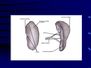
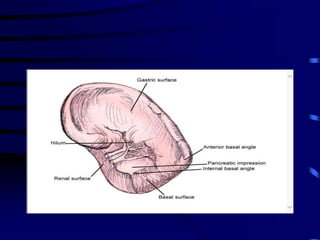
The lymphatic system consists of lymphatic vessels, lymph nodes, and lymphoid organs that work together to drain fluid from tissues, absorb fats, and fight infection. Lymphatic vessels carry lymph fluid and drain into lymph nodes which filter the lymph before returning it to the bloodstream. Major lymphoid organs include the spleen, thymus and tonsils, which help produce immune cells and filter blood and lymph respectively. The lymphatic system is essential for fluid balance, fat absorption, and immune defense.


![CASE_PRESENTATION_ON_subdural_hematoma(SDH)[1 FINAL PPT]-1.pptx](https://cdn.slidesharecdn.com/ss_thumbnails/casepresentationonsubduralhematomasdh1finalppt-1-260129172522-d405d375-thumbnail.jpg?width=640&height=640&fit=bounds)


