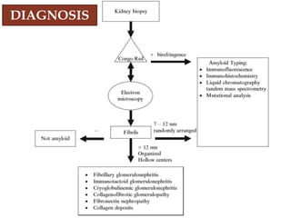
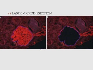
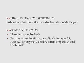
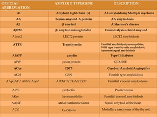
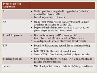
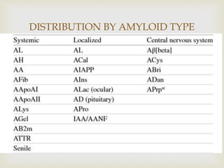
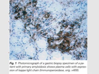
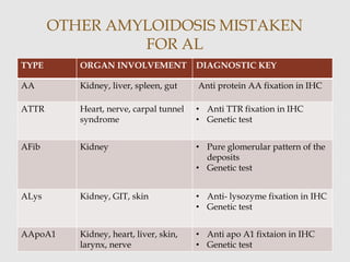
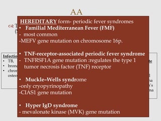
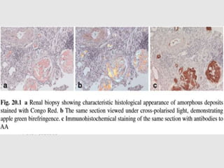
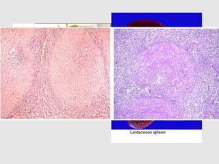
2. “Amyloid”- starch like
In 1854 Rudolph Virchow named it amyloid based of
appearance of violet color after staining these
proteins with iodine and sulfuric acid.
AMYLOID
3. Disorder of protein folding
Deposition of a
• pathologic
• acellular proteinaceous substance
• in extracellular space
• Organ tropism
AMYLOIDOSIS
4. PROPERTIES
Physical properties ( Electron microscopy)
• masses of extracellular, non-branched filaments
• random orientation.
• each fibril consists of two electron-dense filaments
2.5–3.5 nm in diameter, separated by a 2.5 nm space,
giving a total diameter of 8–10 nm.
• X- ray crystallography- beta pleated sheet
5. Chemical properties
Nomenclature: prefix “A” for amyloid, followed by
an abbreviation derived from the name of the protein.
PROTEIN GLYCOPROTEIN-P
85% 15%
7. Recent evidence suggests “fibril deposition may not
be necessary for cellular toxicity rather exposure to
the precursor protein alone is sufficient for
cytotoxicity”
PATHOGENESIS
11. APPEARANCE
STAIN APPEARANCE
H and E Acellular, amorphous, eosinophilic,
refractile, extracellular substance.
CONGO RED On H&E – pink red
On polarizing microscopy – apple green
birefringence
GOLD STANDARD
THIOFLAVIN S Green
12. STAIN APPEARANCE
ALCIAN BLUE Green amorphous
CRYSTAL VIOLET Rose pink
SIRIUS RED Pink red
NOTE:
Amyloid deposits do not stain with periodic acid Schiff
(PAS) or silver stain, but spicules can be seen along the
glomerular basement membranes on silver stain
13. MATERIALS
• frozen tissue is preferred for amyloid typing
• most sensitive -biopsy of a clinically affected organ
• Screening test- abdominal fat aspirate
• Other sites-
*gastrointestinal tract: rectal or gastroduodenal-
deep
*labial salivary gland (LSG)
Recently, a semiquantitative grading system ranging
from 0 (negative) to 4+, for the amount of amyloid in
fat tissue has been proposed by Hazenberg et al
AMYLOID TYPING
Establishing the type of amyloidosis has become a
central issue as the current treatment of systemic
amyloidosis directly depends on the molecular
type of the amyloid protein
14. PRE- REQUISITES FOR CONGO RED
• strong light source
• rotating table
• darkened room for reading slides
• thicker sections (5–10 micro m)
• serial sections
PITFALL: autofluorescence of collagen under
polarization microscopy
15. IMMUNOHISTOCHEMISTRY
• Historically, potassium permanganate was used to
distinguish AA from other forms of amyloid. Applying
potassium permanganate to the tissue prevents Congo red
from binding to AA fibrils but not AL.
• commercially available antisera directed against amyloid
proteins
PITFALLS-
• directed against constant region
• miss a number of hereditary amyloidosis
• serum proteins in background
‘overlay technique’’
Congo red stain and immunohistochemistry are performed
on the same slide
16. LASER MICRODISSECTION
• laser microbe
• melts a thermoplastic ethyl vinyl acetate membrane
that overlays the tissue
• sticks to the selected cells, which can then be lifted
• secured in a microfuge tube containing the
appropriate extraction solution
• Gold standard
PITFALL- Availability
17. FIBRIL TYPING BY PROTEOMICS
Advances allow detection of a single amino acid change
GENE SEQUENCING
• Hereditary amyloidosis
• For-transthyretin, fibrinogen alfa chain, Apo-A1,
Apo-A2, Lysozyme, Gelsolin, serum amyloid A and
Cystatin-C
18. CLASSIFICATION
ORGAN
DISTRIBUTION
• Systemic- > 1
organ
involved
• Localised-
only single
organ
ASSOCIATED
CLINICAL
FINDINGS
• Primary
• Secondary
TUMOR
ASSOCIATED
MYELOMA
ASSOCIATED
HISTORICAL
19. Based on the chemical composition of the deposited
amyloid protein and not neccesarily on the clinical
phenotype.
> 25 types
RECENT CLASSIFICATION
OFFICIAL
ABBREVIATION
AMYLOID TYPE/GENE DESCRIPTION
AL Amyloid light chain (λ) AL amyloidosis/ Multiple myeloma
AA Serum amyloid A protein AA amyloidosis
Aβ β amyloid Alzheimer’s disease
Aβ2M β2 amyloid microglobulin Hemodialysis related amyloid
ALect2 LECT2 protein LECT2 amyloidosis
ATTR Transthyretin Familial amyloid polyneuropathies,
Wild type transthyretin amyloidosis,
leptomeningeal amyloidosis
AIAPP amylin Type II diabetes
APrP prion protein CJD, BSE
ACys CST3 Cerebral Amyloid Angiopathy
AGel GSN Finnish type amyloidosis
AApoA1¹/ Afib²/ Alys³ APOA1¹/ FGA²/LYZ³ Familial visceral amyloidosis
APro prolactin Prolactinoma
AKer keratoepithilin Familial corneal amyloidosis
AANF Atrial natriuretic factor Senile amyloid of the heart
ACal Calcitonin Medullary carcinoma of the thyroid
20.
Types of protein
component
AL • Made up of immunoglobulin light chains (λ chains)
secreted by plasma cells.
• Found in plasma cell tumors
AA • Made from proteolysis of SAA synthesized in liver,
circulates in association with HDL.
• Deposited in inflammatory states as a part of acute
phase response - acute phase protein
β amyloid protein • Derived from Amyloid Precursor protein.
• Core of cerebral plaques found in Alzheimer’s.
• Also deposited in walls of cerebral blood vessels
TTR • Bound to thyroxin and retinol, helps in transporting
them
• Wild TTR -Senile systemic amyloidosis
• Mutant TTR - Familial amyloidotic polyneuropathy
β 2 microglobulin • It’s a component of MHC class I A β 2 m, deposited in
patients on hemodialysis
Prions • Misfolded proteins accumulate in CNS in prion disease
22. most aggressive -13 mnths
kidney and heart are most commonly affected
Histologically, AL deposits can be found in all three
compartments of the kidney
-glomerulus:minimal to massive
-interstitium
-the wall of blood vessels.
Treatment of AL - advanced considerably
- first effective treatment :melphalan and prednisone (MP).
- high dose melphalan followed by autologous STC
-renal transplantation
AL
23. OTHER AMYLOIDOSIS MISTAKEN
FOR AL
TYPE ORGAN INVOLVEMENT DIAGNOSTIC KEY
AA Kidney, liver, spleen, gut Anti protein AA fixation in IHC
ATTR Heart, nerve, carpal tunnel
syndrome
• Anti TTR fixation in IHC
• Genetic test
AFib Kidney • Pure glomerular pattern of the
deposits
• Genetic test
ALys Kidney, GIT, skin • Anti- lysozyme fixation in IHC
• Genetic test
AApoA1 Kidney, heart, liver, skin,
larynx, nerve
• Anti apo A1 fixtaion in IHC
• Genetic test
24. The fibrils in AA are derived from serum amyloid A
(SAA) protein
Secondary to
AA
Infection
• TB,
• bronchiectasis,
• chronic
osteomyelitis
Inflammatory
conditions
• Rheumatoid
arthritis,
• Ankylosing
spondylitis,
• inflammatory
bowel disease,
• Crohn’s and
ulcerative
colitis
skin
popping in
heroin
abusers
non
immunocyte
derived
tumors
• renal cell
carcinoma
• Hodgkin’s
lymphoma
HEREDITARY form- periodic fever syndromes
• Familial Mediterranean Fever (FMF)
- most common
-MEFV gene mutation on chromosome 16p.
• TNF-receptor-associated periodic fever syndrome
- TNFRSF1A gene mutation :regulates the type 1
tumor necrosis factor (TNF) receptor
• Muckle–Wells syndrome
-only cryopyrinopathy
-CIAS1 gene mutation
• Hyper IgD syndrome
- mevalonate kinase (MVK) gene mutation
25. Histologically, one feature that is particular but not
unique to AA is the formation of crescents -most
commonly in AA secondary to rheumatoid arthritis.
Often associated with rapidly progressive
glomerulonephritis and rapid loss in renal function.
Corticosteroids- effective at stabilizing and reversing the
rapid loss in renal function
Treatment - eliminating or controlling the underlying
disease
Reversible
26. AD inheritance
most commonly- transthyretin gene
Familial amyloidotic polyneuropathy
Familial amyloidotic cardiomyopathy
>130 mutations-Val30Met
Microscopy
- amyloid deposition in endoneurium of nerve trunks,
plexuses and sensory and autonomic ganglia
- destruction of the myelin sheath
Treatment- liver transplantation
FAMILIAL AMYLOIDOSIS
27. HEREDITARY RENAL
AMYLOIDOSIS
FEATURES AFib ALys Aapo AI and II
Incidence Most common
Heritance AD AD
Mutations 6 4 13 and 4
Organs Kidney, liver, sleen,
perpheral nerves
and heart
GIT, kidney Kidney, liver, skin,
gonads
Features Glomerular
Sparing of
extraglomerular
compartments
Liver rupture
Bleeding
Visual impairment
Extraglomerular-
medullary deposits
Treatment OLT Renal transplant -
Recurrence + + +
28. LECT2 Amyloidosis (ALECT2)
• Native protein is leukocyte chemotactic factor 2
(LECT2)
• Mean age of presentation - 67 years
• Tandem mass spectrometry
• Kidney,liver, spleen, colon, and adrenal gland
• In the kidney, ALECT2 is highly congophilic and is
deposited extensively in all compartments
• No treatment is currently available
LATEST AMYLOIDOSIS
30. THYROID
• Medullary carcinoma
• Amyloid goitre
• Deposition in stroma, glandular and periglandular
blood vessels
CUTANEOUS
• Localised- macular
-reticular
-nodular
• Primary systemic
• first basement membranes of the blood vessels and
adnexas followed by the replacement of the
collagen of the dermal papilla .
31. CARDIAC
• restrictive cardiomyopathy
• congestive heart failure remains refractory to maximal medical
therapy
• low-voltage changes on electrocardiogram associated with marked
LV hypertrophy
• myocardial dysfunction in the setting of known plasma cell
dyscrasia or connective tissue disorder.
• MICROSCOPY
-interstitial and perimyocyte deposition
-myocyte attenuation
-nodular aggregates
-walls of blood vessels: subendocardial
32.
33. GIT
• Primary (AL)- blood vessel walls and muscularis
propria
• Secondary(AA)- blood vessel wall and lamina propria
lacks the surface epithelial damage and epithelial
lymphocytosis
34. SPLEEN
Two patterns
Deposition in follicles Deposition in walls of sinuses
(SAGO SPLEEN) In red pulp
- LARDACEOUS
SPLEEN
Later fusion of both areas gives the appearance of map like
areas in the spleen
35. RENAL AMYLOIDOSIS
Can involve- the glomeruli(earliest, commonest),
- the blood vessels,
-the interstitium, and
-the tubular basement membranes
40. Despite the similarity in the physical characteristics of the
fibrils, amyloidosis is a tremendously heterogeneous group
of diseases with differences ranging from amyloidogenesis
to organ tropism.
Amyloid typing is important as a number of novel
approaches directly targeting the amyloid fibrils or the
processes of amyloidogenesis have shown very promising
early clinical or pre-clinical results.
Given the complexity inherent in the diagnosis of
amyloidosis, as well as the current need for accurate and
early diagnosis, “loss of chance” doctrine might be
employed in determining liability in some amyloidosis-
related medical malpractice lawsuits
CONCLUSION
41.
Sternberg’s Diagnostic Surgical Pathology 16th edition
Bancroft’s Theory and Practice of Histological Techniques
Robbins and Cotran Pathologic Bais of Disease 8th edition
Recent Advances in Transthyretin Evolution, Structure
and Biological functions-Samantha J. Richardson
Protein Aggregation and Fibrillogenesis in Cerebral and
Systemic Amyloid Diseases-J. Robin Harris
Amyloid journal
REFERENCES