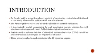
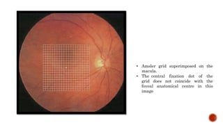
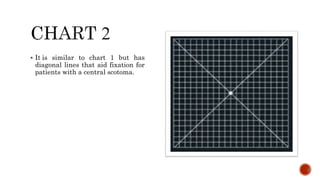
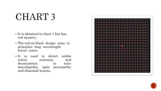
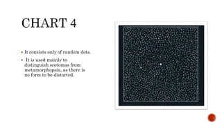
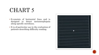
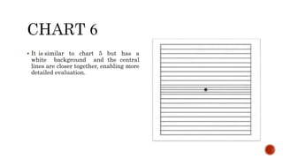
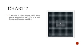
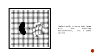
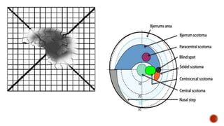
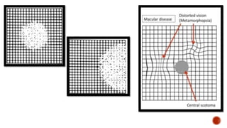
By
Dr. Alshymaa moustafa
Ophthalmology specialist
 An Amsler grid is a simple and easy method of monitoring central visual field and
is commonly abnormal in patients with macular disease.
 The Amsler grid evaluates the 20° of the visual field centred on fixation.
 It is principally useful in screening for and monitoring macular disease, but will
also demonstrate central visual field defects originating elsewhere.
 Patients with a substantial risk of choroidal neovascularization (CNV) should be
provided with an Amsler grid for regular use at home.
 There are seven charts, each consisting of a 10-cm outer square.
• Amsler grid superimposed on the
macula.
• The central fixation dot of the
grid does not coincide with the
foveal anatomical centre in this
image
 consists of a white grid on a black
background.
 The outer grid enclosing 400
smaller 5-mm squares.
 When viewed at about one-third of a
metre, each small square subtends
an angle of 1°.
 It is similar to chart 1 but has
diagonal lines that aid fixation for
patients with a central scotoma.
 It is identical to chart 1 but has
red squares.
 The red-on black design aims to
stimulate long wavelength
foveal cones.
 It is used to detect subtle
colour scotomas and
desaturation in toxic
maculopathy, optic neuropathy
and chiasmal lesions.
 It consists only of random dots.
 It is used mainly to
distinguish scotomas from
metamorphopsia, as there is
no form to be distorted.
 It consists of horizontal lines and is
designed to detect metamorphopsia
along specific meridians.
 It is of particular use in the evaluation of
patients describing difficulty reading.
 It is similar to chart 5 but has a
white background and the central
lines are closer together, enabling more
detailed evaluation.
 It includes a fine central grid, each
square subtending an angle of a half
degree, and is more sensitive.
 The pupils should not be dilated.
 In order to avoid a photostress effect, the eyes should not yet have been examined
on the slit lamp.
 A presbyopic refractive correction should be worn if appropriate.
 The chart should be well illuminated and held at a comfortable reading distance,
optimally around 33 cm.
 One eye is covered.
 The patient is asked to look directly at the central dot with the uncovered eye, to keep
looking at this, and to report any distortion or waviness of the lines on the grid.
 Reminding the patient to maintain fixation on the central dot, he or she is asked if
there are blurred areas or blank spots anywhere on the grid.
 Patients with macular disease often report that the lines are wavy whereas those with
optic neuropathy tend to remark that some of the lines are missing or faint but not
distorted.
 The patient is asked if he or she can see all four corners and all four sides of the
square – a missing corner or border should raise the possibility of causes other than
macular disease, such as glaucomatous field defects or retinitis pigmentosa.
 The patient may be provided with a recording sheet and pen and asked to draw any
anomalies.
 Scotoma may be :
1. Positive : patient aware of it.
2. Negative : patient not aware of it .
 Also may be :
1. Relative : just blurring of certain area in field.
2. Absolute : complete absence of certain area in visual field .
 Metamorphopsia : distorted image.
 Micropsia: smaller image size.
 Macropsia: larger image size.
Stylized Amsler recording sheet shows
wavy lines indicating
metamorphopsia, and a dense
scotoma

Amsler grid
Amsler grid

Amsler grid

  • 1.
  • 2.
     An Amslergrid is a simple and easy method of monitoring central visual field and is commonly abnormal in patients with macular disease.  The Amsler grid evaluates the 20° of the visual field centred on fixation.  It is principally useful in screening for and monitoring macular disease, but will also demonstrate central visual field defects originating elsewhere.  Patients with a substantial risk of choroidal neovascularization (CNV) should be provided with an Amsler grid for regular use at home.  There are seven charts, each consisting of a 10-cm outer square.
  • 3.
    • Amsler gridsuperimposed on the macula. • The central fixation dot of the grid does not coincide with the foveal anatomical centre in this image
  • 4.
     consists ofa white grid on a black background.  The outer grid enclosing 400 smaller 5-mm squares.  When viewed at about one-third of a metre, each small square subtends an angle of 1°.
  • 5.
     It issimilar to chart 1 but has diagonal lines that aid fixation for patients with a central scotoma.
  • 6.
     It isidentical to chart 1 but has red squares.  The red-on black design aims to stimulate long wavelength foveal cones.  It is used to detect subtle colour scotomas and desaturation in toxic maculopathy, optic neuropathy and chiasmal lesions.
  • 7.
     It consistsonly of random dots.  It is used mainly to distinguish scotomas from metamorphopsia, as there is no form to be distorted.
  • 8.
     It consistsof horizontal lines and is designed to detect metamorphopsia along specific meridians.  It is of particular use in the evaluation of patients describing difficulty reading.
  • 9.
     It issimilar to chart 5 but has a white background and the central lines are closer together, enabling more detailed evaluation.
  • 10.
     It includesa fine central grid, each square subtending an angle of a half degree, and is more sensitive.
  • 11.
     The pupilsshould not be dilated.  In order to avoid a photostress effect, the eyes should not yet have been examined on the slit lamp.  A presbyopic refractive correction should be worn if appropriate.  The chart should be well illuminated and held at a comfortable reading distance, optimally around 33 cm.
  • 12.
     One eyeis covered.  The patient is asked to look directly at the central dot with the uncovered eye, to keep looking at this, and to report any distortion or waviness of the lines on the grid.  Reminding the patient to maintain fixation on the central dot, he or she is asked if there are blurred areas or blank spots anywhere on the grid.  Patients with macular disease often report that the lines are wavy whereas those with optic neuropathy tend to remark that some of the lines are missing or faint but not distorted.  The patient is asked if he or she can see all four corners and all four sides of the square – a missing corner or border should raise the possibility of causes other than macular disease, such as glaucomatous field defects or retinitis pigmentosa.  The patient may be provided with a recording sheet and pen and asked to draw any anomalies.
  • 13.
     Scotoma maybe : 1. Positive : patient aware of it. 2. Negative : patient not aware of it .  Also may be : 1. Relative : just blurring of certain area in field. 2. Absolute : complete absence of certain area in visual field .  Metamorphopsia : distorted image.  Micropsia: smaller image size.  Macropsia: larger image size.
  • 14.
    Stylized Amsler recordingsheet shows wavy lines indicating metamorphopsia, and a dense scotoma
  • 15.