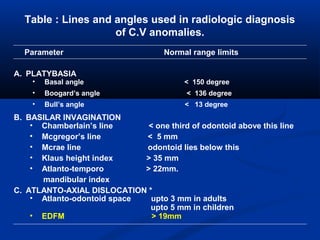
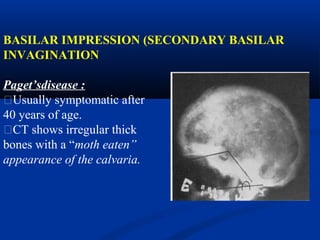
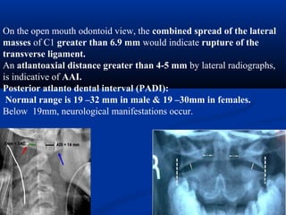
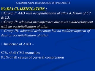
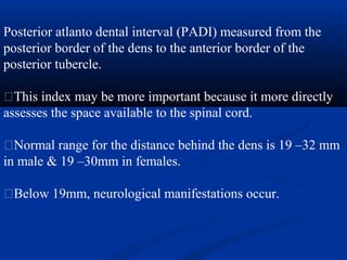
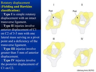
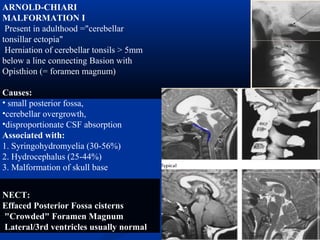
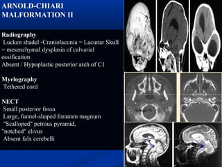
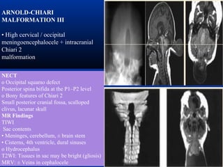
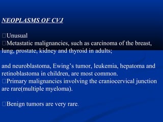
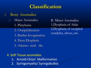
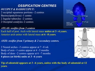
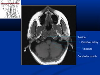
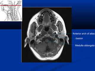
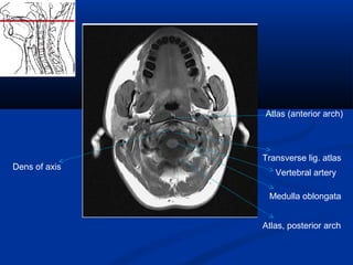
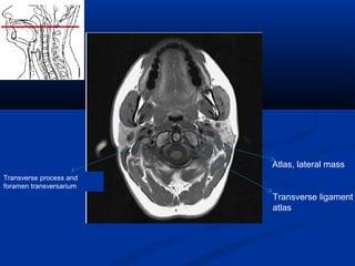
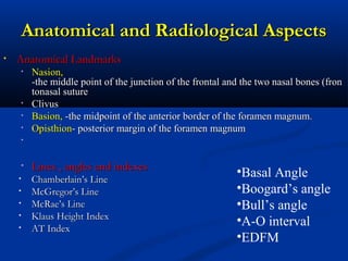
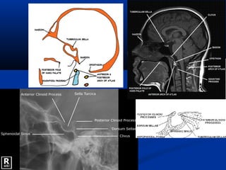
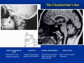
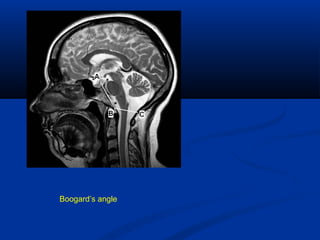
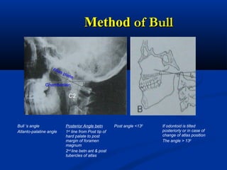
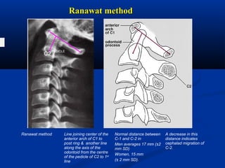
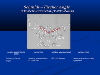
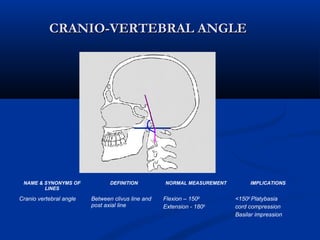
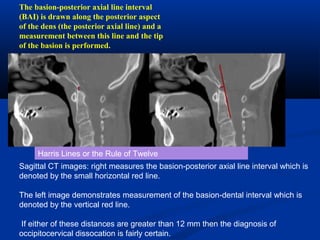
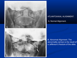
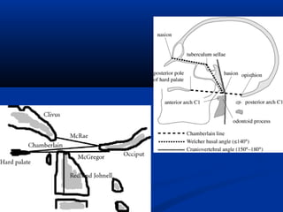
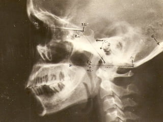
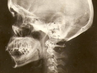
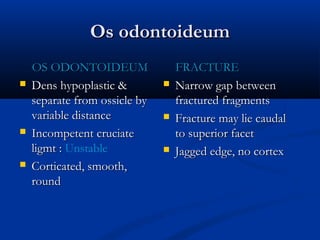
This document discusses cranio-vertebral anomalies and their classification. It begins by classifying bony and soft tissue anomalies. It then discusses the ossification centers of various bones including the occiput, atlas, and axis. Next, it covers the anatomical landmarks and radiological lines used to evaluate the cranio-vertebral junction, such as Chamberlain's line, McGregor's line, and the basilar angle. It provides the normal measurements for these lines. The document concludes by discussing radiological evaluation techniques including CT and MRI measurements.
This document discusses the craniovertebral junction (CVJ), which refers anatomically to the occiput, atlas, and axis vertebrae and their articulations. It describes the anatomy, embryology, biomechanics, imaging modalities including conventional radiography and MRI, and various measurements used to evaluate the CVJ. Common congenital and acquired abnormalities are also discussed, including basilar invagination, platybasia, assimilation of the atlas, and atlantoaxial instability. Imaging plays an important role in identifying bony and soft tissue anomalies, instability, and spinal canal compromise or cord compression.
1) The patellofemoral joint is complex with requirements for normal function dependent on the congruent relationship between the patella and trochlear groove. Developmental or acquired alterations to the joint surface geometry are associated with patellar instability, chondromalacia patella, and anterior knee pain.
2) Trochlear dysplasia, patella alta (high riding patella), and excessive lateralization of the tibial tuberosity are the most important factors predisposing to patellar instability. Trochlear dysplasia can be classified into types A through D based on the shape of the trochlear groove.
3) Measurements of the patella, tro
The document discusses the craniovertebral junction (CVJ) including its embryology, anatomy, and radiology. Regarding embryology, the CVJ develops from the occipital somites which form parts of the occiput, atlas, and axis. Anatomically, the CVJ includes synovial joints between the occiput-atlas and atlas-axis that allow rotation. It is stabilized by ligaments like the transverse ligament. Radiologically, plain films and CT are used to assess the CVJ. Measurements like the Chamberlain's line evaluate for abnormalities like basilar invagination.
The craniovertebral junction (CVJ) refers to the occiput, atlas, axis, and supporting ligaments. It develops from the occipital somites and proatlas in utero. Key ligaments stabilizing the CVJ include the transverse atlantal ligament, alar ligaments, and apical ligament. Neural structures like the medulla, lower cranial nerves, and vertebral artery pass through the CVJ.
1. The document discusses the anatomy, imaging, and pathologies of the craniovertebral junction. It covers bones, ligaments, embryology, radiographic views and measurements, imaging modalities like CT and MRI, craniometry, congenital anomalies, acquired lesions, and traumatic injuries of this region.
2. Various classifications of fractures and abnormalities are mentioned along with normal variations and measurements. Common conditions discussed include Chiari malformations, basilar invagination, rheumatoid arthritis, and traumatic injuries.
3. Detailed anatomy, measurements, and assessments are provided to evaluate abnormalities seen on imaging of the craniovertebral junction.
This document discusses various methods of craniometry used to diagnose craniovertebral junction (CVJ) anomalies through multimodality radiological assessment. It describes several important cranial landmarks and reference lines used to evaluate the CVJ, including Chamberlain's line, McRae's line, McGregor's line, and Wackenheim's line. It then classifies common congenital CVJ anomalies such as atlanto-occipital assimilation, platybasia, basilar invagination, occipital condyle hypoplasia, atlas anomalies, axis anomalies, and discusses associated conditions like Chiari malformation.
This document discusses the craniovertebral junction (CVJ), which refers collectively to the occiput, atlas, axis, and supporting ligaments. It transitions between the mobile cranium and rigid spinal column, enclosing soft tissues of the cervicomedullary junction. The document covers the embryology and development of the CVJ, anatomy including articulations, ligaments, muscles, neural and vascular structures. It also discusses the kinetics, radiological evaluation including craniometry measurements, and common anomalies seen at the CVJ.
CRANIOVERTEBRAL JUNCTION ANATOMY, CRANIOMETRY, ANAMOLIES AND RADIOLOGY dr sum...SUMIT KUMAR
The craniovertebral junction (CVJ) refers anatomically to the occiput, atlas, axis, and their articulations and ligaments. It is a complex region forming the transition between the skull and upper cervical spine.
The document describes the normal anatomy of the CVJ bones including the occiput, atlas, and axis. It discusses the important ligaments including the occipitoatlantoaxial ligaments. Key radiological measurements and lines used to evaluate the CVJ are presented, along with classification of various congenital and acquired CVJ anomalies and their imaging appearance. Basilar invagination, basilar impression, and platybasia are distinguished.
This document contains medical images and descriptions of various musculoskeletal signs and pathologies. It discusses imaging findings and classifications for conditions like:
- Adhesive capsulitis showing thickened ligaments.
- SLAC and SNAC wrist classifications.
- Femoroacetabular impingement presentations.
- Charcot neuroarthropathy acute and chronic stages.
- Various tendon injuries and ligamentous injuries patterns.
It provides comparisons of imaging findings between similar conditions like fibrous dysplasia and osteofibrous dysplasia. Assessment techniques for foot deformities like clubfoot and flatfoot are also outlined.
Presentation1.pptx, ultrasound examination of the elbow joint.Abdellah Nazeer
This document provides an ultrasound summary of the normal elbow anatomy and various pathologies that can be assessed using ultrasound. It describes the lateral, anterior, medial and posterior aspects of the elbow and identifies structures like tendons, ligaments, nerves and bursae. Common pathologies discussed include tendinosis, tears, bursitis, synovitis and effusions. The role of ultrasound in evaluating muscular, tendinous and ligamentous injuries is outlined. Proper scanning technique and ultrasound equipment selection are also reviewed.
The document discusses the craniovertebral junction (CVJ) anatomy. It describes the key components of the CVJ including the occipital bone, atlas, axis, ligaments, and neurovascular structures. It provides a brief overview of CVJ development from somites. Various CVJ abnormalities are also summarized such as atlantoaxial dislocation (AAD) classifications, basilar invagination, and os odontoideum.
Presentation1, radiological imaging of developmental dysplasia of the hip joint.Abdellah Nazeer
Developmental dysplasia of the hip (DDH) is an abnormal development of the hip joint where the femoral head does not properly fit into the acetabulum. It is more common in females and with certain risk factors like breech presentation. Ultrasound is used to evaluate the hip in infants under 6 months by measuring angles like the alpha angle. Once the femoral epiphysis ossifies around 6 months, plain x-rays are used which analyze features like the acetabular angle and Shenton's line to diagnose DDH. DDH can lead to late complications like osteoarthritis if not treated properly.
This document discusses loss of height or collapse of one or more vertebral bodies, which can be caused by several conditions. It provides 10 figures showing examples of vertebral body collapse or loss of height due to severe osteoporosis, multiple myeloma, tuberculous osteomyelitis, brucellosis, fracture, Scheuermann's disease, Langerhans cell histiocytosis, Morquio syndrome, spondyloepiphyseal dysplasia, sickle cell anemia, and osteogenesis imperfecta. Each figure illustrates the characteristic radiographic findings of vertebral body abnormalities associated with each condition.
Spinal trauma can result from automobile accidents and sports activities. Approximately 20% of spinal fractures are associated with fractures elsewhere in the body. Spinal cord injuries occur in 10-14% of spinal fractures and dislocations, with higher rates of neurological damage when fractures affect both the vertebral body and neural arch. Flexion is the most common mechanism of spinal injury. Fractures are most common in the lower cervical and upper thoracic regions. Imaging plays a key role in evaluating spinal trauma and classifying fracture patterns.
1) The document describes the MRI anatomy of the shoulder, highlighting key supporting structures like the rotator cuff muscles and tendons.
2) It examines the shoulder in different planes including axial, coronal, and sagittal views, and provides a checklist of structures and pathologies to evaluate in each view.
3) Special attention is given to evaluating common shoulder injuries like labral tears and rotator cuff tears using specialized views like the ABER position.
Presentation1, radiological imaging of degenerative and inflammatory disease ...Abdellah Nazeer
This document discusses radiological imaging findings of degenerative and inflammatory spine diseases. It provides detailed descriptions and images to illustrate various abnormalities that can be seen, including disc degeneration, herniations, fractures, spinal stenosis, and infections. Key findings are organized by specific pathologies such as disc bulges, protrusions, extrusions, sequestrations, migrating fragments, and vertebral bone marrow changes. Imaging features of conditions like osteoarthritis, synovial cysts, ligamentous thickening, and spinal infections are also reviewed. The document emphasizes the importance of accurate terminology in radiological descriptions and clinical diagnosis of spinal abnormalities.
Craniovertebral juction 1 by dr mohammad mushtaqdrmushtaq22
This document discusses abnormalities of the craniovertebral junction (CVJ). It begins by defining the anatomical structures that make up the CVJ. It then covers the embryology and normal anatomy, including range of motion measurements. Radiological assessment criteria are outlined. Various congenital and acquired disorders are described such as basilar invagination and atlantoaxial subluxation. Specific conditions involving the occiput, atlas, and odontoid are also detailed.
This document provides an overview of MRI techniques for evaluating the shoulder joint and common shoulder pathologies. It begins with normal shoulder anatomy as seen on MRI and descriptions of impingement syndrome, rotator cuff tears, labral tears, instability, biceps tendon injuries, and other conditions. For each pathology, the document describes MRI appearance and features that should be included in reports. In summary, the document is a guide for radiologists to understand MRI of the shoulder and identify and characterize various shoulder injuries and diseases.
The document describes the anatomy of the larynx based on a radiology report. It discusses the boundaries and divisions of the larynx and describes the cartilages that make up its framework, including the thyroid, cricoid, and arytenoid cartilages. It also summarizes the imaging appearance of the larynx on computed tomography (CT) and magnetic resonance imaging (MRI).
Carpal tunnel syndrome is a common compressive neuropathy caused by increased volume in the carpal tunnel or reduced space, compressing the median nerve. MRI is useful for assessing the carpal tunnel at three levels and evaluating the median nerve and flexor retinaculum. Findings of carpal tunnel syndrome include thickening, flattening, or angulation of the median nerve and increased volar bowing of the flexor retinaculum. Rheumatoid arthritis can also cause carpal tunnel syndrome due to synovial thickening, erosions, and tenosynovitis in the wrist. Ultrasound is also used to evaluate the median nerve and fluid along the flexor tendons in a non-
Diagnostic Imaging of Bilateral Abnormalities of the Basal Ganglia & ThalamusMohamed M.A. Zaitoun
The document discusses abnormalities of the basal ganglia and thalamus seen on MRI. It begins by describing the normal anatomy and blood supply of these structures. It then discusses various pathological changes that can be seen, including those from toxins, metabolic diseases, inherited metabolic diseases, vascular causes, infections, tumors and other miscellaneous etiologies. Specific conditions mentioned include Wilson's disease, Leigh's disease, NBIA, hepatic encephalopathy, kernicterus, hypoglycemia and nonketotic hyperglycemia. Radiologic signs for many of these conditions are also described.
This document contains radiographic images and descriptions of various bubbly or lytic bone lesions. It shows examples of lesions caused by conditions such as fibrous dysplasia, giant cell tumor, aneurysmal bone cyst, enchondroma, central chondrosarcoma, plasmacytoma, renal cell carcinoma metastases, lymphoma, Langerhans cell histiocytosis, osteoblastoma, chondroblastoma, intraosseous ganglion cyst, coccidioidomycosis, hemophilia, hemangioendothelioma, cystic angiomatosis, sarcoidosis, and pigmented villonodular synovitis. The images demonstrate the location, appearance,
This document provides information on classifying primary bone tumors based on location and radiographic appearance. Key points include:
- Location within the bone (epiphyseal, diaphyseal, metaphyseal) and age of the patient help classify tumors.
- Features like margins, extent of bone destruction/formation, and presence of a matrix provide clues about tissue type and aggressiveness.
- Common sites for different tumors are listed to aid diagnosis.
- Patterns of bone destruction (lytic, motheaten) and periosteal reactions further characterize lesions.
This document provides an overview of pelvic anatomy and normal pelvic radiology. It describes the bones of the pelvis, ligaments, muscles, blood vessels and lymph nodes. Examples of normal anatomy are shown on plain radiographs, CT scans and MRI images in axial, sagittal and coronal views. Key structures like the sacrum, hip bones, bladder and reproductive organs are labeled on the images.
Here are the key points about rotator interval tears:
- The rotator interval is the space between the supraspinatus and subscapularis tendons through which the long head of the biceps tendon passes.
- Rotator interval tears refer to tears in the capsule in this space between the two tendons.
- They are often associated with instability or repetitive microtrauma and overuse.
- On MRI, they appear as abnormal high signal within the rotator interval capsule on fluid sensitive sequences like T2 or STIR. The torn edges may also enhance with contrast.
- Ultrasound can also identify fluid within the torn interval capsule but MRI is usually better for full
This document discusses MR imaging of the knee. It describes common knee pathologies like meniscal tears, ligament injuries, and cartilage lesions. It provides details on MR imaging techniques and protocols for the knee. Specific meniscal anatomy and grading of meniscal signal are reviewed. Various types of meniscal tears, ligament injuries like ACL and PCL tears are demonstrated with images. Other findings like cartilage lesions, bony lesions, tendon injuries are also described. Potential pitfalls in interpreting MR images of the knee like pseudo meniscal tears are discussed to improve diagnostic accuracy.
This document discusses lumbosacral transitional vertebrae (LSTV), which are congenital spinal anomalies involving sacralization of the lowest lumbar segment or lumbarization of the highest sacral segment. LSTVs can be classified based on imaging features and involve fusion of the transverse process to the sacrum. Numbering the vertebrae can be challenging with LSTVs. Prediction of LSTVs on MRI is possible based on increased A and B angles on sagittal images, which measure the inclination of the sacrum and lumbar lordosis. LSTVs can cause low back pain known as Bertolotti syndrome through various mechanisms.
The document describes various structures of the shoulder joint that provide stability, including the labrum, biceps tendon, and glenohumeral ligaments. It discusses common labral injuries like SLAP tears and Bankart lesions caused by anterior dislocation of the humeral head. It also describes variants like Buford complex and sublabral recesses that should not be confused with pathology.
This document summarizes a case of spastic quadriparesis in an 18-year-old male. Imaging revealed complex craniovertebral junction anomalies including basilar invagination, platybasia, os odontoideum, and atlantoaxial subluxation causing spinal cord compression. The patient was diagnosed with Klippel-Feil syndrome based on clinical features including short stature, low hairline, and restricted neck movement. Treatment options discussed include medical management, traction, and posterior fusion surgery to stabilize and decompress the craniocervical junction.
This document discusses imaging of the cranio-vertebral junction. It begins by defining the cranio-vertebral junction and its key anatomical parts. It then discusses various imaging modalities for evaluating the CV junction, including plain radiographs, CT, MRI, and angiography. Specific abnormalities that can be seen include basilar invagination, atlantoaxial subluxation, occipital anomalies, and os odontoideum. Measurement lines and angles used in evaluation are also defined.
The cranio-vertebral junction includes the occiput, atlas, axis, and connecting ligaments. It is the transition between the mobile cranium and rigid spinal column. The atlanto-axial joint is the most active joint in the body. Craniometry uses lines, planes, and angles to define the normal anatomical relationships at the cranio-vertebral junction that can be measured on imaging. Key measurements include the Chamberlain's line, McRae's line, and basal angle, which are used to diagnose conditions like basilar invagination.
This document discusses various bony and soft tissue anomalies of the craniocervical junction that can cause compression of the brainstem and spinal cord. It outlines different classification systems and measurement lines used to evaluate these anomalies on imaging. Common anomalies discussed include platybasia, basilar invagination, occipito-atlantal fusion, Arnold-Chiari malformation and syringomyelia. Clinical features and approaches to management are also summarized.
This document discusses cranio-vertebral (CV) anomalies, including their classification, anatomical aspects, specific anomalies like atlanto-axial dislocation and dens dysplasia, Arnold-Chiari malformation (ACM), syringomyelia, clinical features, and management. CV anomalies can involve bony or soft tissue structures and are classified accordingly. Radiological evaluation utilizes various lines and angles to assess anomalies like platybasia or basilar invagination. Specific anomalies like atlanto-axial dislocation and dens dysplasia are further classified. ACM and syringomyelia are discussed in detail along with their diagnosis and presentation. Clinical features vary depending on the underlying anomaly. Treatment involves conservative management or
The craniovertebral junction consists of the occiput, atlas, axis, and connecting ligaments. It is a transition zone between the mobile cranium and spinal column. Common anomalies include occipitalization of the atlas, basilar invagination, atlantoaxial dislocation, and platybasia. Radiological assessment involves measurements of angles and distances on plain radiographs and CT/MRI to evaluate for abnormalities. Key measurements include the Chamberlain's line, McGregor's line, and posterior atlantodental interval.
This document provides an overview of the anatomy, classification, and clinical features of craniovertebral junction anomalies. It begins by describing the normal anatomy of the occiput-C1-C2 articulations and ligaments. It then discusses various congenital and acquired bony and soft tissue anomalies that can affect the craniovertebral junction, including occipitalization, basilar invagination, atlantoaxial instability, and dens dysplasia. The document also outlines methods for evaluating craniovertebral junction anomalies using radiography, CT, MRI, and craniometric measurements. It concludes by describing clinical signs and symptoms as well as treatments for different types of craniovertebral junction abnormalities.
This document discusses definitions and classifications of subcortical lesions seen on MRI brain imaging. It defines perivascular spaces, lacunes, subcortical white matter changes, and microbleeds. For each type of lesion, it provides histopathological definitions as well as MRI-based definitions in terms of appearance and severity scales. It also discusses the vascular supply of the brain and implications for the pathogenesis of these subcortical lesions.
This document defines several types of brain lesions that can be seen on MRI scans:
1. Perivascular spaces seen as punctiform dilatations in white matter and basal ganglia.
2. Lacunes appear as small hyperintense lesions on T2WI and hypointense on T1WI, up to 10mm in size.
3. Subcortical white matter changes range from focal lesions to diffuse involvement, graded from 0-3 in severity.
4. Microbleeds appear as homogeneous, round signal losses up to 5mm on gradient echo imaging.
It also provides definitions for assessing the severity and location of intracranial vessel stenosis seen on MRI and methods for measuring
This document provides guidelines for analyzing various spinal x-rays, including cervical, lumbar, and lateral views. Key points analyzed on x-rays include stress lines, vertebral angles and positions, pelvic tilt, and spinal curvatures. Deviations from normal values are measured in degrees or millimeters and can indicate loss of motion, ligament instability, or subluxations. The document establishes a standardized method for interpreting spinal x-rays and identifying areas of dysfunction.
This document discusses lumbosacral transitional vertebrae (LSTV), which are congenital spinal anomalies involving sacralization of the lowest lumbar segment or lumbarization of the highest sacral segment. LSTVs can be classified based on imaging features and involve fusion of the transverse process to the sacrum. Numbering the vertebrae can be challenging with LSTVs. Prediction of LSTVs on MRI is possible based on increased A and B angles on sagittal images, which measure the inclination of the sacrum and lumbar lordosis. LSTVs can cause low back pain known as Bertolotti syndrome through various mechanisms.
This document discusses various methods used to study bone growth, including both measurement and experimental approaches. Measurement approaches involve directly measuring bones and skulls without manipulation, using techniques like craniometry, anthropometry, and radiography/imaging. Experimental approaches analyze bone growth by inducing changes and making observations, such as using fluorescent labels, microelectrodes, or metallic implants in animals. Both approaches involve collecting data through methods like cross-sectional or longitudinal studies, analyzing the data, and presenting it using tables, graphs or diagrams.
1) The document discusses the possibility that cranio-vertebral junction (CVJ) anomalies have been overlooked as a cause of vertebro-basilar insufficiency (VBI).
2) A prospective study was conducted on 19 patients with fixed atlanto-axial dislocation (AAD) using brain SPECT scans to check for signs of posterior circulation ischemia.
3) The results showed decreased cerebellar perfusion in 75% of patients with VBI symptoms, compared to 14% without symptoms. After surgery, 55% of symptomatic patients showed improved perfusion and reduction of VBI symptoms.
1. The document provides detailed anatomical descriptions and diagrams of the craniovertebral junction and foramen magnum region. It describes key landmarks such as the hypoglossal canal, jugular tubercle, pharyngeal tubercle, and their relationships to surrounding structures.
2. Surgical approaches to this region are discussed, including a transoral approach where the soft palate and portions of bone can be removed to fully expose the clivus, atlas, and axis.
3. Important vascular structures like the vertebral artery and its relation to nearby muscles are depicted. The document serves as a comprehensive reference for the intricate anatomy of the lower skull base.
The Indian Dental Academy is the Leader in continuing dental education , training dentists in all aspects of dentistry and
offering a wide range of dental certified courses in different formats.for more details please visit
www.indiandentalacademy.com
This document discusses various measurements and anatomical relationships that can be assessed on radiographs of the skull and cervical spine. It describes measurements such as the basilar angle, Chamberlain's line, and McGregor's line that can help detect abnormalities related to basilar impression. It also discusses measurements of the sella turcica size and the atlantodental interspace that provide information about conditions affecting the pituitary gland or atlantoaxial joint. Finally, it outlines methods for assessing cervical lordosis and the occipitoatlantal alignment on lateral radiographs.
Klippel-Feil syndrome is characterized by a short neck, restricted movement, and low posterior hairline due to blocked or fused vertebrae in the cervical spine. It can cause scoliosis and other spinal abnormalities. Sprengel's deformity, where one or both scapulae are elevated, is seen in about 25% of Klippel-Feil cases. Other associations include heart defects and renal anomalies. The condition results from failed segmentation of the cervical vertebrae during fetal development.
The craniovertebral junction (CVJ) refers collectively to the occiput, atlas, axis, and supporting ligaments. It is a transition zone between the mobile cranium and spinal column, enclosing the soft tissue structures of the cervicomedullary junction. The CVJ has important implications for embryology, anatomy, classification of anomalies, investigations, and clinical management. Anomalies can involve bony, soft tissue, arterial, and neural structures in this region. A variety of imaging modalities like X-rays, CT, MRI are used to classify and characterize CVJ anomalies.
This document discusses C.N.S. vascular malformations, specifically arteriovenous malformations (AVMs) and dural arteriovenous fistulas (DAVF). It covers the definition, types, clinical presentation, radiographic features, grading systems, complications and treatment options for each condition. Key points include that AVMs are congenital lesions with direct connections between arteries and veins, while DAVFs are acquired lesions resulting from damage to venous structures. Presentations can include hemorrhage, seizures, and neurological deficits. Diagnosis is made through CT, MRI, and catheter angiography. Management depends on the size, location, and severity of the lesion.
This document discusses atypical parkinsonism (AP), which accounts for 15-20% of all cases of parkinsonism. It begins by classifying AP into primary, multisystem degenerations, hereditodegenerative, and secondary types. It then focuses on the major AP syndromes - Progressive Supranuclear Palsy (PSP), Multiple System Atrophy (MSA), and Corticobasal Degeneration (CBD). For PSP and MSA, it provides details on clinical presentation, diagnostic criteria, investigations, pathology, and treatment approaches. The document emphasizes the importance of differentiating AP from Parkinson's disease to allow for accurate prognosis and management of patients.
The craniovertebral junction (CVJ) refers to the occiput, atlas, axis, and supporting ligaments. It is a transition zone between the mobile cranium and spinal column. The CVJ encloses important neural and vascular structures.
Anomalies of the CVJ can be congenital due to malformations during embryological development, or acquired through conditions like trauma, infection, or tumors. Common congenital anomalies include occipitalization, basilar invagination, dens dysplasia, and atlantoaxial dislocation. Radiological evaluation is important for assessing abnormalities. Measurements of angles and distances between anatomical structures help characterize anomalies.
The craniovertebral junction (CVJ) refers to the occiput, atlas, axis, and supporting ligaments. It is a transition zone between the mobile cranium and spinal column. The CVJ encloses important neural and vascular structures.
Anatomically, the CVJ includes bony structures like the occiput, atlas, and axis along with their articulations and connecting ligaments. It also has muscles, neural elements like the medulla and lower cranial nerves, lymphatics, arteries and veins. Congenital anomalies of the CVJ can occur due to malformations during embryological development.
Radiological evaluation of the CVJ involves measurements and angles on X
CVJ.Ghty.24thoct2010.ppt sms medical collegedineshdandia
This document discusses imaging of the cranio-vertebral junction. It begins by describing the key anatomical structures in the CV junction, including the occiput, atlas, axis, and their ligaments. It then covers the embryology and normal anatomy and biomechanics of the atlantoaxial joint. The document discusses various imaging modalities for evaluating the CV junction, including plain radiographs, CT, MRI, and angiography. It also outlines common congenital and acquired disorders of the CV junction, including basilar invagination, atlantoaxial subluxation, and atlantoaxial rotatory fixation. Key diagnostic measurements and classifications of abnormalities are provided.
This document discusses the embryology, anatomy, and craniometric evaluation of the craniovertebral junction (CVJ). It describes the development of the atlas and axis from sclerotomes. It outlines the anatomy of the joints, ligaments, and muscles around the CVJ. It then introduces several craniometric lines used to evaluate the CVJ on lateral and anteroposterior radiographs, including Chamberlain's line, McGregor's line, McRae's line, and the Klauss height index. It provides the normal measurements for these lines and their clinical implications when abnormal.
This document discusses the anatomy, imaging, classification, and treatment of various cervical vertebral anomalies. It begins with an overview of the normal anatomy of the atlantoaxial joint and landmarks seen on imaging. It then discusses various congenital and acquired bony and soft tissue anomalies that can occur in this region, including platybasia, basilar invagination, occipitalization of the atlas, and atlantoaxial dislocation. Imaging criteria and classifications for these conditions are provided. Common associated findings like Chiari malformation and syringomyelia are also mentioned. The document concludes with sections on clinical presentation and evaluation of these cervical vertebral anomalies.
This document discusses the craniovertebral junction (CVJ) anatomy and various imaging measurements used to evaluate the CVJ. It describes common CVJ abnormalities like atlantoaxial dislocation, basilar invagination, occipitalization of the atlas, and os odontoideum. It also discusses fractures like Jefferson's fracture and odontoid fractures. The document provides definitions and normal measurements for various craniometric lines and angles used to assess the CVJ on imaging studies.
This document discusses the anatomy, landmarks, measurements, common anomalies, syndromes, and injuries of the craniovertebral junction. It begins with a brief description of the craniovertebral junction's development and components. It then outlines several key anatomical landmarks and measurements used to evaluate the region on imaging. The remainder of the document details various congenital anomalies, developmental abnormalities, syndromes, and acquired conditions that can affect the craniovertebral junction.
Imaging in CranioVertebral Junction AnomaliesBharath J
This document discusses various anatomical structures and anomalies related to the craniovertebral junction. It begins with the normal anatomy and embryology of the central pillar and ringed structures. It then describes various craniometric measurements and their clinical implications for assessing conditions like basilar invagination. Several congenital anomalies are discussed in detail, including os odontoideum, basilar impression, occipital condyle hypoplasia, atlas assimilation, and anomalies of the anterior and posterior atlantal arches.
1) Os Odontoideum is a congenital anomaly where the odontoid process is separated from the body of C2 as a separate ossicle with no bony connection. It can be asymptomatic or cause neck pain and myelopathy.
2) Management depends on symptoms and degree of instability on imaging. Conservative treatment is used if asymptomatic with no instability. Surgical options include anterior release and posterior fusion or resection of the ossicle with fusion.
3) The document describes the embryology, classification, clinical features, imaging, and various surgical management strategies for Os Odontoideum including anterior and posterior releasing techniques and resection with fusion. Case examples are also presented.
Congenital anomalies and Normal skeletal variants- Cervical spineSanal Kumar
The document discusses several congenital anomalies and normal variants of the cervical spine, including:
- Platybasia, which is flattening of the skull base angle. It can occur alone or with skeletal dysplasias. Most cases are asymptomatic.
- Basilar invagination, where the upper cervical vertebrae are positioned too far superiorly in relation to the skull base. It can be primary/congenital or secondary due to bone disease. Symptoms typically begin in the third to fourth decade of life.
- Occipitalization of the atlas, the most common craniocervical junction anomaly, is failure of segmentation of the atlas from the occiput.
The document provides an overview of the anatomy and pathologies of the craniovertebral junction (CVJ). It begins with the normal anatomy and embryological development of the CVJ. It then discusses the most common congenital malformations including assimilation, platybasia, basilar invagination, and abnormalities of the occiput, atlas, and axis. Finally, it reviews acquired disorders, associated syndromes like Chiari malformations and Klippel-Feil syndrome, and imaging techniques for evaluating the CVJ.
The document discusses the anatomy, classification, and imaging of cervical vertebral anomalies. It describes the following key points:
1. The cervical vertebrae have complex articulations and ligaments that allow flexion, extension, and rotation. Common anomalies involve bony fusions or ligamentous laxity.
2. Anomalies are classified as congenital bony anomalies, soft tissue anomalies like Chiari malformation, or developmental/acquired issues. Atlantoaxial instability and basilar invagination are frequently discussed anomalies.
3. Imaging plays an important role in evaluation. Measurements like Chamberlain's line and Atlanto-dens interval on radiographs help characterize anomalies and risk of cord compression.
The document provides guidance on interpreting x-rays for trauma patients, focusing on the cervical spine, chest, pelvis, maxillofacial region, and wrist/hand. It describes what to look for on each image such as alignment, bone fractures, soft tissue swelling, and anatomical landmarks. Proper visualization and positioning is important to detect injuries and abnormalities like pneumothorax, spine fractures, and bone displacements.
The craniovertebral junction refers to the occiput, atlas, and axis vertebrae and their articulations and ligaments. It is a complex anatomical region forming the transition between the skull and cervical spine. Common craniovertebral junction anomalies include occipitalization of the atlas, basilar invagination, atlantoaxial dislocation, and dens dysplasia. These anomalies can be developmental, post-traumatic, or acquired. Imaging studies including X-rays, CT, MRI are used to classify and evaluate the anomalies. Treatment may involve surgery such as fusion if the anomaly causes spinal cord compression.
This document provides an overview of various radiographic views and techniques used to image the foot and ankle. It describes standard ankle x-rays such as AP, mortise, and lateral views. It also discusses stress views used to evaluate ankle instability. Other imaging modalities such as CT, ultrasound, MRI, bone scanning are summarized. Common foot and ankle pathologies and how they appear on different tests are outlined. The purpose is to serve as a reference for foot and ankle imaging evaluations.
This document discusses femoro-acetabular impingement (FAI), which occurs when there is reduced range of motion of the hip due to uneven surfaces of the femoral head or acetabulum. It can be caused by congenital or acquired factors. FAI is classified into cam, pincer, and mixed types. Cam FAI involves a bump on the femoral head-neck junction, while pincer FAI is due to overcoverage of the acetabulum. Clinical features include groin pain exacerbated by activity. Imaging can identify bone abnormalities, and treatments range from activity modification to surgical procedures like arthroscopy or osteotomy.
This document discusses common osseous variants of the spine that can mimic trauma findings on imaging. It outlines normal spinal development and highlights variants that can present as pseudotrauma, with a focus on CT findings. Variants described include incomplete fusion of C1-C2, partially ossified posterior atlantooccipital membranes, persistent os terminale of C2, os odontoideum of C2, anterior wedging of cervical vertebrae in children, and limbus vertebrae in the lumbar spine. Recognition of normal variants is important to avoid an incorrect diagnosis of injury based on imaging alone.
The document discusses the Wits appraisal method for assessing sagittal jaw disharmony. It describes how the Wits appraisal aims to eliminate variations caused by rotations and vertical dimensions seen in measurements like ANB. The Wits appraisal involves drawing perpendiculars from points A and B on the maxilla and mandible to the occlusal plane, and measuring the distance between where they intersect. Studies have found the average distance in males to be 1mm forward for B point, and in females the points generally coincide. The Wits appraisal is said to better reflect the severity of class II and III malocclusions compared to ANB alone. It is influenced less by variations in the cranial base and rotations.
This document discusses upper C-spine fractures, including epidemiology, anatomy, common injuries, and management approaches. It provides details on the anatomy of the upper cervical spine and ligaments. Common injuries discussed include atlanto-occipital dislocations, occipital condyle fractures, C1 fractures such as Jefferson fractures, C2 fractures including odontoid fractures. Odontoid fractures are classified using the Anderson and D'Alonzo system. Non-surgical and surgical management approaches are outlined for different upper cervical spine fractures and injuries.
The document discusses scaphoid fractures, including:
- Anatomy of the scaphoid bone and its blood supply.
- Mechanisms of injury typically involve falls on an outstretched hand causing hyperextension and radial deviation of the wrist.
- Classification systems for scaphoid fractures include Russe's, Mayo, Herbert's, and AO.
- Treatment depends on fracture displacement and stability, ranging from cast immobilization for nondisplaced fractures to surgery for displaced or unstable fractures.
Similar to Amol cranio vertebralanomalies-21-10-14 (20)
The document discusses various imaging modalities used to evaluate peripheral arterial disease (PAD), including duplex ultrasonography, ankle brachial pressure index (ABPI) measurements, digital subtraction angiography (DSA), computed tomography angiography (CTA), and magnetic resonance angiography (MRA). DSA is considered the gold standard but is invasive, while CTA and MRA provide non-invasive alternatives but have limitations such as exposure to radiation or contrast dye. Ultrasonography is useful first-line but not adequate for surgical planning. Overall, the optimal imaging choice depends on each patient's clinical situation and risk factors.
The document discusses the embryogenesis and development of the gastrointestinal tract. It begins by describing how the endoderm-lined yolk sac is incorporated into the embryo to form the primitive gut during folding of the embryo. It then describes the specific regions of the gut - the pharyngeal gut, foregut, midgut and hindgut. It notes that the endoderm forms the gut lining and associated glands while mesoderm forms the muscle, connective tissue and peritoneal components. The mesentery is also described. Common causes of gastrointestinal obstruction by age are listed. The document then focuses specifically on the development and common abnormalities of the esophagus.
The bone scan is a nuclear medicine procedure that uses radiopharmaceuticals like technetium-99m to generate images of the skeletal system. It is useful for evaluating bone disorders and can detect abnormalities earlier than other imaging modalities. The scan involves injection of the radiotracer and acquisition of blood flow, blood pool, and bone scan phase images over hours. It is helpful for detecting fractures, metastases, infections and other bone abnormalities. Precise diagnosis may require correlation with clinical history and other imaging tests.
This document discusses imaging of prostate cancer. It begins with an introduction to prostate cancer epidemiology and symptoms. Transrectal ultrasound is described as the primary imaging method for diagnosis and staging, allowing visualization of the prostate zones and measurement of volume. Appearances of normal variants, benign conditions like BPH and prostatitis, and prostate cancer on ultrasound are presented. The role of MRI in accurate staging of extracapsular extension and seminal vesicle invasion is also summarized.
Cardiac tumors can be primary tumors originating from the heart or secondary tumors from metastases. Primary tumors are more commonly benign myxomas found in the left atrium of middle aged patients, while secondary tumors are more prevalent. Imaging with echocardiography, CT, and MRI can characterize tumors and determine if they are resectable. MRI provides the best evaluation of tumor extent, involvement of surrounding structures, and differentiation of tumor types.
Amol toxic and metabolic encephalopathy syndromeAmol Gulhane
This document discusses various toxic and metabolic encephalopathies and their imaging findings on MRI. It describes conditions such as hyperammonemic encephalopathy, hepatic encephalopathy, osmotic myelinolysis, metronidazole induced encephalopathy, alcoholic encephalopathy, Wernicke's encephalopathy, hypoglycemic encephalopathy, and posterior reversible encephalopathy syndrome. For each condition, it provides details on clinical presentation, characteristic imaging patterns on different MRI sequences, and important differential diagnoses.
Muktapishti is a traditional Ayurvedic preparation made from Shoditha Mukta (Purified Pearl), is believed to help regulate thyroid function and reduce symptoms of hyperthyroidism due to its cooling and balancing properties. Clinical evidence on its efficacy remains limited, necessitating further research to validate its therapeutic benefits.
TEST BANK For Community Health Nursing A Canadian Perspective, 5th Edition by...Donc Test
TEST BANK For Community Health Nursing A Canadian Perspective, 5th Edition by Stamler, Verified Chapters 1 - 33, Complete Newest Version Community Health Nursing A Canadian Perspective, 5th Edition by Stamler, Verified Chapters 1 - 33, Complete Newest Version Community Health Nursing A Canadian Perspective, 5th Edition by Stamler Community Health Nursing A Canadian Perspective, 5th Edition TEST BANK by Stamler Test Bank For Community Health Nursing A Canadian Perspective, 5th Edition Pdf Chapters Download Test Bank For Community Health Nursing A Canadian Perspective, 5th Edition Pdf Download Stuvia Test Bank For Community Health Nursing A Canadian Perspective, 5th Edition Study Guide Test Bank For Community Health Nursing A Canadian Perspective, 5th Edition Ebook Download Stuvia Test Bank For Community Health Nursing A Canadian Perspective, 5th Edition Questions and Answers Quizlet Test Bank For Community Health Nursing A Canadian Perspective, 5th Edition Studocu Test Bank For Community Health Nursing A Canadian Perspective, 5th Edition Quizlet Test Bank For Community Health Nursing A Canadian Perspective, 5th Edition Stuvia Community Health Nursing A Canadian Perspective, 5th Edition Pdf Chapters Download Community Health Nursing A Canadian Perspective, 5th Edition Pdf Download Course Hero Community Health Nursing A Canadian Perspective, 5th Edition Answers Quizlet Community Health Nursing A Canadian Perspective, 5th Edition Ebook Download Course hero Community Health Nursing A Canadian Perspective, 5th Edition Questions and Answers Community Health Nursing A Canadian Perspective, 5th Edition Studocu Community Health Nursing A Canadian Perspective, 5th Edition Quizlet Community Health Nursing A Canadian Perspective, 5th Edition Stuvia Community Health Nursing A Canadian Perspective, 5th Edition Test Bank Pdf Chapters Download Community Health Nursing A Canadian Perspective, 5th Edition Test Bank Pdf Download Stuvia Community Health Nursing A Canadian Perspective, 5th Edition Test Bank Study Guide Questions and Answers Community Health Nursing A Canadian Perspective, 5th Edition Test Bank Ebook Download Stuvia Community Health Nursing A Canadian Perspective, 5th Edition Test Bank Questions Quizlet Community Health Nursing A Canadian Perspective, 5th Edition Test Bank Studocu Community Health Nursing A Canadian Perspective, 5th Edition Test Bank Quizlet Community Health Nursing A Canadian Perspective, 5th Edition Test Bank Stuvia
Basavarajeeyam is a Sreshta Sangraha grantha (Compiled book ), written by Neelkanta kotturu Basavaraja Virachita. It contains 25 Prakaranas, First 24 Chapters related to Rogas& 25th to Rasadravyas.
Cell Therapy Expansion and Challenges in Autoimmune DiseaseHealth Advances
There is increasing confidence that cell therapies will soon play a role in the treatment of autoimmune disorders, but the extent of this impact remains to be seen. Early readouts on autologous CAR-Ts in lupus are encouraging, but manufacturing and cost limitations are likely to restrict access to highly refractory patients. Allogeneic CAR-Ts have the potential to broaden access to earlier lines of treatment due to their inherent cost benefits, however they will need to demonstrate comparable or improved efficacy to established modalities.
In addition to infrastructure and capacity constraints, CAR-Ts face a very different risk-benefit dynamic in autoimmune compared to oncology, highlighting the need for tolerable therapies with low adverse event risk. CAR-NK and Treg-based therapies are also being developed in certain autoimmune disorders and may demonstrate favorable safety profiles. Several novel non-cell therapies such as bispecific antibodies, nanobodies, and RNAi drugs, may also offer future alternative competitive solutions with variable value propositions.
Widespread adoption of cell therapies will not only require strong efficacy and safety data, but also adapted pricing and access strategies. At oncology-based price points, CAR-Ts are unlikely to achieve broad market access in autoimmune disorders, with eligible patient populations that are potentially orders of magnitude greater than the number of currently addressable cancer patients. Developers have made strides towards reducing cell therapy COGS while improving manufacturing efficiency, but payors will inevitably restrict access until more sustainable pricing is achieved.
Despite these headwinds, industry leaders and investors remain confident that cell therapies are poised to address significant unmet need in patients suffering from autoimmune disorders. However, the extent of this impact on the treatment landscape remains to be seen, as the industry rapidly approaches an inflection point.
Does Over-Masturbation Contribute to Chronic Prostatitis.pptxwalterHu5
In some case, your chronic prostatitis may be related to over-masturbation. Generally, natural medicine Diuretic and Anti-inflammatory Pill can help mee get a cure.
Histololgy of Female Reproductive System.pptxAyeshaZaid1
Dive into an in-depth exploration of the histological structure of female reproductive system with this comprehensive lecture. Presented by Dr. Ayesha Irfan, Assistant Professor of Anatomy, this presentation covers the Gross anatomy and functional histology of the female reproductive organs. Ideal for students, educators, and anyone interested in medical science, this lecture provides clear explanations, detailed diagrams, and valuable insights into female reproductive system. Enhance your knowledge and understanding of this essential aspect of human biology.
Promoting Wellbeing - Applied Social Psychology - Psychology SuperNotesPsychoTech Services
A proprietary approach developed by bringing together the best of learning theories from Psychology, design principles from the world of visualization, and pedagogical methods from over a decade of training experience, that enables you to: Learn better, faster!
Local Advanced Lung Cancer: Artificial Intelligence, Synergetics, Complex Sys...Oleg Kshivets
Overall life span (LS) was 1671.7±1721.6 days and cumulative 5YS reached 62.4%, 10 years – 50.4%, 20 years – 44.6%. 94 LCP lived more than 5 years without cancer (LS=2958.6±1723.6 days), 22 – more than 10 years (LS=5571±1841.8 days). 67 LCP died because of LC (LS=471.9±344 days). AT significantly improved 5YS (68% vs. 53.7%) (P=0.028 by log-rank test). Cox modeling displayed that 5YS of LCP significantly depended on: N0-N12, T3-4, blood cell circuit, cell ratio factors (ratio between cancer cells-CC and blood cells subpopulations), LC cell dynamics, recalcification time, heparin tolerance, prothrombin index, protein, AT, procedure type (P=0.000-0.031). Neural networks, genetic algorithm selection and bootstrap simulation revealed relationships between 5YS and N0-12 (rank=1), thrombocytes/CC (rank=2), segmented neutrophils/CC (3), eosinophils/CC (4), erythrocytes/CC (5), healthy cells/CC (6), lymphocytes/CC (7), stick neutrophils/CC (8), leucocytes/CC (9), monocytes/CC (10). Correct prediction of 5YS was 100% by neural networks computing (error=0.000; area under ROC curve=1.0).
- Video recording of this lecture in English language: https://youtu.be/kqbnxVAZs-0
- Video recording of this lecture in Arabic language: https://youtu.be/SINlygW1Mpc
- Link to download the book free: https://nephrotube.blogspot.com/p/nephrotube-nephrology-books.html
- Link to NephroTube website: www.NephroTube.com
- Link to NephroTube social media accounts: https://nephrotube.blogspot.com/p/join-nephrotube-on-social-media.html
Basavarajeeyam is an important text for ayurvedic physician belonging to andhra pradehs. It is a popular compendium in various parts of our country as well as in andhra pradesh. The content of the text was presented in sanskrit and telugu language (Bilingual). One of the most famous book in ayurvedic pharmaceutics and therapeutics. This book contains 25 chapters called as prakaranas. Many rasaoushadis were explained, pioneer of dhatu druti, nadi pareeksha, mutra pareeksha etc. Belongs to the period of 15-16 century. New diseases like upadamsha, phiranga rogas are explained.
2. ClassificationClassification
I.I. Bony AnomaliesBony Anomalies
A.A. Major AnomaliesMajor Anomalies
1. Platybasia1. Platybasia
2. Occipitalization2. Occipitalization
3. Basilar Invagination3. Basilar Invagination
4. Dens Dysplasia4. Dens Dysplasia
5. Atlanto- axial dis.5. Atlanto- axial dis.
B. Minor Anomalies
1.Dysplasia of Atlas
2.Dysplasia of occipital
condyles, clivus, etc.
II. Soft Tissue anomalies
1. Arnold-Chiari Malformation
2. Syringomyelia/ Syringobulbia
3. OSSIFICATION CENTRES
OCCIPUT & BASIOCCIPUT:
2 occipital squamous portions –2 centres
Basiocciput(clivus) -1 centre
2 Jugular tubercles –2 centres
2 Occipital condyles–2 centres
ATLAS: ossifies from 3 centres
Each half of post. Arch with lateral mass unites at 3 –4 years.
Anterior arch unites with lateral mass at 6 –8 years.
AXIS: ossifies from 5 primary & 2 secondary centres.
2 Neural arches –2 centres appear at 7 –8 wk
Body of axis –1 centre appear at 4 –5 months
Body of dens –2 centres appear at 6 –7 months
4 pieces (at birth) unite at 3 –6 years
Tip of odontoid appears at 3 –6 years, unites with the body of odontoid at 12
years.
11. Anatomical and Radiological AspectsAnatomical and Radiological Aspects
• Anatomical LandmarksAnatomical Landmarks
• Nasion,Nasion,
-the middle point of the junction of the frontal and the two nasal bones (fron-the middle point of the junction of the frontal and the two nasal bones (fron
tonasal suturetonasal suture
• ClivusClivus
• Basion,Basion, --the midpoint of the anterior border of the foramen magnum.the midpoint of the anterior border of the foramen magnum.
• OpisthionOpisthion-- posterior margin of the foramen magnumposterior margin of the foramen magnum
•
• Lines , angles and indexesLines , angles and indexes
• Chamberlain’s LineChamberlain’s Line
• McGregor’s LineMcGregor’s Line
• McRae’s LineMcRae’s Line
• Klaus Height IndexKlaus Height Index
• AT IndexAT Index
•Basal Angle
•Boogard’s angle
•Bull’s angle
•A-O interval
•EDFM
12.
13.
14. CRANIOMETRY:
Craniometry of the CVJ uses a series of lines, planes
& angles to define the normal anatomic relationships
of the CVJ.
These measurements can be taken on plain X rays,
3D CT or on MRI.
15. The Chamberlain’s lineThe Chamberlain’s line
NAME & SYNONYMS OF
LINES
DEFINITION NORMAL MEASUREMENT IMPLICATIONS
Chamberlain ‘s line
Palato-occipital line
Posterior pole of hard
palate to the Opisthion.
Tip of the dens usually
below and upto 3 mm
above this line.
Dens > 6mm in basilar
impression.
H O
16.
17. Significance
An abnormal superior position of the odontoid indicates
basilar impression.
Common precipitating causes include platybasia, atlas
occipitalization, and bone-softening diseases of the skull
base (e.g., Paget’s disease, osteomalacia, and fibrous
dysplasia).
Occasionally, rheumatoid arthritis may also precipitate this
deformity.
18. A drawn from the posterosuperior margin of the
hard palate to the most inferior surface of the
occipital bone.
The McGregor’s line
19. The McGregor’s lineThe McGregor’s line
H
Low occiput
NAME & SYNONYMS OF
LINES
DEFINITION NORMAL MEASUREMENT IMPLICATIONS
Mc Gregor’s line
Basal line
MOST ACCURATE
Postero-superior margin
of Hard palate – most
inferior surface of
occipital bone.
Odontoid apex shouldn’t
lie above.
< 5mm
Superior lie of odontoid
indicates basilar
impression.(>5mm)
Low occiput
20. The McRae’s lineThe McRae’s line
B O
Mc Rae’s line
Formen magnum line
Anterior and posterior
ends of formen
magnum.
(Basion and Opisthion)
Inf margin of occiput
should lie at / below this
line. Tip of dens does
not exceed this line.
Perpendicular line along
odontoid intersects 1st
line in its anterior
quadrant.
Inf margin of occiput lies
superior – Basilar imp.
If sagittal diameter
< 20mm neurological
symptoms (+) (foramen
magnum stenosis)
21. Significance
If the inferior margin of the occipital bone is convex in a
superior direction and/or lies above this line, then basilar
impression is present.
Predisposing causes include platybasia, occipitalization,
rheumatoid arthritis, and bone-softening diseases (e.g.,
Paget’s disease, osteomalacia, and fibrous dysplasia).
If the odontoid apex does not lie in the ventral quarter of this
line, a dislocation of the atlanto-occipital joint or a fracture
or dysplasia of the dens may be present.
22. Clivus-Canal lineClivus-Canal line
C
B
OC2
H
N
NAME & SYNONYMS OF
LINES
DEFINITION NORMAL MEASUREMENT IMPLICATIONS
Wackenhie’s line
Clivus canal line
Drawn along clivus into
cervical canal
Odontoid tip is ventral
and tangential to line
Odontoid transects the
line in basilar imp
23. Basilar Angle/Welcker’s basilar angle/ Martin’s
basilar angle/ sphenobasilar angle.
Landmarks
Three points are located and joined together by two
lines; the subsequent angle is measured.
The three points are the nasion (frontal-nasal junction),
the center of the sella turcica (midpoint between the
clinoid processes), and the basion (anterior margin of the
foramen magnum).
The average normal angle subtended by these two lines
is 137°, with a normal variation of 123-152°
24. Significance
The measurement is an index of the relationship between
the anterior skull and its base.
The angle will increase beyond 152° in platybasia, in
which the base is elevated in relation to the rest of the
skull.
This may or may not be associated with basilar
impression.
The deformity may be congenital (isolated impression,
occipitalization) or acquired (Paget’s disease, rheumatoid
arthritis, fibrous dysplasia).
25. Basilar AngleBasilar Angle
Mid sella
B
N
S
B
N
NAME & SYNONYMS OF
LINES
DEFINITION NORMAL MEASUREMENT IMPLICATIONS
Basilar angle
Welcker’s / Martin’s /
Spheno-BA
Nasion – Centre of the
sella – Basion.
Angle 1370
(123-1520
)
>1520
Platybasia
(Base is elevated)
+/- Basilar impression
26. Modified MRI technique
This technique described by Koenigsbert et al yields a normal value range (95% C.I)
116° - 118° for adults and 113° - 115° for children.
Angle formed by :
line extending across the anterior cranial fossa to the tip to the dorsum sellae
line drawn along the posterior margin of the clivus
27. The Boogard’s lineThe Boogard’s line
N
O
NAME & SYNONYMS OF
LINES
DEFINITION NORMAL MEASUREMENT IMPLICATIONS
Boogard ‘s Line Nasion to Opisthion Basion should lie below
this line
Altered in basilar
impression
28. Boogard’s angleBoogard’s angle
Tuberculum sellaTuberculum sella
B o
N
Mc Ray
s
C
NAME & SYNONYMS OF
LINES
DEFINITION NORMAL MEASUREMENT IMPLICATIONS
Boogard ‘s Angle Angle intersected by
1st
line between Dorsum
sellae to Basion &
Mc Rae’s line.
119-1350
Average - 1220
> 1350
Basillar impression
30. MethodMethod of Bullof Bull
C2
Atlas plane
Chamberlain
Bull ‘s angle
Atlanto-palatine angle
Posterior Angle betn
1st
line from Post tip of
hard palate to post
margin of foramen
magnum
2nd
line betn ant & post
tubercles of atlas
Post angle <130
If odontoid is tilted
posteriorly or in case of
change of atlas position
The angle > 130
31. Ranawat methodRanawat method
C2
C1
Ranawat method Line joining center of the
anterior arch of C1 to
post ring & another line
along the axis of the
odontoid from the centre
of the pedicle of C2 to 1st
line
Normal distance between
C-1 and C-2 in
Men averages 17 mm (±2
mm SD)
Women, 15 mm
(± 2 mm SD).
A decrease in this
distance indicates
cephalad migration of
C-2.
C2
C1
C2
C1
PEDICLE
32. Schmidt – Fischer AngleSchmidt – Fischer Angle
(ATLANTO-OCCIPITAL JT AXIS ANGLE)(ATLANTO-OCCIPITAL JT AXIS ANGLE)
O
C2
AA JT
AO JT
C1 C1
NAME & SYNONYMS OF
LINES
DEFINITION NORMAL MEASUREMENT IMPLICATIONS
Schmidt – Fischer
Angle
Angle of axis of Atlanto-
Occipital joint
125 +/- 2 degrees Angle is wider in condylar
hypoplasia
33. CRANIO-VERTEBRAL ANGLECRANIO-VERTEBRAL ANGLE
ax
C
NAME & SYNONYMS OF
LINES
DEFINITION NORMAL MEASUREMENT IMPLICATIONS
Cranio vertebral angle Between clivus line and
post axial line
Flexion – 1500
Extension - 1800
<1500
Platybasia
cord compression
Basilar impression
34. •BDI less than 8.5 mm compared
with 12 mm on data from plain
radiographs.
•An ADI less than 2 mm, compared
with 3 mm previously accepted.
Midsagittal MDCT image of the
craniocervical junction demonstrates
the BDI(basion-dental interval) as
the distance from the most inferior
portion of the basion to the closest
point of the superior aspect of the
dens.
MDCT VS PLAIN RADIOGRAPHY
IN CRANIOMETRY…..
35. MDCT image of the craniocervical junction
demonstrates the ADI, which is calculated
by drawing a line from the posterior aspect
of the anterior arch of C1 to the most
anterior aspect of the dens at the midpoint
of the thickness of the arch in
craniocaudal dimension
Atlantodental Interspace (ADI)
Synonyms.
Atlas-odontoid space, predental interspace,
atlas-dens interval
Flexion is the optimum view to assess the
interspace, because in this position the
most stress is placed on the transverse
ligament of the atlas
36. Normal Values for Atlantodental Interspace
Adults: 1 to 3 mm
Children:1 to 5 mm
Significance.
A decreased space is to be expected with advancing age because of
degenerative joint disease of the atlantodental joint.
A more significant change is an abnormally widened space with
reduction in the neural canal size.
The most frequent causes include trauma, occipitalization, Down’s
syndrome, pharyngeal infections, and inflammatory arthropathies
(e.g., ankylosing spondylitis, rheumatoid arthritis, psoriatic arthritis,
and Reiter’s syndrome).
37. Sagittal CT images: right measures the basion-posterior axial line interval which is
denoted by the small horizontal red line.
The left image demonstrates measurement of the basion-dental interval which is
denoted by the vertical red line.
If either of these distances are greater than 12 mm then the diagnosis of
occipitocervical dissocation is fairly certain.
The basion-posterior axial line interval
(BAI) is drawn along the posterior aspect
of the dens (the posterior axial line) and a
measurement between this line and the tip
of the basion is performed.
Harris Lines or the Rule of Twelve
38. The Powers ratio can be measured to
determine if there is anterior
occipitoatlantal dissociation. The Powers
ratio is the distance between the basion
and the posterior spinolaminar line of C1
(BC) divided by the distance between the
anterior arch of C1 and the opisthion (AO).
If the Power's Rule (BC)/(AO) is greater
than 1 then anterior occipitoatlantal
dissocation has likely occurred.
The accuracy of the Powers ratio is
controversial since it can be difficult to
locate the position of the basion and the
opisthion on the lateral skull radiograph. It
is easier to obtain on sagittal CT..
39. Sagittal MDCT image of the craniocervical
junction demonstrates the AOI, which is
calculated by drawing a line perpendicular
to the articular surfaces of the occipital
condyle and the lateral mass of C1. This
line is drawn at the center of the
articulation by correlating the sagittal and
coronal images.
•The AOI demonstrated 95% of the population
ranged between 0.5 mm and 1.4 mm.
40. AP open mouth
If the lateral margin of the atlas lateral mass lies
lateral to the lateral axis margin, this may be a
radiologic sign of Jefferson’s fracture, odontoid
fracture, alar ligament instability, or rotatory
atlantoaxial subluxation
Atlantoaxial Alignment
41. ATLANTOAXIAL ALIGNMENT.
A. Normal Alignment.
B. Abnormal Alignment. The
abnormality (arrow) is the result of
a Jefferson’s fracture of the atlas.
42.
43.
44.
45. Table : Lines and angles used in radiologic diagnosis
of C.V anomalies.
Parameter Normal range limits
A. PLATYBASIA
B. BASILAR INVAGINATION
C. ATLANTO-AXIAL DISLOCATION *
• Basal angle < 150 degree
• Boogard’s angle < 136 degree
• Bull’s angle < 13 degree
• Chamberlain’s line < one third of odontoid above this line
• Mcgregor’s line < 5 mm
• Mcrae line odontoid lies below this
• Klaus height index > 35 mm
• Atlanto-temporo > 22mm.
mandibular index
• Atlanto-odontoid space upto 3 mm in adults
upto 5 mm in children
• EDFM > 19mm
46. RADIOLOGY OF CVJ
(NORMAL VARIANTS
& ANOMALIES)
THE OCCIPUT :
�The basiocciput forms the lower portion
of the clivus.
�The upper portion of the clivusis formed by the basisphenoid, separated
from the basiocciput by the sphenooccipital synchondrosis
�The age at which this synchondrosis fuses, ranges from “after the twelvth
year”to 14-16 years for girls and 16-18.5 years for boys.
�Most occipital anomalies are associated with decreased skull base height
and basilar invagination.
47. Condylus Tertius
Anomalies and malformations of the most caudal of the occipital
sclerotomes are collectively termed ‘ ‘manifestations of occipital vertebrae”
When the hypochordal bow of the fourth occipital sclerotome (proatlas)
persists or when the proatlas fails to integrate, an ossifled
remnant may be present at the distal end of the clivus, called the condylus
tertius or third occipital condyle .
This third condyle may form a joint or pseudojoint with the
odontoid process or with the anterior arch of the atlas and may lead to
limitation in the
range of motion of the CVJ .
There is an increased prevalence of os odontoideum associated
with this abnormality
The occipital bone is composed of asioccipital, exoccipital, and
supraoccipital portions enclosing the foramen magnum
48. Condylus tertius and platybasia. Midsagittal Ti-weighted MR image reveals
marked skull base flattening, with a Welcher basal angle of 150 dotted line). Note the
marked bowstring deformity of the cervicomedullary junction.
The C-1 arch (A) lies directly above the tip of the odontoid process (0). Marrow within
accessory ossification centers (condylus tertius)
49.
50. Condylar Hypoplasia
In condylar hypoplasia, the occipital condyles are
underdeveloped and have a flattened appearance, leading to
basilar invagination (violation of the Chamberlain line) and
widening of the atlantooccipital joint axis angle
The tip of the odontoid process and the lateral masses of the
atlas typically lie below a line connecting the mastoid tips
(bimastoid line), this relationship is violated in condylar
hypoplasia.
51. The lateral masses of the atlas may be fused to the hypoplastic
condyles,
further accentuating the basilar invagination.
Clinically, condylar hypoplasia limits, or may even abolish,
movements at the atlantooccipital joint and may occasionally lead to
compression of the vertebral artery secondary
to excessive posterior gliding of the occiput in
relation to the atlas
54. PLATYBASIA/Martin’s anamoly
Flattening of angle between the clivus and
the body of the sphenoid
C/F
PRIMARY
- Isolated or in conjunction with other
dysplasias like Achondroplasia,
Osteogenesis imperfecta
SECONDARY - Paget’s disease / bone
softening / degenerative disease
Basilar angle > 152°(N=123-1520)
Craniovertebral = clivus-canal angle
becomes acute (<150°)
MC associated changes - Basilar
invagination, anomalies of
C1(occipitalisation)block vertebra,
Klippel-Feil syndrome.
55. BASILAR INVAGINATION
�Basilar invagination implies that the floor of the skull is
indented by the upper cervical spine, & hence the tip of
odontoid is more cephalad protruding into the FM.
�There are two types of basilar invagination: primary
invagination, which is developmental and more common
secondary invagination, which is acquired.
�Primary invagination can be associated with
occipitoatlantal fusion, hypoplasia of the atlas, a bifid
posterior arch of the atlas, odontoid anomalies.
56. In BI, all three parts of the occipital bone (basiocciput,
exoccipital& squamous occipital bone) are deformed.
Topographic types of BI :
�Anterior BI : hypoplasia of the basilar process of the
occipital bone.
�BI of the occipital condyles(ParamedianBI)–Condylar
hypoplasia
�BI in the lateral condylar area.
�Posterior BI: posterior margin of the FM is invaginated.
�Unilateral BI.
�Generalised BI
57. �BI is associated with high incidence of vertebral artery
anomalies.
�Abnormal curvature of VA is due to the fact that they are of
normal length & course through a reduced bone space (wide
angle b/w 3rd
& 4th
part of VA).
�SIGNS / SYMPTOMS: usually occur in 2nd
or 3rd
decade.
�Short neck(78%),torticollis (68%)
�s/s of associated ACM (cerebellar& vestibular disturbances)
& syringomyelia(25 to 35%).
58. �Motor & sensory disturbances (85%).
�Lower cranial nerves involvement
�Headache & pain in the nape of neck (greater occipital N)
�s/s of raised ICP (HCP) due to posterior encroachment which causes
blockage of aqueduct of sylvius.
�Compression of cerebellum & vestibular apparatus leading to vertical
or lateral nystagmus(65%) (not due to direct pressure from post rim of
FM but rather due to a thickened band of dura).
�Vertebral artery insufficiency s/s.
59. •BASILAR INVAGINATION
•Floor of the skull is indented by the upper
cervical spine, & hence the tip of odontoid
is more cephalad protruding into the FM.
•Primary invagination can be associated
with occipito atlantal fusion, hypoplasia of
the atlas, a bifid posterior arch of the atlas,
odontoid anomalies.
•BI is associated with high incidence of
vertebral artery anomalies.
Chamberlain’s line- tip of dens is >6mm above this line
Mc Gregor’s line- tip of dens is > 5mm above this line
Mc Rae’s line- tip of dens is above this line
Boogard’s line- basion is above this line
60. BASILAR IMPRESSION (SECONDARY BASILAR
INVAGINATION
• Basilar impression refers to secondary or acquired forms of
BI
• due to softening of the bone & is seen in conditions such as
rickets, hyperparathyroidism, osteogenesis imperfecta, Pagets
disease, neurofibromatosis, skeletal dysplasias, and RA &
infection producing bone destruction with or without
ligamentous laxity.
•May be associated with developmental cervical canal stenosis
& also fibrous bands & dural adhesions at the dorsal
cervicomedullary junction.
61. BASILAR IMPRESSION (SECONDARY BASILAR
INVAGINATION
Paget’sdisease :
�Usually symptomatic after
40 years of age.
�CT shows irregular thick
bones with a “moth eaten”
appearance of the calvaria.
62. Achondroplasia:
�Genetically dominant disorder characterized by inhibition of
endochondral bone formation.
�The base of the skull is affected but the membraneous
convexity skull bone grows normally.
�This differential bone growth results in large calvarium on a
small base.
�The mortality is high in the 1st
year of life due to
cervicomedullary dysfunction at the FM.
�A small FM with hypertrophic bone & a posterior dural shelf
results in compression of neural structures.
63.
64. Atlantooccipital Assimilation
failure of segmentation between the skull and first cervical
vertebra results in assimilation of the atlas.
The assimilation may be cornplete or partial.
It invariably results in basilar invagination.
Although the Wackenheim clivus baseline may be normal,
the clivus-canal angle may be decreased.
65. When incompletely assimilated, the atlas arches appear
too high on the lateral plain radiograph
or, when completely assimilated, are not visible
at all
There is an increased prevalence of associated fusion of the axis
and third cervical vertebra in association with atlantooccipital
Assimilation .
When this is present, gradual loosening of the atlantodental joint
with progressive atlantoaxial subluxation may
occur, reported in approximately 50% of cases
66. TOPOGRAPHIC FORMS (WACKENHEIM):
�Type I: Occipitalization(generally subtotal) associated with
BI.
�Type II: Occipitalization(generally subtotal) associated with
BI & fusion of axis & 3rdcervical vertebrae.
�Type III: Total or subtotal occipitalizationwith BI &
maldevelopment of the transverse ligament.
�Type III may be associated with various malformations like
C2-C3 fusion, hemivertebra, dens aplasia, tertiary condyle, etc.
67. The neurological symptoms are not caused by occipitalization proper
but rather by the fact that in the absence of a free atlas, TL fails to
develop which causes posterior displacement of axis & compression
of the spinal cord.
�Instability b/w atlas & axis is reducible in patients <15 yr’s but after
that irreducible state occurs.
68.
69.
70. ATLAS :
�With the exception of the various atlantooccipital
assimilations, most atlas anomalies, when isolated,
produce no abnormal CVJ relationships and are not
associated with basilar invagination.
�The vast majority of anomalies consist of various arch
clefts, aplasias, and hypoplasias.
�Arch anomalies are frequently mistaken for fractures in
the evaluation of plain radiographs of patients with a
history of cervical spine trauma.
71. The irregular fragments located inferior to
the Anterior arch of the atlas may be
mistaken for fracture fragments . The clear
cortical margins and characteristic location
help to differentiate this variant from a
fracture.
Accessory ossification centre for the anterior arch of
the atlas……
72. PONTICULUS POSTICUS /
KIMMERLE’S DEFORMITY :
�It is a bony ridge projecting posteriorly from the
articular edge of the atlas superior articular facet.
�The bony projection may be only a few mm long or
may elongate to unite with the adjacent neural arch of
the atlas to produce an “ARCUATE CANAL”through
which the vertebral artery passes.
�This is due to ossification of a portion of the oblique
A-O ligament.
73.
74.
75. Posterior Arch Anomalies (MC atlas anomaly) :
�Total or partial aplasia of the posterior atlas arch
is rare.
�Although absence of the posterior arch, when
isolated, is usually asymptomatic, but may be
associated with anterior atlantoaxial subluxation.
�Bilateral atlantoaxial subluxation may be
associated with both total and partial aplasias,
simulating the Jefferson fracture.
76. In contrast to the aplasias and hypoplasias, clefts of the
atlas arches are much more common.
�Posterior rachischisis, most common, is observed in 4%
of adults.
�The majority of posterior atlas clefts (97%) are midline,
whereas lateral clefts, through the sulcus of the vertebral
artery, account for the remaining 3%.
�Posterior arch rachischisis may be superimposed on the
odontoid process or the axis body on the open-mouth
odontoid view, simulating a fracture.
78. Split Atlas :
�In contrast to posterior arch rachischisis, anterior arch
rachischisisis quite rare (0. 1 %).
�It is typically encountered in association with posterior
rachischisis-“split atlas”.
�Normally, on a lateral radiograph, the anterior arch of the
atlas appears crescentic or half-moon-shaped, with dense
cortical bone surrounding the medullary cavity and a well-
defined predental space.
79. In anterior arch rachischisis, the anterior arch appears fat or
plump and rounded in configuration, appearing to ‘‘overlap’‘
the odontoid process
(making identification of
the predental space impossible);
the arch may have unsharp,
duplicated anterior margins
80. CONGENITAL ODONTOID ANOMALIES OR DYSPLASIA
Types of dens dysplasia
Type 1 (Os odontoideum) separate odontoid process
Type 2 (Ossiculum terminale) failure of fusion of .
apical segment with its base
Type 3 – Agenesis of odontoid base & apical segment . .
lies separately.
Type 4 – Agenesis of odontoid apical segment
Type 5 –Total agenesis of odontoid process.
81. Persistent OssiculumTerminale:
�Also called Bergman ossicle,
results from failure of fusion of the
terminal ossicle to the remainder of
the odontoid process.
�The fusion typically is
accomplished by 12 years of age.
� Bergman ossicle may be confused with a type 1 odontoid fracture
(avulsion of the terminal ossicle), and absolute differentiation between
the two diagnoses may be difficult.
�Whether traumatic or congenital in origin, this anomaly is stable
when isolated and of relatively little clinical significance.
�The odontoid process is usually normal in height.
82. the normal dense secondary ossification center for the odontoid process tip,
which exhibits a characteristic symmetrical V-shaped lucent zone of separation
from the body of the dens. COMMENT: This is a normal finding of the odontoid
seen in 25% of patients < 12 years of age; but it is usually not seen after this
age, at which time it constitutes non-union (ossiculum terminale of Bergmann).
83. OdontoidAplasia:
�Total aplasia of the odontoid process is extremely rare.
�A true aplasia is associated with an excavation defect into
the body of axis.
�may simulate os odontoideum, as the os fragment may be
perfectly projected over the atlas arch on the open mouth
odontoid view.
84. AP Open Mouth. Here the odontoid
process exists as an abbreviated remnant
stump; therefore, it is not true agenesis.
The lateral shift of the atlas relative to the
axis, C1 on C2, indicates instability
85. OS ODONTOIDEUM
�This term first introduced by Giacominiin 1886, refers to an
independent osseous structure lying cephalad to the axis
body in the location of the odontoid process.
�The anterior arch of the atlas is rounded and hypertrophic
but the posterior arch is hypoplastic.
�As the gap between the os odontoideum and the axis body
usually extends above the level of the superior articular facet
of the axis, cruciate ligament incompetence and A-A
instability are common.
86. The margins of the axis body, the os, and anterior arch are
all well corticated.
�Type 2 odontoid fracture is typically associated with a
flattened, sharp, uncorticated margin to the upper axis body
and a normal, half moon-shaped appearance to the anterior
atlas arch with a narrow gap in b/w # segments.
�Etiology –Embryologic, Traumatic &/or Vascular.
�Types –Orthotopic& Dystopic.
�Instability is more common with dystopic type.
87. � Reducible–on flexion, dorsal compression of the cord
occurs
on extension ventral compression occurs secondary to
increased angulation anteriorly.
�Irreducible–due to displacement of TL ventral to the
ossicle.
Treatment:
Reducible –Occipitocervical PF in neutral position.
Irreducible –Transoral resection of the os, odontoid
remnant & surrounding granulation tissue.
88. Os odontoideum is defined as
non-union of the dens with the
axis body. A transverse,
radiolucent cleft separates an
ossicle of variable size from
the axis body
89. Os odontoideumOs odontoideum
OS ODONTOIDEUMOS ODONTOIDEUM
Dens hypoplastic &Dens hypoplastic &
separate from ossicle byseparate from ossicle by
variable distancevariable distance
Incompetent cruciateIncompetent cruciate
ligmt :ligmt : Unstable
Corticated, smooth,Corticated, smooth,
roundround
FRACTUREFRACTURE
Narrow gap betweenNarrow gap between
fractured fragmentsfractured fragments
Fracture may lie caudalFracture may lie caudal
to superior facetto superior facet
Jagged edge, no cortexJagged edge, no cortex
91. Atlanto-Axial Instability
•A: Rotational
–Around the dens
•B: Translational
–Translation between C1–C2, where transverse lig is disrupted
•C: Distraction:
–Indicating craniocervical dissociation
92. Non-traumatic conditions associated with increase in the atlanto axial
distance:
�Down syndrome
�Due to laxity of the transverse ligament
�Grisel syndrome
�Atlantoaxial subluxation associated with inflammation of adjacent soft
tissues of the neck
�Rheumatoid arthritis
�From laxity of the ligaments and destruction of the articular cartilage
�Osteogenesis imperfecta
�Neurofibromatosis
�Morquio syndrome
�Secondary to odontoid hypoplasia or aplasia
�Other arthridities (Psoriasis,Lupus)
93. On the open mouth odontoid view, the combined spread of the lateral
masses of C1 greater than 6.9 mm would indicate rupture of the
transverse ligament.
An atlantoaxial distance greater than 4-5 mm by lateral radiographs,
is indicative of AAI.
Posterior atlanto dental interval (PADI):
Normal range is 19 –32 mm in male & 19 –30mm in females.
Below 19mm, neurological manifestations occur.
94. WADIA CLASSIFICATION :
�Group I: AAD with occipitalization of atlas & fusion of C2
& C3.
�Group II: odontoid incompetence due to its maldevelopment
with no occipitalization of atlas.
�Group III: odontoid dislocation but no maldevelopment of
dens or occipitalization of atlas.
�Incidence of AAD –
57% of all CVJ anomalies.
8.3% of all causes of cervical compression
ATLANTO-AXIAL DISLOCATION OR INSTABILITY
95. Posterior atlanto dental interval (PADI) measured from the
posterior border of the dens to the anterior border of the
posterior tubercle.
�This index may be more important because it more directly
assesses the space available to the spinal cord.
�Normal range for the distance behind the dens is 19 –32 mm
in male & 19 –30mm in females.
�Below 19mm, neurological manifestations occur.
96. Rotatory displacement
(Fielding and Hawkins
classification):
�Type I is simple rotatory
displacement with an intact
transverse ligament.
�Type II injuries involve
anterior displacement of C1
on C2 of 3-5 mm with one
lateral mass serving as a pivot
point and a deficiency of the
transverse ligament.
�Type III injuries involve
greater than 5 mm of anterior
displacement.
�Type IV injuries involve
the posterior displacement of
C1 on C2.
97. Type I injuries (stable subluxations) –Collar.
�Type II injuries may be potentially unstable.
�Type III and IV rotatory displacements that are
unstable are treated surgically with a reduction
and C1-2 fusion.
98. Non-traumatic conditions associated with increase in the atlantoaxial distance:
� Down syndrome
�Due to laxity of the transverse ligament
�Grisel syndrome
�Atlantoaxial subluxation associated with inflammation of
adjacent soft tissues of the neck
�Rheumatoid arthritis
�From laxity of the ligaments and destruction of the articular
cartilage
�Osteogenesis imperfecta
�Neurofibromatosis
�Morquio syndrome
�Secondary to odontoid hypoplasia or aplasia
�Other arthridities(Psoriasis,Lupus)
99. RHEUMATOID ARTHRITIS & CVJ
�First described by Garrodin 1890.
�20% of the patients with RA have AAD.
�AAD is due to loss of tensile strength & stretching of TL
due to destructive inflammatory changes as well as secondary
degenerative changes in tissues from vasculitis.
�Similar changes occur in the median & lateral joints which
result in erosive changes in adjacent bone & formation of
granulation tissue in the synovial joints.
�Odontoid process –osteoporosis, angulation/ #.
100. OCCIPITO-ATLANTAL
INSTABILITY:Traumatic / non
traumatic
Traumatic usually fatal, 8%
incidence in RTA.
Seen with cardiorespiratory
arrest, quadriplegia, loss of
autonomic function, VA
insufficiency, etc
Traynelis classification:
Type I : anterior displacement of occiput on atlas.
Type II : vertical displacement b/w occiput & cervical spine
Type III : posterior displacement of occiput on atlas.
101. TRAUMATIC LESIONS OF CVJ
�# OF ATLAS:
�Posterior arch #: 2/3rdof all #, occur at the junction of
posterior arch & lateral mass (hyperextension injury).
�Anterior arch #: rare
�Jefferson s # : burst # of atlas,
1st
described by Geoffrey jefferson in 1920.
Axial loading –downward displacement of condyles with
separation of lateral mass of C1.
Classically 4 part # -2 # each in ant & post arch.
neck pain & stiffness
Cervical collar / Halo immobilization
Non union –occiputto C2 fusion
102.
103. open-mouth show lateral spine
dispalcement of lateral masses of C1
CT scan of C1 shows fratures
through anterior and
posterior rings of C1
104. HANGMAN’S # ( TRAUMATIC SPONDYLOLISTHESIS
OF AXIS ):
�“Judicial Hanging”-submental knots causes # dislocation of
neural arch of axis.
�Today majority due to RTA.
�Two basic mechanisms :
Hyperextension & distraction
Hyperextension & compression
105. Type I: # are either non-displaced or have no angulation&
<3mm of displacement (stable injury with uncommon
neurological deficits).
�Type II: # with significant angulation& translation of
anterior fragment.
�Type III: # with severe angulation& displacement along
with concomitant U/L or B/L facet dislocation.
�Neck pain but neurological deficits less.
�Surgical Rx seldom required due to high chances of
spontaneous interbody fusion & # healing.
�Most # managed by reduction & external immobilization.
106. Radiographic features: (best seen on lateral view)
1. Prevertebral soft tissue swelling.
2. Avulsion of anterior inferior corner of C2 associated with rupture of the anterior
longitudinal ligament.
3. Anterior dislocation of the C2 vertebral body.
4. Bilateral C2 pars interarticularis fractures.
Hangman's Fracture- # through pars of c2
107. Hangman's Fracture
* Traumatic spondylolisthesis of C2
* Fractures of the lamina, articular facets,
pedicles or pars interarticulares of C2 with
disruption of C2-C3 junction
108. Axial CT image of the cervical spin
at C2 level shows fractures of
bilateral C2 pars interarticulares.
The fracture on the right extends to
the transverse foramen where the
right vertebral artery is located.
109. ODONTOID #:
�Constitute about 7 –14 % of cervical spine #.
�Flexion is the MC mechanism of injury causing anterior
displacement of C1 on C2.
Anderson & D’Alonzo classification–
�Type I: oblique avulsion # through the upper part of the
odontoid process at the point of alar ligament attachment.
�Type II: # occur at the junction of the odontoid process &
the body of axis.
�Type III: # extend down in to the body of axis.
110. ODONTOID #
�Type I # are stable & heal well if immobilised in a collar or
brace.
�Type III are usually stable #, skull traction f/b halo or
brace for 3 –4 months results in fusion.
�Type II # are prone to non union, with a failure rate of 30
-60 % with conservative measures.
�Indications for Sx–displacement >= 5mm, nonunion, age
>7 years / disruption of the TL.
�Odontoid compression screws (acute type II #) / C1-2
arthrodesis(wiring / fusion, transarticular screws)
111. CHIARI MALFORMATION
�The Chiari malformations are a group of hindbrain
herniation syndromes initially described by Austrian
pathologist Hans Chiari in 1891.
�Types of Chiari malformations :
�Type I: Caudal descent of cerebellar tonsils in cervical
spine. Osseous anomalies of posterior skull base and spine.
It presents in early adulthood rather than at birth.
Associated with syringomyelia in 50 to 70%.
112. Type II: Caudal descent of cerebellar vermis and brain stem
into cervical spine.
Open spinal dysraphism
Hydrocephalus
Multiple neuroaxis anomalies
�Type III: Craniocervical encephalocele containing
portions of cerebellum and brain stem.
Hydrocephalus
�Type IV: (Controversial: not commonly accepted as a
Chiarimalformation)
Aplasia/hypoplasia cerebellum
113. ARNOLD-CHIARI
MALFORMATION I
Present in adulthood ="cerebellar
tonsillar ectopia"
Herniation of cerebellar tonsils > 5mm
below a line connecting Basion with
Opisthion (= foramen magnum)
Causes:
• small posterior fossa,
•cerebellar overgrowth,
•disproportionate CSF absorption
Associated with:
1. Syringohydromyelia (30-56%)
2. Hydrocephalus (25-44%)
3. Malformation of skull base
NECT:
Effaced Posterior Fossa cisterns
"Crowded" Foramen Magnum
Lateral/3rd ventricles usually normal
114. ARNOLD-CHIARI
MALFORMATION II
Radiography
Lucken shadel -Craniolacunia = Lacunar Skull
= mesenchymal dysplasia of calvarial
ossification
Absent / Hypoplastic posterior arch of C1
Myelography
Tethered cord
NECT
Small posterior fossa
Large, funnel-shaped foramen magnum
"Scalloped" petrous pyramid,
"notched" clivus
Absent falx cerebelli
115. ARNOLD-CHIARI
MALFORMATION III
• High cervical / occipital
meningoencephalocele + intracranial
Chiari 2
malformation
NECT
o Occipital squamo defect
Posterior spina bifida at the P1–P2 level
o Bony features of Chiari 2
Small posterior cranial fossa, scalloped
clivus, lacunar skull
MR Findings
TIWI
Sac contents
• Meninges, cerebellum, ± brain stem
• Cisterns, 4th ventricle, dural sinuses
o Hydrocephalus
T2WI: Tissues in sac may be bright (gliosis)
MRV: ± Veins in cephalocele
116. NEOPLASMS OF CVJ
�Unusual
�Metastatic malignancies, such as carcinoma of the breast,
lung, prostate, kidney and thyroid in adults;
and neuroblastoma, Ewing’s tumor, leukemia, hepatoma and
retinoblastoma in children, are most common.
�Primary malignancies involving the craniocervical junction
are rare(multiple myeloma).
�Benign tumors are very rare.
Figure 11.4 The Chamberlain line. This line is drawn from the posterior margin of the foramen magnum (opisthion) to the dorsal (posterior) margin of the hard palate. The odontoid process should not project above this line more than 3 mm; a projection of 6.6 mm (±2 SD) above this line strongly indicates cranial settling.
Figure 11.6 The McGregor line. This line connects the posterosuperior margin of the hard palate to the most caudal part of the occipital curve of the skull. The tip of the odontoid normally does not extend more than 4.5 mm above the line.
Figure 11.5 The McRae line. This line defines the opening of the foramen magnum and connects the anterior margin (basion) with posterior margin (opisthion) of the foramen magnum. The odontoid process should be just below this line or the line may intersect only at the tip of the odontoid process. In addition, a perpendicular line drawn from the apex of the odontoid to this line should intersect it in its ventral quarter.
Figure 11.7 Ranawat method. Ranawat and associates developed a method for determining the extent of the superior margin of the odontoid process, since the hard palate often is not identifiable on radiographs of the cervical spine. The coronal axis of C-1 is determined by connecting the center of the anterior arch of the first cervical vertebra with its posterior ring. The center of the sclerotic ring in C-2, representing the pedicles, is marked. The line is drawn along the axis of the odontoid process to the first line. The normal distance between C-1 and C-2 in men averages 17 mm (±2 mm SD), and in women, 15 mm (± 2 mm SD). A decrease in this distance indicates cephalad migration of C-2.