This document defines several types of brain lesions that can be seen on MRI scans:
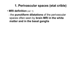
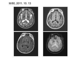
1. Perivascular spaces seen as punctiform dilatations in white matter and basal ganglia.
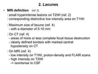
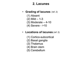
2. Lacunes appear as small hyperintense lesions on T2WI and hypointense on T1WI, up to 10mm in size.
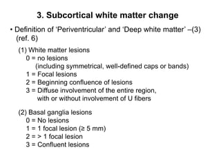
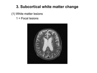
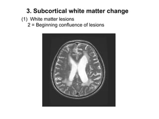
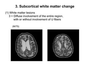
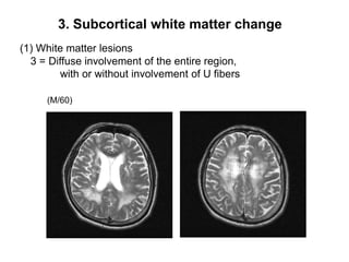
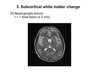
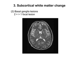
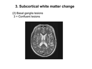
3. Subcortical white matter changes range from focal lesions to diffuse involvement, graded from 0-3 in severity.
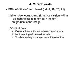
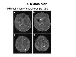
4. Microbleeds appear as homogeneous, round signal losses up to 5mm on gradient echo imaging.
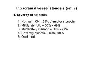
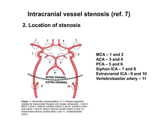
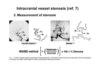
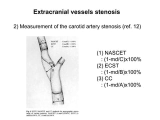
It also provides definitions for assessing the severity and location of intracranial vessel stenosis seen on MRI and methods for measuring