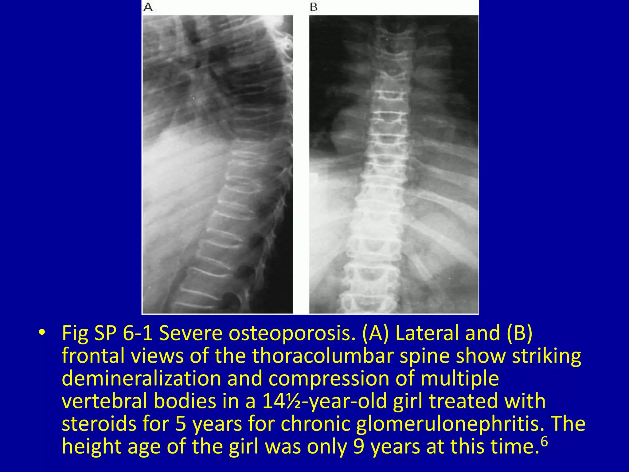
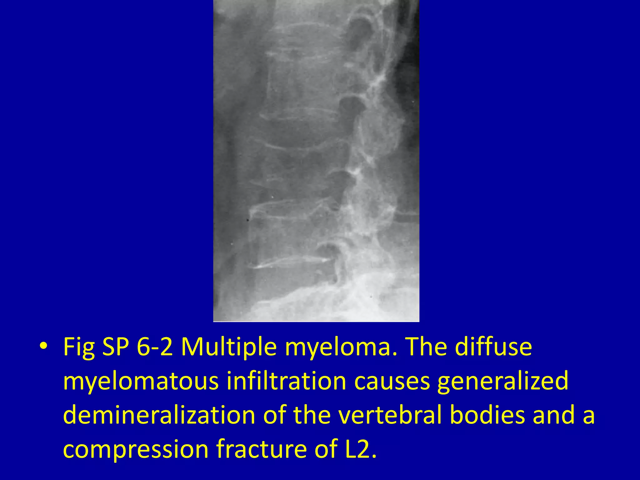
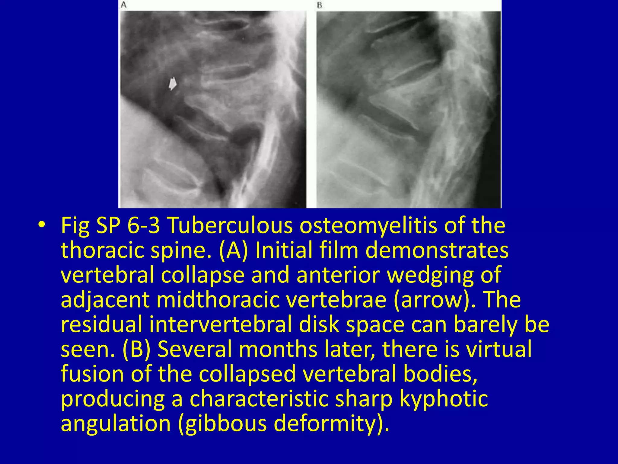
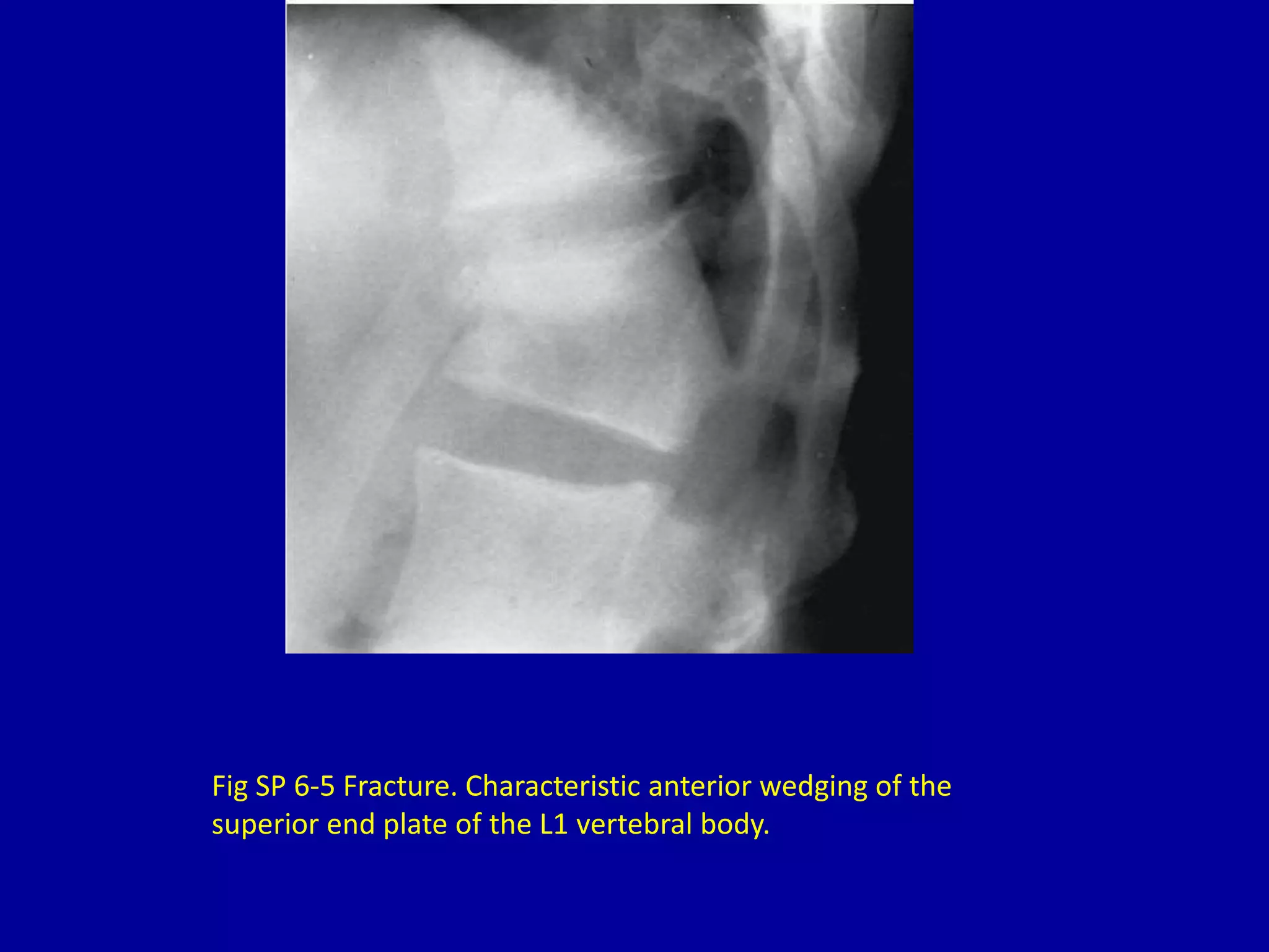
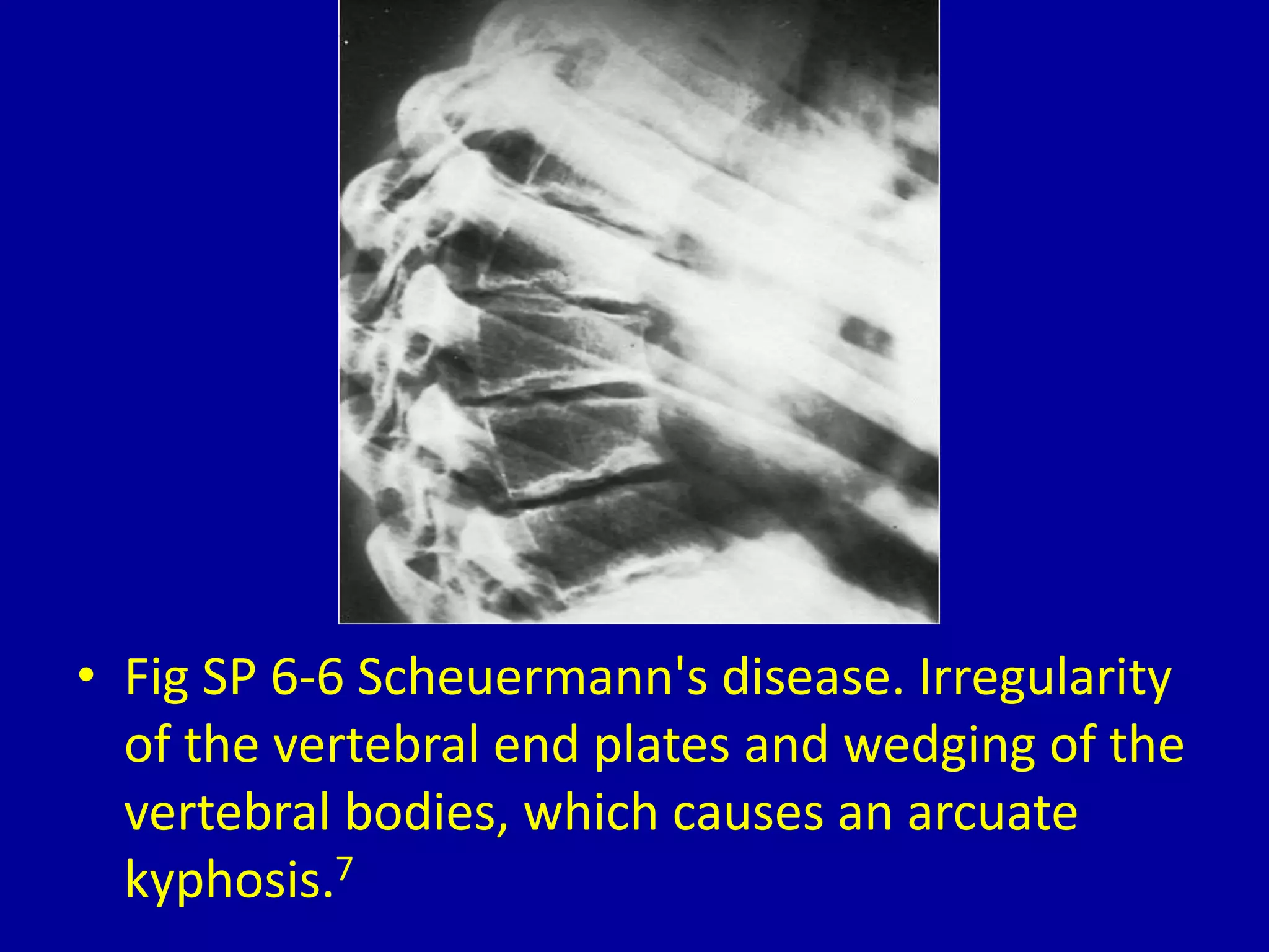
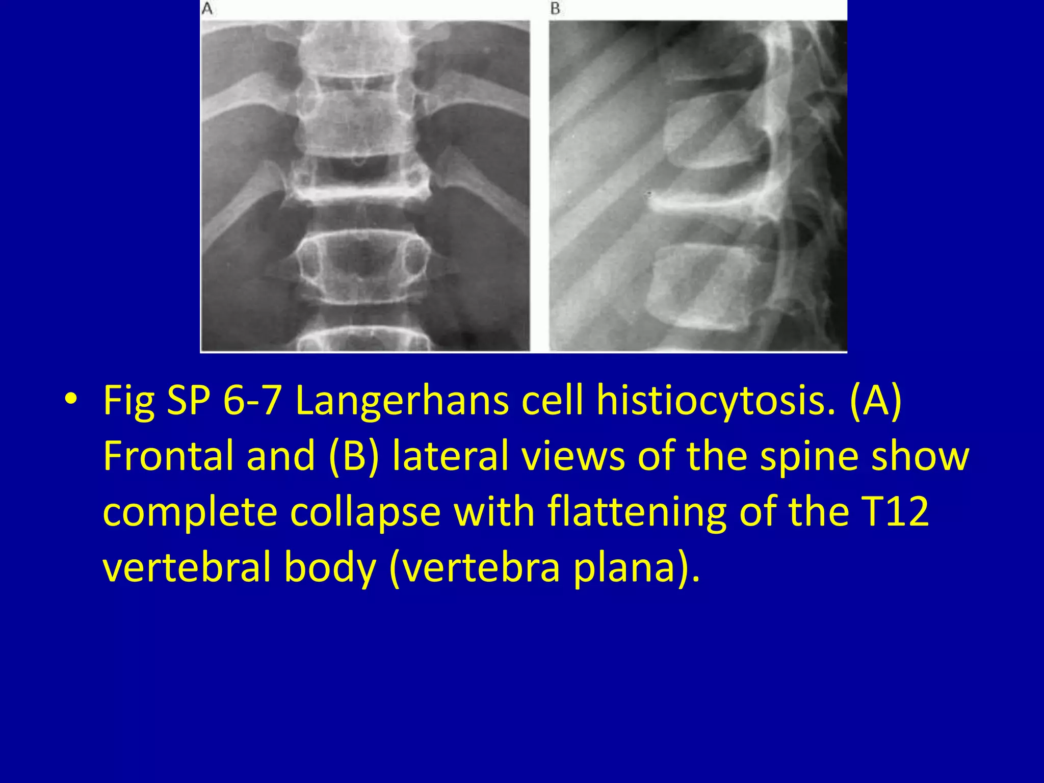
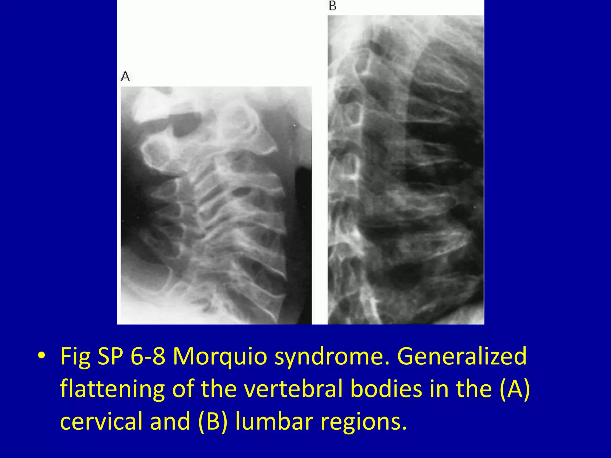
SlideShare a Scribd company logo
6Loss of Height of One or More
Vertebral Bodies
CLINICAL IMAGAGING
AN ATLAS OF DIFFERENTIAL DAIGNOSIS
EISENBERG
DR. Muhammad Bin Zulfiqar
PGR-FCPS III SIMS/SHL
• Fig SP 6-1 Severe osteoporosis. (A) Lateral and (B)
frontal views of the thoracolumbar spine show striking
demineralization and compression of multiple
vertebral bodies in a 14½-year-old girl treated with
steroids for 5 years for chronic glomerulonephritis. The
height age of the girl was only 9 years at this time.6
• Fig SP 6-2 Multiple myeloma. The diffuse
myelomatous infiltration causes generalized
demineralization of the vertebral bodies and a
compression fracture of L2.
• Fig SP 6-3 Tuberculous osteomyelitis of the
thoracic spine. (A) Initial film demonstrates
vertebral collapse and anterior wedging of
adjacent midthoracic vertebrae (arrow). The
residual intervertebral disk space can barely be
seen. (B) Several months later, there is virtual
fusion of the collapsed vertebral bodies,
producing a characteristic sharp kyphotic
angulation (gibbous deformity).
• Fig SP 6-4 Brucellosis. (A) Frontal plain film of the lower
thoracic spine demonstrates loss of height of the T11
and T12 vertebral bodies with destruction of the end
plates and swelling of the paravertebral soft tissues
(arrows). (B) A lateral tomogram of the lower thoracic
spine demonstrates cortical destruction with sclerosis
of the inferior end plate of T11 and the superior end
plate of T12 (arrows). There is a mild degree of anterior
wedging. The overall radiographic appearance is
indistinguishable from that of tuberculous spondylitis.
Fig SP 6-5 Fracture. Characteristic anterior wedging of the
superior end plate of the L1 vertebral body.
• Fig SP 6-6 Scheuermann's disease. Irregularity
of the vertebral end plates and wedging of the
vertebral bodies, which causes an arcuate
kyphosis.7
• Fig SP 6-7 Langerhans cell histiocytosis. (A)
Frontal and (B) lateral views of the spine show
complete collapse with flattening of the T12
vertebral body (vertebra plana).
• Fig SP 6-8 Morquio syndrome. Generalized
flattening of the vertebral bodies in the (A)
cervical and (B) lumbar regions.
• Fig SP 6-9 Spondyloepiphyseal dysplasia.
Generalized flattening of the vertebral bodies
(platyspondyly).
• Fig SP 6-10 Sickle cell anemia. (A) Biconcave
indentations on both the superior and inferior
margins of the soft vertebral bodies produce
the characteristic “fish vertebrae.” (B)
Localized step-like central depressions of
multiple vertebral end plates.
• Fig SP 6-11 Osteogenesis imperfecta.
Generalized flattening of vertebral bodies
associated with fractures of multiple ribs and
long bones in an infant.
6 loss of height of one or more

More Related Content

What's hot

Presentation1, radiological imaging of developmental dysplasia of the hip joint.
Presentation1, radiological imaging of developmental dysplasia of the hip joint.Presentation1, radiological imaging of developmental dysplasia of the hip joint.
Presentation1, radiological imaging of developmental dysplasia of the hip joint.
Abdellah Nazeer
 
MRI of Shoulder anatomy
MRI of Shoulder anatomyMRI of Shoulder anatomy
MRI of Shoulder anatomy
Dr. Muhammad Bin Zulfiqar
 
Mri shoulder joint with common pathologies
Mri shoulder joint with common pathologiesMri shoulder joint with common pathologies
Mri shoulder joint with common pathologies
Gobardhan Thapa
 
An approach to limping child
An approach to limping childAn approach to limping child
An approach to limping child
manoj das
 
DDH
DDHDDH
DDH
DDHDDH
Imaging in arthritis
Imaging in arthritisImaging in arthritis
Imaging in arthritis
Navni Garg
 
Craniovertebral anomalies
Craniovertebral anomaliesCraniovertebral anomalies
Craniovertebral anomalies
Shivshankar Badole
 
Shoulder labral tears MRI
Shoulder labral tears MRIShoulder labral tears MRI
Shoulder labral tears MRI
Dr. Mohit Goel
 
Patello femoral joint - MRI
Patello femoral joint - MRIPatello femoral joint - MRI
Patello femoral joint - MRI
Dr. Mohit Goel
 
Imaging of lumbar spondylosis
Imaging of lumbar spondylosisImaging of lumbar spondylosis
Imaging of lumbar spondylosis
hazem youssef
 
DDH
DDHDDH
Mri imaging finding in wilson disease
Mri imaging finding in wilson diseaseMri imaging finding in wilson disease
Mri imaging finding in wilson disease
gulabsoni
 
Ultrasound of Developmental dysplasia of hip Joint ..Dr.Mohamed Soliman
Ultrasound of Developmental dysplasia of hip Joint ..Dr.Mohamed SolimanUltrasound of Developmental dysplasia of hip Joint ..Dr.Mohamed Soliman
Ultrasound of Developmental dysplasia of hip Joint ..Dr.Mohamed Soliman
Mohamed Soliman
 
MRI KNEE JOINT ANATOMY
MRI KNEE JOINT ANATOMYMRI KNEE JOINT ANATOMY
MRI KNEE JOINT ANATOMY
Nikhil Bansal
 
Legg calve perthes
Legg calve perthesLegg calve perthes
Legg calve perthes
airwave12
 
Diagnostic Imaging of Pediatric Hip Lesions
Diagnostic Imaging of Pediatric Hip LesionsDiagnostic Imaging of Pediatric Hip Lesions
Diagnostic Imaging of Pediatric Hip Lesions
Mohamed M.A. Zaitoun
 
Avascular necrosis of Hip Xray
Avascular necrosis of Hip XrayAvascular necrosis of Hip Xray
Avascular necrosis of Hip Xray
Gaurav Singh
 
Madelung deformity
Madelung deformityMadelung deformity
Madelung deformity
Dr. Anurag Mittal
 
Wilson radiology
Wilson radiologyWilson radiology
Wilson radiology
laxmikant joshi
 

What's hot (20)

Presentation1, radiological imaging of developmental dysplasia of the hip joint.
Presentation1, radiological imaging of developmental dysplasia of the hip joint.Presentation1, radiological imaging of developmental dysplasia of the hip joint.
Presentation1, radiological imaging of developmental dysplasia of the hip joint.
 
MRI of Shoulder anatomy
MRI of Shoulder anatomyMRI of Shoulder anatomy
MRI of Shoulder anatomy
 
Mri shoulder joint with common pathologies
Mri shoulder joint with common pathologiesMri shoulder joint with common pathologies
Mri shoulder joint with common pathologies
 
An approach to limping child
An approach to limping childAn approach to limping child
An approach to limping child
 
DDH
DDHDDH
DDH
 
DDH
DDHDDH
DDH
 
Imaging in arthritis
Imaging in arthritisImaging in arthritis
Imaging in arthritis
 
Craniovertebral anomalies
Craniovertebral anomaliesCraniovertebral anomalies
Craniovertebral anomalies
 
Shoulder labral tears MRI
Shoulder labral tears MRIShoulder labral tears MRI
Shoulder labral tears MRI
 
Patello femoral joint - MRI
Patello femoral joint - MRIPatello femoral joint - MRI
Patello femoral joint - MRI
 
Imaging of lumbar spondylosis
Imaging of lumbar spondylosisImaging of lumbar spondylosis
Imaging of lumbar spondylosis
 
DDH
DDHDDH
DDH
 
Mri imaging finding in wilson disease
Mri imaging finding in wilson diseaseMri imaging finding in wilson disease
Mri imaging finding in wilson disease
 
Ultrasound of Developmental dysplasia of hip Joint ..Dr.Mohamed Soliman
Ultrasound of Developmental dysplasia of hip Joint ..Dr.Mohamed SolimanUltrasound of Developmental dysplasia of hip Joint ..Dr.Mohamed Soliman
Ultrasound of Developmental dysplasia of hip Joint ..Dr.Mohamed Soliman
 
MRI KNEE JOINT ANATOMY
MRI KNEE JOINT ANATOMYMRI KNEE JOINT ANATOMY
MRI KNEE JOINT ANATOMY
 
Legg calve perthes
Legg calve perthesLegg calve perthes
Legg calve perthes
 
Diagnostic Imaging of Pediatric Hip Lesions
Diagnostic Imaging of Pediatric Hip LesionsDiagnostic Imaging of Pediatric Hip Lesions
Diagnostic Imaging of Pediatric Hip Lesions
 
Avascular necrosis of Hip Xray
Avascular necrosis of Hip XrayAvascular necrosis of Hip Xray
Avascular necrosis of Hip Xray
 
Madelung deformity
Madelung deformityMadelung deformity
Madelung deformity
 
Wilson radiology
Wilson radiologyWilson radiology
Wilson radiology
 

Viewers also liked

End plate damage score (SPINE 2010)
End plate damage score (SPINE 2010)End plate damage score (SPINE 2010)
End plate damage score (SPINE 2010)
Prof. Dr. Mohamed Mohi Eldin
 
The modic vertebral endplate and marrow changes (spine 2010)
The modic vertebral endplate and marrow changes (spine 2010)The modic vertebral endplate and marrow changes (spine 2010)
The modic vertebral endplate and marrow changes (spine 2010)
Prof. Dr. Mohamed Mohi Eldin
 
TL Spine Injury 2
TL Spine Injury 2TL Spine Injury 2
TL Spine Injury 2
Mahmood Hassan
 
MIA in Thoraco-lumbar Trauma (SPINE 2009)
MIA in Thoraco-lumbar Trauma (SPINE 2009)MIA in Thoraco-lumbar Trauma (SPINE 2009)
MIA in Thoraco-lumbar Trauma (SPINE 2009)
Prof. Dr. Mohamed Mohi Eldin
 
Anatomy of thoraco lumbar vertabrae (www.Am-medicine.com)
Anatomy of thoraco lumbar vertabrae (www.Am-medicine.com)Anatomy of thoraco lumbar vertabrae (www.Am-medicine.com)
Anatomy of thoraco lumbar vertabrae (www.Am-medicine.com)
Ammedicine Medicine
 
metabolic bone disorders
metabolic bone disordersmetabolic bone disorders
metabolic bone disorders
Sindhu Gowdar
 
Thoraco lumbar injuries
Thoraco lumbar injuriesThoraco lumbar injuries
Thoraco lumbar injuries
dralizameer
 
X-ray spine
X-ray spineX-ray spine

Viewers also liked (8)

End plate damage score (SPINE 2010)
End plate damage score (SPINE 2010)End plate damage score (SPINE 2010)
End plate damage score (SPINE 2010)
 
The modic vertebral endplate and marrow changes (spine 2010)
The modic vertebral endplate and marrow changes (spine 2010)The modic vertebral endplate and marrow changes (spine 2010)
The modic vertebral endplate and marrow changes (spine 2010)
 
TL Spine Injury 2
TL Spine Injury 2TL Spine Injury 2
TL Spine Injury 2
 
MIA in Thoraco-lumbar Trauma (SPINE 2009)
MIA in Thoraco-lumbar Trauma (SPINE 2009)MIA in Thoraco-lumbar Trauma (SPINE 2009)
MIA in Thoraco-lumbar Trauma (SPINE 2009)
 
Anatomy of thoraco lumbar vertabrae (www.Am-medicine.com)
Anatomy of thoraco lumbar vertabrae (www.Am-medicine.com)Anatomy of thoraco lumbar vertabrae (www.Am-medicine.com)
Anatomy of thoraco lumbar vertabrae (www.Am-medicine.com)
 
metabolic bone disorders
metabolic bone disordersmetabolic bone disorders
metabolic bone disorders
 
Thoraco lumbar injuries
Thoraco lumbar injuriesThoraco lumbar injuries
Thoraco lumbar injuries
 
X-ray spine
X-ray spineX-ray spine
X-ray spine
 

Similar to 6 loss of height of one or more

4 generalized vertebral otosclerosis
4 generalized vertebral otosclerosis4 generalized vertebral otosclerosis
4 generalized vertebral otosclerosis
Dr. Muhammad Bin Zulfiqar
 
Diagnostic Imaging of Spondyloarthropathies
Diagnostic Imaging of SpondyloarthropathiesDiagnostic Imaging of Spondyloarthropathies
Diagnostic Imaging of Spondyloarthropathies
Mohamed M.A. Zaitoun
 
Transient Osteoporosis of Hip
Transient Osteoporosis of HipTransient Osteoporosis of Hip
Transient Osteoporosis of Hip
vinod naneria
 
chronic pathological changes
chronic     pathological         changeschronic     pathological         changes
chronic pathological changes
Mohamed Alashram
 
spine chronic pathological changes.
spine chronic    pathological     changes.spine chronic    pathological     changes.
spine chronic pathological changes.
Mohamed Alashram
 
2 lytic lesion of a vertebral body or
2 lytic lesion of a vertebral body or2 lytic lesion of a vertebral body or
2 lytic lesion of a vertebral body or
Dr. Muhammad Bin Zulfiqar
 
Case record...Lumbar spondylosis
Case record...Lumbar spondylosisCase record...Lumbar spondylosis
Case record...Lumbar spondylosis
Professor Yasser Metwally
 
Imaging findings of metabolic bone diseases
Imaging findings of metabolic bone diseases Imaging findings of metabolic bone diseases
Imaging findings of metabolic bone diseases
Pankaj Kaira
 
1 generalized vertebral osteoporosis
1 generalized vertebral osteoporosis1 generalized vertebral osteoporosis
1 generalized vertebral osteoporosis
Dr. Muhammad Bin Zulfiqar
 
7 narrowing of the intervertebral disk space and
7 narrowing of the intervertebral disk space and7 narrowing of the intervertebral disk space and
7 narrowing of the intervertebral disk space and
Dr. Muhammad Bin Zulfiqar
 
Spine disease e srs update version
Spine disease  e srs update versionSpine disease  e srs update version
Spine disease e srs update version
sr sumon (sohail)
 
3 osteosclerotic vertebral lesions
3 osteosclerotic vertebral lesions3 osteosclerotic vertebral lesions
3 osteosclerotic vertebral lesions
Dr. Muhammad Bin Zulfiqar
 
Cervical spondylosis
Cervical spondylosisCervical spondylosis
Cervical spondylosis
ranjan mishra
 
Presentation1, radiological film reading of the hip joint.
Presentation1, radiological film reading of the hip joint.Presentation1, radiological film reading of the hip joint.
Presentation1, radiological film reading of the hip joint.
Abdellah Nazeer
 
Ct spine fractures ppt
Ct spine fractures pptCt spine fractures ppt
Ct spine fractures ppt
BipulBorthakur
 
Case record...Spinal multiple sclerosis
Case record...Spinal multiple sclerosisCase record...Spinal multiple sclerosis
Case record...Spinal multiple sclerosis
Professor Yasser Metwally
 
Paraplegia and spinal cord syndromes
Paraplegia and spinal cord syndromesParaplegia and spinal cord syndromes
Paraplegia and spinal cord syndromes
ramtinyoung
 
THE SPINE IN THE COURSE OF SPONDYLOARTHROPATHIES.pptx
THE SPINE IN THE COURSE OF SPONDYLOARTHROPATHIES.pptxTHE SPINE IN THE COURSE OF SPONDYLOARTHROPATHIES.pptx
THE SPINE IN THE COURSE OF SPONDYLOARTHROPATHIES.pptx
kavitjetly
 
35 DAVID SUTTON PICTURES THE CONGENITAL SKELETAL ANOMALIES: SKELETAL DYSPLAS...
35 DAVID SUTTON PICTURES  THE CONGENITAL SKELETAL ANOMALIES: SKELETAL DYSPLAS...35 DAVID SUTTON PICTURES  THE CONGENITAL SKELETAL ANOMALIES: SKELETAL DYSPLAS...
35 DAVID SUTTON PICTURES THE CONGENITAL SKELETAL ANOMALIES: SKELETAL DYSPLAS...
Dr. Muhammad Bin Zulfiqar
 
Presentation3, radiological imaging of vanshing bone tumour.
Presentation3, radiological imaging of vanshing bone tumour.Presentation3, radiological imaging of vanshing bone tumour.
Presentation3, radiological imaging of vanshing bone tumour.
Abdellah Nazeer
 

Similar to 6 loss of height of one or more (20)

4 generalized vertebral otosclerosis
4 generalized vertebral otosclerosis4 generalized vertebral otosclerosis
4 generalized vertebral otosclerosis
 
Diagnostic Imaging of Spondyloarthropathies
Diagnostic Imaging of SpondyloarthropathiesDiagnostic Imaging of Spondyloarthropathies
Diagnostic Imaging of Spondyloarthropathies
 
Transient Osteoporosis of Hip
Transient Osteoporosis of HipTransient Osteoporosis of Hip
Transient Osteoporosis of Hip
 
chronic pathological changes
chronic     pathological         changeschronic     pathological         changes
chronic pathological changes
 
spine chronic pathological changes.
spine chronic    pathological     changes.spine chronic    pathological     changes.
spine chronic pathological changes.
 
2 lytic lesion of a vertebral body or
2 lytic lesion of a vertebral body or2 lytic lesion of a vertebral body or
2 lytic lesion of a vertebral body or
 
Case record...Lumbar spondylosis
Case record...Lumbar spondylosisCase record...Lumbar spondylosis
Case record...Lumbar spondylosis
 
Imaging findings of metabolic bone diseases
Imaging findings of metabolic bone diseases Imaging findings of metabolic bone diseases
Imaging findings of metabolic bone diseases
 
1 generalized vertebral osteoporosis
1 generalized vertebral osteoporosis1 generalized vertebral osteoporosis
1 generalized vertebral osteoporosis
 
7 narrowing of the intervertebral disk space and
7 narrowing of the intervertebral disk space and7 narrowing of the intervertebral disk space and
7 narrowing of the intervertebral disk space and
 
Spine disease e srs update version
Spine disease  e srs update versionSpine disease  e srs update version
Spine disease e srs update version
 
3 osteosclerotic vertebral lesions
3 osteosclerotic vertebral lesions3 osteosclerotic vertebral lesions
3 osteosclerotic vertebral lesions
 
Cervical spondylosis
Cervical spondylosisCervical spondylosis
Cervical spondylosis
 
Presentation1, radiological film reading of the hip joint.
Presentation1, radiological film reading of the hip joint.Presentation1, radiological film reading of the hip joint.
Presentation1, radiological film reading of the hip joint.
 
Ct spine fractures ppt
Ct spine fractures pptCt spine fractures ppt
Ct spine fractures ppt
 
Case record...Spinal multiple sclerosis
Case record...Spinal multiple sclerosisCase record...Spinal multiple sclerosis
Case record...Spinal multiple sclerosis
 
Paraplegia and spinal cord syndromes
Paraplegia and spinal cord syndromesParaplegia and spinal cord syndromes
Paraplegia and spinal cord syndromes
 
THE SPINE IN THE COURSE OF SPONDYLOARTHROPATHIES.pptx
THE SPINE IN THE COURSE OF SPONDYLOARTHROPATHIES.pptxTHE SPINE IN THE COURSE OF SPONDYLOARTHROPATHIES.pptx
THE SPINE IN THE COURSE OF SPONDYLOARTHROPATHIES.pptx
 
35 DAVID SUTTON PICTURES THE CONGENITAL SKELETAL ANOMALIES: SKELETAL DYSPLAS...
35 DAVID SUTTON PICTURES  THE CONGENITAL SKELETAL ANOMALIES: SKELETAL DYSPLAS...35 DAVID SUTTON PICTURES  THE CONGENITAL SKELETAL ANOMALIES: SKELETAL DYSPLAS...
35 DAVID SUTTON PICTURES THE CONGENITAL SKELETAL ANOMALIES: SKELETAL DYSPLAS...
 
Presentation3, radiological imaging of vanshing bone tumour.
Presentation3, radiological imaging of vanshing bone tumour.Presentation3, radiological imaging of vanshing bone tumour.
Presentation3, radiological imaging of vanshing bone tumour.
 

More from Dr. Muhammad Bin Zulfiqar

Dislocations of joint. Joint Dislocation
Dislocations of joint. Joint DislocationDislocations of joint. Joint Dislocation
Dislocations of joint. Joint Dislocation
Dr. Muhammad Bin Zulfiqar
 
Role of color doppler ultrasound in rvhtn
Role of color doppler ultrasound in rvhtnRole of color doppler ultrasound in rvhtn
Role of color doppler ultrasound in rvhtn
Dr. Muhammad Bin Zulfiqar
 
Bone age assessment
Bone age assessmentBone age assessment
Bone age assessment
Dr. Muhammad Bin Zulfiqar
 
Trauma axial skeleton Dr. Muhammad Bin Zulfiqar
Trauma axial skeleton Dr. Muhammad Bin ZulfiqarTrauma axial skeleton Dr. Muhammad Bin Zulfiqar
Trauma axial skeleton Dr. Muhammad Bin Zulfiqar
Dr. Muhammad Bin Zulfiqar
 
21 non ischaemic acquired Dr.Muhammad Bin Zulfiqar
21 non ischaemic acquired Dr.Muhammad Bin Zulfiqar21 non ischaemic acquired Dr.Muhammad Bin Zulfiqar
21 non ischaemic acquired Dr.Muhammad Bin Zulfiqar
Dr. Muhammad Bin Zulfiqar
 
Mri anatomy of knee Dr. Muhammad Bin Zulfiqar
Mri anatomy of knee Dr. Muhammad Bin ZulfiqarMri anatomy of knee Dr. Muhammad Bin Zulfiqar
Mri anatomy of knee Dr. Muhammad Bin Zulfiqar
Dr. Muhammad Bin Zulfiqar
 
20 congenital heart disease Dr. Muhammmad Bin Zulfiqar
20 congenital heart disease Dr. Muhammmad Bin Zulfiqar20 congenital heart disease Dr. Muhammmad Bin Zulfiqar
20 congenital heart disease Dr. Muhammmad Bin Zulfiqar
Dr. Muhammad Bin Zulfiqar
 
An approach to cardiac xray Dr. Muhammad Bin Zulfiqar
An approach to cardiac xray Dr. Muhammad Bin ZulfiqarAn approach to cardiac xray Dr. Muhammad Bin Zulfiqar
An approach to cardiac xray Dr. Muhammad Bin Zulfiqar
Dr. Muhammad Bin Zulfiqar
 
19 cardiac anatomy and Imaging techniques Dr. Muhammad Bin Zulfiqar
19 cardiac anatomy and Imaging techniques Dr. Muhammad Bin Zulfiqar19 cardiac anatomy and Imaging techniques Dr. Muhammad Bin Zulfiqar
19 cardiac anatomy and Imaging techniques Dr. Muhammad Bin Zulfiqar
Dr. Muhammad Bin Zulfiqar
 
Pediatric brain tumors Dr. Muhammad Bin Zulfiqar
Pediatric brain tumors Dr. Muhammad Bin Zulfiqar Pediatric brain tumors Dr. Muhammad Bin Zulfiqar
Pediatric brain tumors Dr. Muhammad Bin Zulfiqar
Dr. Muhammad Bin Zulfiqar
 
Prostate Biopsy--New Emerging Trends, Dr. Muhammad Bin Zulfiqar
Prostate Biopsy--New Emerging Trends, Dr. Muhammad Bin Zulfiqar Prostate Biopsy--New Emerging Trends, Dr. Muhammad Bin Zulfiqar
Prostate Biopsy--New Emerging Trends, Dr. Muhammad Bin Zulfiqar
Dr. Muhammad Bin Zulfiqar
 
Eponymous fractures name Dr. muhammad Bin Zulfiqar
Eponymous fractures name Dr. muhammad Bin ZulfiqarEponymous fractures name Dr. muhammad Bin Zulfiqar
Eponymous fractures name Dr. muhammad Bin Zulfiqar
Dr. Muhammad Bin Zulfiqar
 
18 Airspace Diseases Dr. Muhammad Bin Zulfiqar
18 Airspace Diseases Dr. Muhammad Bin Zulfiqar18 Airspace Diseases Dr. Muhammad Bin Zulfiqar
18 Airspace Diseases Dr. Muhammad Bin Zulfiqar
Dr. Muhammad Bin Zulfiqar
 
17 Thoracic Trauma and Related Topics
17 Thoracic Trauma andRelated Topics17 Thoracic Trauma andRelated Topics
17 Thoracic Trauma and Related Topics
Dr. Muhammad Bin Zulfiqar
 
Basic approach to brain CT Dr. Muhammad Bin Zulfiqar
Basic approach to brain CT Dr. Muhammad Bin ZulfiqarBasic approach to brain CT Dr. Muhammad Bin Zulfiqar
Basic approach to brain CT Dr. Muhammad Bin Zulfiqar
Dr. Muhammad Bin Zulfiqar
 
16 High Resolution Computed Tomography of Interstitial and Occupational Lung ...
16 High Resolution Computed Tomography of Interstitial and Occupational Lung ...16 High Resolution Computed Tomography of Interstitial and Occupational Lung ...
16 High Resolution Computed Tomography of Interstitial and Occupational Lung ...
Dr. Muhammad Bin Zulfiqar
 
15 Pulmonary Neoplasms Dr. Muhammad Bin Zulfiqar
15 Pulmonary Neoplasms Dr. Muhammad Bin Zulfiqar15 Pulmonary Neoplasms Dr. Muhammad Bin Zulfiqar
15 Pulmonary Neoplasms Dr. Muhammad Bin Zulfiqar
Dr. Muhammad Bin Zulfiqar
 
Ultrasound of spinal cord in neonates Dr. Muhammad Bin Zulfiqar
Ultrasound of spinal cord in neonates Dr. Muhammad Bin ZulfiqarUltrasound of spinal cord in neonates Dr. Muhammad Bin Zulfiqar
Ultrasound of spinal cord in neonates Dr. Muhammad Bin Zulfiqar
Dr. Muhammad Bin Zulfiqar
 
Intervention radiology— an introduction Dr. Muhammad Bin Zulfiqar
Intervention radiology— an introduction Dr. Muhammad Bin ZulfiqarIntervention radiology— an introduction Dr. Muhammad Bin Zulfiqar
Intervention radiology— an introduction Dr. Muhammad Bin Zulfiqar
Dr. Muhammad Bin Zulfiqar
 
Hepatocellular carcinoma—role of interventional radiologist Dr. Muhammad Bin ...
Hepatocellular carcinoma—role of interventional radiologist Dr. Muhammad Bin ...Hepatocellular carcinoma—role of interventional radiologist Dr. Muhammad Bin ...
Hepatocellular carcinoma—role of interventional radiologist Dr. Muhammad Bin ...
Dr. Muhammad Bin Zulfiqar
 

More from Dr. Muhammad Bin Zulfiqar (20)

Dislocations of joint. Joint Dislocation
Dislocations of joint. Joint DislocationDislocations of joint. Joint Dislocation
Dislocations of joint. Joint Dislocation
 
Role of color doppler ultrasound in rvhtn
Role of color doppler ultrasound in rvhtnRole of color doppler ultrasound in rvhtn
Role of color doppler ultrasound in rvhtn
 
Bone age assessment
Bone age assessmentBone age assessment
Bone age assessment
 
Trauma axial skeleton Dr. Muhammad Bin Zulfiqar
Trauma axial skeleton Dr. Muhammad Bin ZulfiqarTrauma axial skeleton Dr. Muhammad Bin Zulfiqar
Trauma axial skeleton Dr. Muhammad Bin Zulfiqar
 
21 non ischaemic acquired Dr.Muhammad Bin Zulfiqar
21 non ischaemic acquired Dr.Muhammad Bin Zulfiqar21 non ischaemic acquired Dr.Muhammad Bin Zulfiqar
21 non ischaemic acquired Dr.Muhammad Bin Zulfiqar
 
Mri anatomy of knee Dr. Muhammad Bin Zulfiqar
Mri anatomy of knee Dr. Muhammad Bin ZulfiqarMri anatomy of knee Dr. Muhammad Bin Zulfiqar
Mri anatomy of knee Dr. Muhammad Bin Zulfiqar
 
20 congenital heart disease Dr. Muhammmad Bin Zulfiqar
20 congenital heart disease Dr. Muhammmad Bin Zulfiqar20 congenital heart disease Dr. Muhammmad Bin Zulfiqar
20 congenital heart disease Dr. Muhammmad Bin Zulfiqar
 
An approach to cardiac xray Dr. Muhammad Bin Zulfiqar
An approach to cardiac xray Dr. Muhammad Bin ZulfiqarAn approach to cardiac xray Dr. Muhammad Bin Zulfiqar
An approach to cardiac xray Dr. Muhammad Bin Zulfiqar
 
19 cardiac anatomy and Imaging techniques Dr. Muhammad Bin Zulfiqar
19 cardiac anatomy and Imaging techniques Dr. Muhammad Bin Zulfiqar19 cardiac anatomy and Imaging techniques Dr. Muhammad Bin Zulfiqar
19 cardiac anatomy and Imaging techniques Dr. Muhammad Bin Zulfiqar
 
Pediatric brain tumors Dr. Muhammad Bin Zulfiqar
Pediatric brain tumors Dr. Muhammad Bin Zulfiqar Pediatric brain tumors Dr. Muhammad Bin Zulfiqar
Pediatric brain tumors Dr. Muhammad Bin Zulfiqar
 
Prostate Biopsy--New Emerging Trends, Dr. Muhammad Bin Zulfiqar
Prostate Biopsy--New Emerging Trends, Dr. Muhammad Bin Zulfiqar Prostate Biopsy--New Emerging Trends, Dr. Muhammad Bin Zulfiqar
Prostate Biopsy--New Emerging Trends, Dr. Muhammad Bin Zulfiqar
 
Eponymous fractures name Dr. muhammad Bin Zulfiqar
Eponymous fractures name Dr. muhammad Bin ZulfiqarEponymous fractures name Dr. muhammad Bin Zulfiqar
Eponymous fractures name Dr. muhammad Bin Zulfiqar
 
18 Airspace Diseases Dr. Muhammad Bin Zulfiqar
18 Airspace Diseases Dr. Muhammad Bin Zulfiqar18 Airspace Diseases Dr. Muhammad Bin Zulfiqar
18 Airspace Diseases Dr. Muhammad Bin Zulfiqar
 
17 Thoracic Trauma and Related Topics
17 Thoracic Trauma andRelated Topics17 Thoracic Trauma andRelated Topics
17 Thoracic Trauma and Related Topics
 
Basic approach to brain CT Dr. Muhammad Bin Zulfiqar
Basic approach to brain CT Dr. Muhammad Bin ZulfiqarBasic approach to brain CT Dr. Muhammad Bin Zulfiqar
Basic approach to brain CT Dr. Muhammad Bin Zulfiqar
 
16 High Resolution Computed Tomography of Interstitial and Occupational Lung ...
16 High Resolution Computed Tomography of Interstitial and Occupational Lung ...16 High Resolution Computed Tomography of Interstitial and Occupational Lung ...
16 High Resolution Computed Tomography of Interstitial and Occupational Lung ...
 
15 Pulmonary Neoplasms Dr. Muhammad Bin Zulfiqar
15 Pulmonary Neoplasms Dr. Muhammad Bin Zulfiqar15 Pulmonary Neoplasms Dr. Muhammad Bin Zulfiqar
15 Pulmonary Neoplasms Dr. Muhammad Bin Zulfiqar
 
Ultrasound of spinal cord in neonates Dr. Muhammad Bin Zulfiqar
Ultrasound of spinal cord in neonates Dr. Muhammad Bin ZulfiqarUltrasound of spinal cord in neonates Dr. Muhammad Bin Zulfiqar
Ultrasound of spinal cord in neonates Dr. Muhammad Bin Zulfiqar
 
Intervention radiology— an introduction Dr. Muhammad Bin Zulfiqar
Intervention radiology— an introduction Dr. Muhammad Bin ZulfiqarIntervention radiology— an introduction Dr. Muhammad Bin Zulfiqar
Intervention radiology— an introduction Dr. Muhammad Bin Zulfiqar
 
Hepatocellular carcinoma—role of interventional radiologist Dr. Muhammad Bin ...
Hepatocellular carcinoma—role of interventional radiologist Dr. Muhammad Bin ...Hepatocellular carcinoma—role of interventional radiologist Dr. Muhammad Bin ...
Hepatocellular carcinoma—role of interventional radiologist Dr. Muhammad Bin ...
 

6 loss of height of one or more

  • 1. 6Loss of Height of One or More Vertebral Bodies
  • 2. CLINICAL IMAGAGING AN ATLAS OF DIFFERENTIAL DAIGNOSIS EISENBERG DR. Muhammad Bin Zulfiqar PGR-FCPS III SIMS/SHL
  • 3. • Fig SP 6-1 Severe osteoporosis. (A) Lateral and (B) frontal views of the thoracolumbar spine show striking demineralization and compression of multiple vertebral bodies in a 14½-year-old girl treated with steroids for 5 years for chronic glomerulonephritis. The height age of the girl was only 9 years at this time.6
  • 4. • Fig SP 6-2 Multiple myeloma. The diffuse myelomatous infiltration causes generalized demineralization of the vertebral bodies and a compression fracture of L2.
  • 5. • Fig SP 6-3 Tuberculous osteomyelitis of the thoracic spine. (A) Initial film demonstrates vertebral collapse and anterior wedging of adjacent midthoracic vertebrae (arrow). The residual intervertebral disk space can barely be seen. (B) Several months later, there is virtual fusion of the collapsed vertebral bodies, producing a characteristic sharp kyphotic angulation (gibbous deformity).
  • 6. • Fig SP 6-4 Brucellosis. (A) Frontal plain film of the lower thoracic spine demonstrates loss of height of the T11 and T12 vertebral bodies with destruction of the end plates and swelling of the paravertebral soft tissues (arrows). (B) A lateral tomogram of the lower thoracic spine demonstrates cortical destruction with sclerosis of the inferior end plate of T11 and the superior end plate of T12 (arrows). There is a mild degree of anterior wedging. The overall radiographic appearance is indistinguishable from that of tuberculous spondylitis.
  • 7. Fig SP 6-5 Fracture. Characteristic anterior wedging of the superior end plate of the L1 vertebral body.
  • 8. • Fig SP 6-6 Scheuermann's disease. Irregularity of the vertebral end plates and wedging of the vertebral bodies, which causes an arcuate kyphosis.7
  • 9. • Fig SP 6-7 Langerhans cell histiocytosis. (A) Frontal and (B) lateral views of the spine show complete collapse with flattening of the T12 vertebral body (vertebra plana).
  • 10. • Fig SP 6-8 Morquio syndrome. Generalized flattening of the vertebral bodies in the (A) cervical and (B) lumbar regions.
  • 11. • Fig SP 6-9 Spondyloepiphyseal dysplasia. Generalized flattening of the vertebral bodies (platyspondyly).
  • 12. • Fig SP 6-10 Sickle cell anemia. (A) Biconcave indentations on both the superior and inferior margins of the soft vertebral bodies produce the characteristic “fish vertebrae.” (B) Localized step-like central depressions of multiple vertebral end plates.
  • 13. • Fig SP 6-11 Osteogenesis imperfecta. Generalized flattening of vertebral bodies associated with fractures of multiple ribs and long bones in an infant.