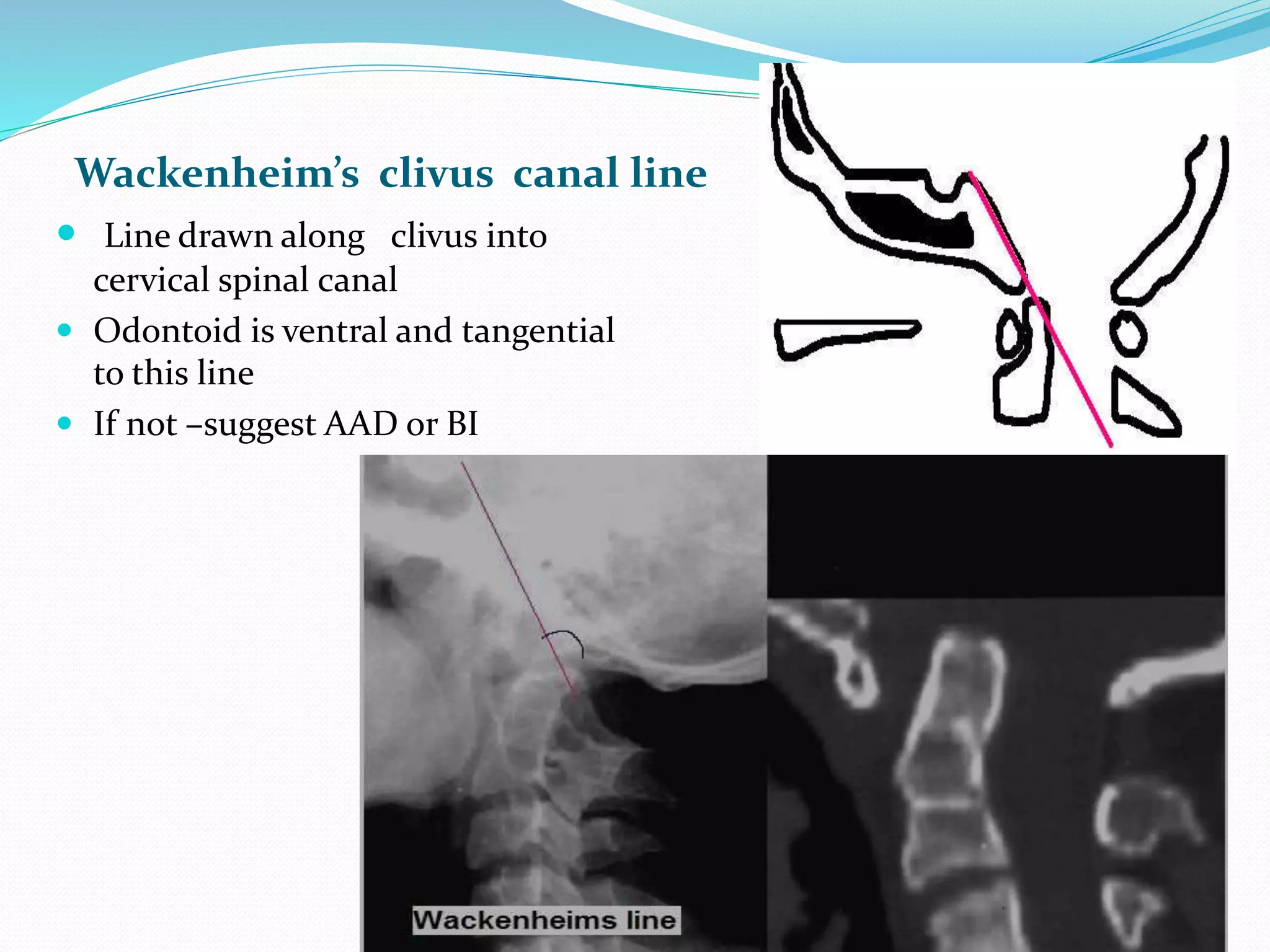
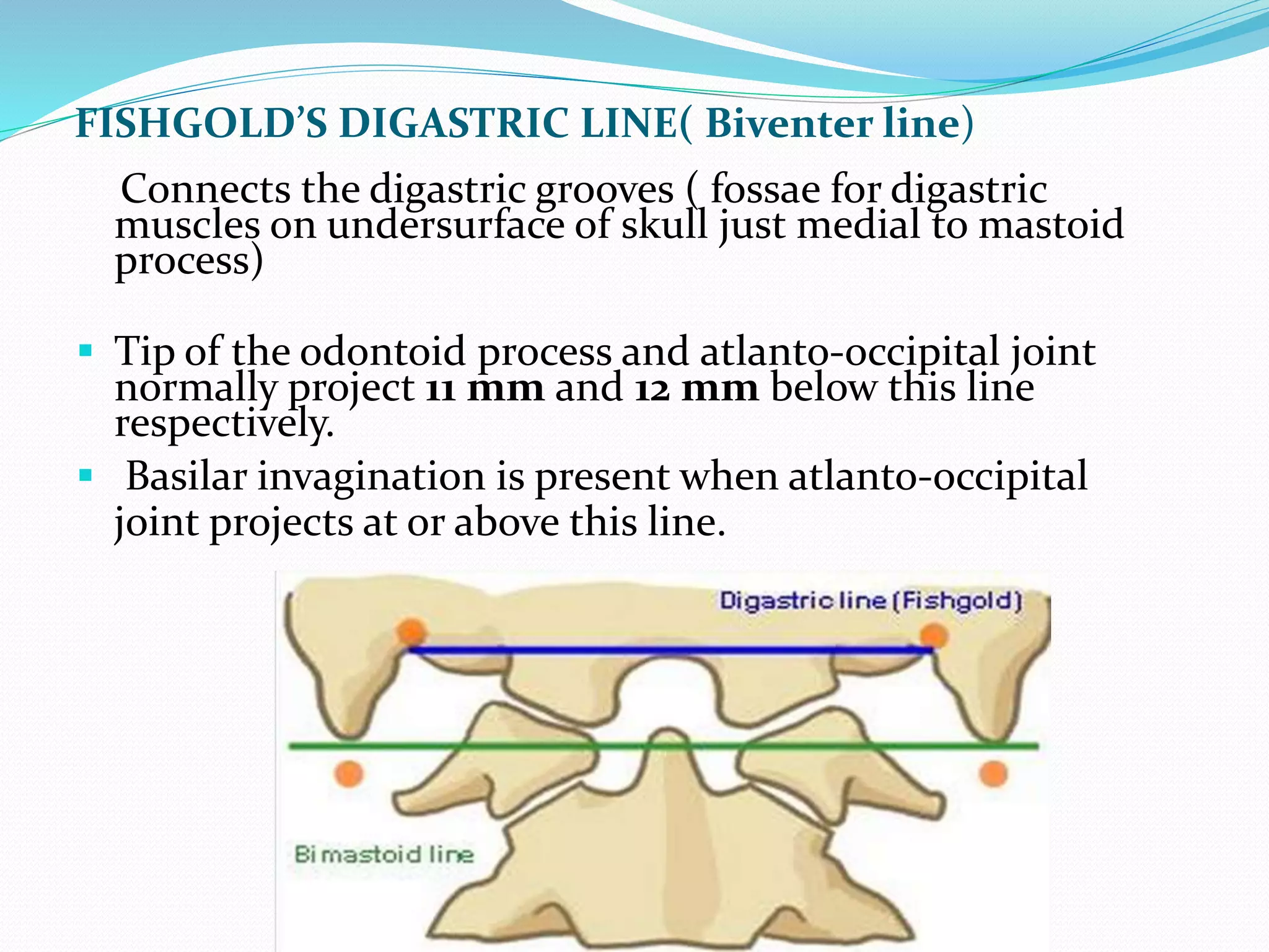
DR. SUMIT KAMBLE
SENIOR RESIDENT
DEPT. OF NEUROLOGY
GMC, KOTA
ANATOMY OF CVJ (ARTICULAR)
Occiput & atlas
 Upper surfaces of C1 lateral masses is cup-like or
concave which fit into the ball & socket configuration
with occipital condyle.( flexion 10*, extension 25*).
Atlas & axis – 4 synovial joints
 2 median –front & back of dens (Pivot variety)
 2 lateral –b/w opposing articular facets (Plane variety)
 Each joint has its own capsule & synovial cavity.
 Rotation is upto 90* & approx ½ occurs at the A-A
joint.
ANATOMY OF CVJ(LIGAMENTOUS)
Principal stabilizing ligaments of C1 -
 -Transverse atlantal ligament
 -Alar ligaments
Secondary stabilizing ligaments of CVJ are more elastic &
weaker than the primary ligaments.
 -Apical ligament
 -Anterior & posterior A-O membranes
 -Tectorial membrane
 -Ligamentum flavum
 -Capsular ligaments
NEURAL
Structures related are –
 Caudal brainstem (Medulla)
 Fourth ventricle
 Rostral part of spinal cord
 Lower cranial (9,10,11 ,12) & upper cervical
nerves (C1,C2, and C3 nerves with both rami).
 In cerebellum, only the tonsils, biventral lobules
& the lower part of the vermis (nodule, uvula &
pyramid)
Classification of CV Anomalies
I. Bony Anomalies
A. Major Anomalies
1. Platybasia
2. Occipitalization of
atlas
3. Basilar Invagination
4. Dens Dysplasia
5. Atlanto- axial
dislocation
B. Minor Anomalies
1. Dysplasia of Atlas
2. Dysplasia of
occipital condyles,
clivus, etc.
II. Soft Tissue anomalies
1. Arnold-Chiari Malformation
2. Syringomyelia/ Syringobulbia
Classification of CV Anomalies
Congenital-
 Malformation of occipital sclerotomes
Clivus segmentation anomalies
Condylar hypoplasia
Assimilation of atlas
 Malformation of atlas
Assimilation of atlas
Atlantoaxial fusion.
Aplasia of atlas arches.
 Malformation of axis.
Irregular Atlantoaxial segmentations.
Dens dysplasia
 Segmentations failure of c2-c3
Developmental and acquired
 At foraman magnum- stenosis
 Sec.basilar invagination- OI, Pagets ds, osteomalacia,
rickets, RA.
 Atlantoaxial instability
1. down syndrome
2. Ehlers danlos syndrome
3. MPS
4. Trauma
5. Infection –TB
6. Tumors- Mets, chordom,osteoblastoma, NF
 Spontaneous rotatory subluxation- Grisel syndrome
Clinical features
A. Cervical symptoms and signs- pain suboccipital region
radiating vertex, stiffness in 85%
B. Myelopathic Features- long tract involvement and
wasting
C. CN involvement- IX, X,XI,XI in 20%
D. Vascular - in 15% Transient Attack of V-B insufficiency
E. Sensory symptom of post. column involvement.
F. Cerebellar symptoms/signs- Nystagmus, Ataxia,
intention tremor, dysarthria
G. Features of Raised ICT- usually seen in Pateints Having
basilar impresssion and/or ACM
INVESTIGATIONS
 X Rays
-Antero-posterior view
-Lateral view
-Open mouth view for dens
 Stress X-Rays (neutral, flexion, extention)
 CT Scan and 3D reconstruction
 MRI conventional and dynamic
 Myelogram & Ventriculogram
 Angiography
CRANIOMETRY:
 Craniometry of the CVJ uses a series of lines, planes &
angles to define the normal anatomic relationships of the
CVJ.
 These measurements can be taken on plain X rays,3D CT or
on MRI.
 No single measurement is helpful.
 disadvantage --anatomic structures and planes vary within
a normal range.
Lines and angles used in radiologic diagnosis of C.V
anomalies.
Parameter Normal range limits
A. PLATYBASIA
B. BASILAR INVAGINATION
C. ATLANTO-AXIAL DISLOCATION *
• Basal angle < 150 degree
• Boogard’s angle < 136 degree
• Bull’s angle < 13 degree
• Chamberlain’s line < one third of odontoid above this line
• Mcgregor’s line < 5 mm
• Mcrae line odontoid lies below this
• Klaus height index > 35 mm
• Atlanto-temporo > 22mm.
mandibular index
• Atlanto-odontoid space upto 3 mm in adults
upto 5 mm in children
Chamberlain’s line
 From tip of hard palate to posterior
tip of Foramen Magnum
(opisthion).
 It helps to recognise basilar
invagination which is said to be
present if the tip of the dens is >3
mm above this line
Mc Gregor’s line (basal line)
 Line drawn from posterior
tip of Hard palate to lowest
part of Occiput
 Odontoid tip >5mm above =
Basilar Invagination
 Position changed with
flexion and extension so not
used.
 Should be used when lowest
part of occipital bone is not
Foramen Magnum.
Wackenheim’s clivus canal line
 Line drawn along clivus into
cervical spinal canal
 Odontoid is ventral and tangential
to this line
 If not –suggest AAD or BI
Mc rae’s line ( foramen magnum line)
 Joins anterior and posterior edges of Foramen magnum
 Tip of odontoid is below this line.
 When sagittal diameter of canal <20mm, in patient of >8
yr of age neurological symptoms occur – Foramen
Magnum Stenosis
Welcher’s Basal Angle
 Nasion to tuberculum sella
 Tuberculum sellae to the basion
along plane of the clivus
 Normal – 1240 - 142
 > 1400 = platybasia
 < 1300 is seen in achondroplasia
Bulls angle
 Line representing
prolongation of hard
palate and line joining
the midpoints of the ant
& post arches of C1.
 Normal : <100
 Basilar invagination -
>130
Boogard ‘s Angle
 1st line between Dorsum sellae to Basion & Mc
Rae’s line.
 Average - 1220
 > 1350
 Basillar impression
Atlantooccipital joint Axis Angle
(Schmidt – Fischer angle)
 Range between 124- 127.
 Wider in occipital condyle hypoplasia.
O
C2
AA JT
AO JT
C1 C1
FISHGOLD’S DIGASTRIC LINE( Biventer line)
Connects the digastric grooves ( fossae for digastric
muscles on undersurface of skull just medial to mastoid
process)
 Tip of the odontoid process and atlanto-occipital joint
normally project 11 mm and 12 mm below this line
respectively.
 Basilar invagination is present when atlanto-occipital
joint projects at or above this line.
FISHGOLD’S BIMASTOID LINE
 Line connecting tip of mastoid process.
 Odontoid process should be less than 10 mm above this line-
BI
HEIGHT INDEX OF KLAUS
 Distance between tip of dens
and tuberculum cruciate
line( line drawn from
tuberculum sella to internal
occipital protruberence)
 40-41mm normal
 In basilar invagination <30
mm
1.nasion
2.tuberculum sella
3.basion (anterior
margin of the
foramen magnum)
4. opisthion
(posterior margin of
the foramen
magnum)
5. posterior pole of
the hard
palate
6. anterior arch of
the atlas
7. posterior arch of
the atlas
8. odontoid process
Platybasia
 Refers only to an abnormally obtuse basal angle, may
be asymptomatic, and is not a measure of basilar
invagination.
 >140 basal angle.
Occipitalization of atlas/assimilation
 50% of all cvj anomaly in india.
 Failure of segmentation btw last
occipital and first spinal
sclerotome.
 Gradual or sudden onset by trauma
 No movement btw OA –leads
increases stress at AA joint –get
instability
 Associated –with basilar
invagination, occipital vertebra, KF
syndrome
Coronal polytomogram demonstrates complete fusion of the lateral
C-i masses (1) to the occipital condyles (0).
 Incidence - 1.4 to 2.5 per 1000 children. It affects both sexes
equally.
 Neurological symptoms usually occur in third and fourth
decades and vary depending on the area of spinal cord
impingement.
 Clinical Findings
 Low hairlines
 Torticollis
 Short necks
 Restricted neck movement.
 Dull, aching pain in the posterior occiput and the neck
 Episodic neck stiffness
TOPOGRAPHIC FORMS (WACKENHEIM):
 Type I: Occipitalization (subtotal) with BI.
 Type II: Occipitalization(subtotal) with BI & fusion of 2nd &
3rd cervical vertebrae.
 Type III: occipitalization (Total or subtotal) with BI &
maldevelopment of the transverse ligament. may be
associated with various malformations like C2-C3 fusion,
hemivertebra, dens aplasia, tertiary condyle, etc
 Symptoms are due to-absence of a free atlas- TL fails to
develop which causes posterior displacement of axis &
compression of the spinal cord
BASILAR INVAGINATION
 Basilar invagination implies that the floor of the skull
is indented by the upper cervical spine, & hence the tip
of odontoidis more cephalad protruding into the FM.
 Two types : primary invagination, which is
developmental and more common, and secondary
invagination, which is acquired.
 Primary invagination can be associated with occipito
atlantal fusion, hypoplasia of the atlas, a bifid
posterior arch of the atlas, odontoid anomalies.
SECONDARY BASILAR INVAGINATION
1. Hyperparathyroidism
2. Hurler's syndrome
3. Rickets/OM/Scurvy
4. Hajdu-Cheney Syndrome.
5. Paget's disease.
6. Cleidocranial dysostosis
7. Osteogenesis Imperfecta
 BI is associated with high incidence of vertebral artery
anomalies.
Topographic types of BI :
 Anterior BI : hypoplasia of the basilar process of the
occipital bone.
 BI of the occipital condyles (Paramedian BI)–Condylar
hypoplasia
 BI in the lateral condylararea.
 Posterior BI: posterior margin of the FM is invaginated.
 Unilateral BI.
 Generalised BI
SIGNS / SYMPTOMS
Usually occur in 2nd or 3rd decade.
 Short neck(78%),torticollis (68%)
 Associated ACM & syringomyelia(25 to 35%).
 Motor & sensory disturbances (85%).
 Lower cranial nerves involvement
 Headache & pain in the nape of neck (greater occipital N)
 Raised ICP due to posterior encroachment which causes
blockage of aqueduct of sylvius.
 Compression of cerebellum & vestibular apparatus leading to
vertical or lateral nystagmus(65%) .
 Vertebral artery insufficiency s/s.
Atlantoaxial Instability
 Atlantoaxial instability (AAI) is characterized by excessive
movement at the junction between the atlas (C1) and axis
(C2) as a result of either a bony or ligamentous
abnormality.
 Neurologic symptoms can occur when the spinal cord or
adjacent nerve roots are involved.
Incidence of AAD –
 57% of all CVJ anomalies.
 8.3% of all causes of cervical compression
 GREENBERG’S CLASSIFICATION :
Incompetence of the odontoid process –
Congenital
Traumatic -# of odontoid
Infections
Tumor –1o/ 2o
Incompetence of the TAL –
Congenital
Traumatic
Inflammatory –Children (pharynx nasopharynx) Adults (RA &
ankylosing spondylitis)
WADIA CLASSIFICATION :
 Group I: AAD with occipitalization of atlas & fusion of C2
& C3.
 Group II: odontoid incompetence due to its
maldevelopment with no occipitalization of atlas.
 Group III: odontoid dislocation but no maldevelopment of
dens or occipitalization of atlas.
Non-traumatic conditions associated with increase in
the atlantoaxial distance:
 Down syndrome -Due to laxity of the transverse ligament
 Grisel syndrome –Atlantoaxial subluxation associated with
inflammation of adjacent soft tissues of the neck
 Rheumatoid arthritis-From laxity of the ligaments and
destruction of the articular cartilage
 Osteogenesis imperfecta
 Neurofibromatosis
 Morquio syndrome -Secondary to odontoid hypoplasiaor
aplasia
 Other arthridities (Psoriasis, Lupus)
Anterior Atlanto-Dental Interval (AADI)
 AAS is + when >3 mm in adults & >5mm in children
 Measured from posteroinferior margin of ant arch of C1 to
the ant surface of odontoid
 AADI 3-6 mm  trans lig. damage
 AADI >6mm  alar lig. damage also
Posterior Atlanto-Dental Interval (PADI) :
 Distance b/w posterior
surface of odontoid &
anterior margin of post
ring of C1
 Considered better method
as it directly measures the
spinal canal
 Normal : 17-29 mm at C1
 PADI <14mm : predicts
cord compression
RISK FACTORS FOR CORD
COMPRESSION IN AAS-
 AADI > 9 mm
 PADI < 14 mm
 Basilar Invagination,
especially if associated
with AAS of any degree
ATLANTO-AXIAL SUBLUXATION (AAS)
Fielding and Hawkins classification:
 Type I- is simple rotatory displacement with an intact
transverse ligament.
 Type II- injuries involve anterior displacement of C1 on C2
of 3-5 mm with one lateral mass serving as a pivot point
and a deficiency of the transverse ligament.
 Type III -injuries involve greater than 5 mm of anterior
displacement.
 Type IV-injuries involve the posterior displacement of C1
on C2.
 Both Type III and IV are highly unstable injuries.
TREATMENT-
 Type I injuries (stable subluxations) –Collar.
 Type II injuries may be potentially unstable.
 Type III and IV rotatory displacements that are
unstable are treated surgically with a reduction and C1-
2 fusion.
 Techniques of fusion vary from sublaminar wiring
techniques like Brooks or Gallie, Halifax clamp, or
transarticular screw of Magerl.
DENS DYSPLASIA
 Type 1 (Os odontoideum)
separate odontoid process
 Type 2 (Ossiculum terminale)
failure of fusion of apical
segment with its base
 Type 3 – Agenesis of odontoid
base & apical segment lies
separately.
 Type 4 – Agenesis of odontoid
apical segment
 Type 5 –Total agenesis of
odontoid process.
OS ODONTOIDEUM
 At birth odontoid base is separate
from the body of axis by a
cartilage which persists until the
age of 8, later -ossified,or may
remain separate as Os-
odontoidium.
 Independent osseous structure
lying cephalad to the axis body in
the location of the odontoid
process.
 Anterior arch of the atlas is
rounded and hypertrophic but the
posterior arch is hypoplastic.
 Cruciate ligament incompetence
and A-A instability are common
Persistent ossiculum terminale: Bergman ossicle
 Failure of fusion of the
terminal ossicle to the
remainder of the odontoid-
normally by 12 years of age.
 Confused with a type 1
odontoid fracture.
 Stable when isolated and
of relatively little clinical
significance.
 Odontoid process is
usually normal in height.
Condylar Hypoplasia:
 Occipital condyles are
underdeveloped and have a
flattened -- and widening of
the AO joint axis angle --
leading to BI.
 Lateral masses of the atlas
may be fused to the
hypoplastic condyles, further
accentuating the BI.
 Limits movements at the A-O
joint.
 Violation of the Chamberlain
line and widening of
atlantooccipital joint axis
angle
Basiocciput Hypoplasia:
 Hypoplasia of the basiocciput
may be mild or severe,
depending on the number of
occipital sclerotomes affected.
 Lead-basilar invagination.
 Clivus-canal angle is typically
decreased
Posterior Arch Anomalies
 Posterior rachischisis > aplasias and hypoplasia
 Total or partial aplasia of the posterior atlas arch.
 Isolated, is usually asymptomatic, but may be associated
with anterior AA subluxation.
 Simulating Jefferson fracture.
SPLIT ATLAS
 Anterior +posterior arch rachischisisis =“split atlas”.
 Usually asymptomatic but wide clefts with only a
fibrous covering may lead to atlas instability
Klippel- Feil Syndrome
Triad
 Decreased range of motion in the
cervical spine m/c
 Short, webbed neck
 Low hairline.
Type
1- Massive fusion of cervical and upper
thoracic vertebra
2 –Fusion of 2 cervical vertebra
,hemivertebra, scoliosis, OA fusion
3-Lower thoracic and upper lumber
spine anomaly.
4-Sacral agenesis
ASSOCIATED CONDITIONS:
 Scoliosis- 60%.
 Genito-urinary- 65%. m/c is absence of kidney.
 Sprengel's deformity- 35%
 Cardio-pulmonary-5-15%, m/c V.S.D.
 Deafness-30%, all types, MC mixed.
 Sykinesis-Mirror motions 20%.
 Cranio-cervical abnormalities- (25%)- Includes C1-C2
hypermobility and instability, BI, Chiari I malformation,
diastematomyelia, & syringomyelia.
 20% of patients may show facial asymmetry, torticollis and
neck webbing (pterygium colli).
 Ptosis of the eye, Duane's eye contracture, lateral rectus
palsy, facial nerve palsy and cleft palate.
 Upper extremity abnormalities, ie. syndactyly, hypoplastic
thumb, supernumary digits and hypoplasia of the upper
extremity.
SYMPTOMS:
 Due to the hypermobility occurring at the open segments,
can lead to either frank instability or osteoarthritis.
 Mechanical symptoms due to joint irritation.
 Neurologic symptoms due to root irritation or spinal cord
compression
Arnold-Chiari Malformation
 Type 1- m/c -caudal displacement of peglike cerebellar tonsils
below the level of the foramen magnum, -congenital tonsillar
herniation, tonsillar ectopia, or tonsillar descent.
Syringomyelia in 50 to 70%.
 Type II -less common and more severe, almost invariably
associated with myelomeningocele. Symptomatic in infancy
or early childhood. -caudal displacement of lower brainstem
(vermis, medulla, pons, 4th ventricle) through the foramen
magnum.
 Type III -herniation of cerebellum into a high cervical
myelomeningocele.
 Type IV -cerebellar agenesis.
type III and IV -exceedingly rare and incompatible with life .
 Chiari type I malformation.
 (white line) down to the level of C1 posterior arch.
TREATMENT:
 No role for prophylactic treatment in an asymptomatic
patient with an incidental CMI.
 All symptomatic patients require surgical treatment.
 In patients with CMI and hydrocephalus, the primary
treatment must be shunting the ventricular system.
 In presence of symptomatic ventral compression from BI or
retroflexion of the odontoid, the treatment is ventral
decompression.
 In patients with a CMI,syrinx with scoliosis, the initial
treatment is posterior cervicomedullary decompression.
OUTCOMES:
 Patients presenting with pain (mainly headache and neck
pain) & weakness without associated atrophy –best results.
 Cranial nerve dysfunction –moderate recovery
 Sensory recovery poor.
 Presence of central cord syndrome due to a syrinx-
indicative of poor recovery.
 Three factors most prognostic of poor outcome are atrophy,
ataxia, and scoliosis.
 Brain stem and cerebellar syndromes -good recovery
TUBERCULOUS AAD
 <1% of all cases of spinal TB.
 Local pain, restriction of neck movements & acute
tenderness of upper C-spine –Cardinal features.
 Compression of CMJ could be due to granulation tissue,
cold abscess or bony instability & displacement.
 Waxing & waning picture .
 Ligaments are extensively infiltrated .
 Hyperaemic decalcification occurs.
Radiological findings in 3 stages–
 Stage I: Retropharyngeal abscess with ligamentous laxity +,
bony architecture of C1-C2 preserved.
 Stage II: Ligamentous disruption with AAD, minimal bone
destruction & retropharyngeal mass +
 Stage III: marked destruction of bone, complete
obliteration of anterior arch of C1 & complete loss of
odontoid process, marked AAD & O-A instability.
TREATMENT:
 Bed rest, cervical traction, evacuation of retropharyngeal abscess
& prolonged external immobilization along with ATT.
Indications of Surgery :
 Gross bony destruction with instability
 Major neurological deficits
 Unstable spine following conservative Tx
Surgery :
 Posterior fusion
 Anterior decompression with or without fusion
RHEUMATOID ARTHRITIS & CVJ
 20% of RA have AAD.
 Osteophyte formation (stabilizing effect) does not occur
secondary to deficient osteogenesis(characteristic of RA).
 Loss of tensile strength & stretching of TL due to
destructive inflammatory changes as well as secondary
degenerative changes in tissues from vasculitis--AAD.
 Granulation tissue in the synovial joints.
 Odontoid process –osteoporosis, angulation/#.
REFFERENCES-
 Textbook of contemporary neurosurgery vol 1 By Vincent A
Thamburaj
 Apley’s Textbook of Orthopaedics
 A Textbook of Neuroanatomy GARTNER and Rhoton
 Youmans textbook of neurosurgury
 Texbook of neurosurgery –rengachary and shetty.
 Management of Congenital Atlanto-Axial Dislocation Neurology
India, Vol. 50, No. 4, Dec, 2002, pp. 386-397 Review Article
 Uptodate.com
 Radiopedia.org

Cranio vertebral anomalies- overview -

  • 1.
    DR. SUMIT KAMBLE SENIORRESIDENT DEPT. OF NEUROLOGY GMC, KOTA
  • 2.
    ANATOMY OF CVJ(ARTICULAR) Occiput & atlas  Upper surfaces of C1 lateral masses is cup-like or concave which fit into the ball & socket configuration with occipital condyle.( flexion 10*, extension 25*). Atlas & axis – 4 synovial joints  2 median –front & back of dens (Pivot variety)  2 lateral –b/w opposing articular facets (Plane variety)  Each joint has its own capsule & synovial cavity.  Rotation is upto 90* & approx ½ occurs at the A-A joint.
  • 3.
    ANATOMY OF CVJ(LIGAMENTOUS) Principalstabilizing ligaments of C1 -  -Transverse atlantal ligament  -Alar ligaments Secondary stabilizing ligaments of CVJ are more elastic & weaker than the primary ligaments.  -Apical ligament  -Anterior & posterior A-O membranes  -Tectorial membrane  -Ligamentum flavum  -Capsular ligaments
  • 5.
    NEURAL Structures related are–  Caudal brainstem (Medulla)  Fourth ventricle  Rostral part of spinal cord  Lower cranial (9,10,11 ,12) & upper cervical nerves (C1,C2, and C3 nerves with both rami).  In cerebellum, only the tonsils, biventral lobules & the lower part of the vermis (nodule, uvula & pyramid)
  • 6.
    Classification of CVAnomalies I. Bony Anomalies A. Major Anomalies 1. Platybasia 2. Occipitalization of atlas 3. Basilar Invagination 4. Dens Dysplasia 5. Atlanto- axial dislocation B. Minor Anomalies 1. Dysplasia of Atlas 2. Dysplasia of occipital condyles, clivus, etc. II. Soft Tissue anomalies 1. Arnold-Chiari Malformation 2. Syringomyelia/ Syringobulbia
  • 7.
    Classification of CVAnomalies Congenital-  Malformation of occipital sclerotomes Clivus segmentation anomalies Condylar hypoplasia Assimilation of atlas  Malformation of atlas Assimilation of atlas Atlantoaxial fusion. Aplasia of atlas arches.  Malformation of axis. Irregular Atlantoaxial segmentations. Dens dysplasia  Segmentations failure of c2-c3
  • 8.
    Developmental and acquired At foraman magnum- stenosis  Sec.basilar invagination- OI, Pagets ds, osteomalacia, rickets, RA.  Atlantoaxial instability 1. down syndrome 2. Ehlers danlos syndrome 3. MPS 4. Trauma 5. Infection –TB 6. Tumors- Mets, chordom,osteoblastoma, NF  Spontaneous rotatory subluxation- Grisel syndrome
  • 9.
    Clinical features A. Cervicalsymptoms and signs- pain suboccipital region radiating vertex, stiffness in 85% B. Myelopathic Features- long tract involvement and wasting C. CN involvement- IX, X,XI,XI in 20% D. Vascular - in 15% Transient Attack of V-B insufficiency E. Sensory symptom of post. column involvement. F. Cerebellar symptoms/signs- Nystagmus, Ataxia, intention tremor, dysarthria G. Features of Raised ICT- usually seen in Pateints Having basilar impresssion and/or ACM
  • 10.
    INVESTIGATIONS  X Rays -Antero-posteriorview -Lateral view -Open mouth view for dens  Stress X-Rays (neutral, flexion, extention)  CT Scan and 3D reconstruction  MRI conventional and dynamic  Myelogram & Ventriculogram  Angiography
  • 11.
    CRANIOMETRY:  Craniometry ofthe CVJ uses a series of lines, planes & angles to define the normal anatomic relationships of the CVJ.  These measurements can be taken on plain X rays,3D CT or on MRI.  No single measurement is helpful.  disadvantage --anatomic structures and planes vary within a normal range.
  • 12.
    Lines and anglesused in radiologic diagnosis of C.V anomalies. Parameter Normal range limits A. PLATYBASIA B. BASILAR INVAGINATION C. ATLANTO-AXIAL DISLOCATION * • Basal angle < 150 degree • Boogard’s angle < 136 degree • Bull’s angle < 13 degree • Chamberlain’s line < one third of odontoid above this line • Mcgregor’s line < 5 mm • Mcrae line odontoid lies below this • Klaus height index > 35 mm • Atlanto-temporo > 22mm. mandibular index • Atlanto-odontoid space upto 3 mm in adults upto 5 mm in children
  • 13.
    Chamberlain’s line  Fromtip of hard palate to posterior tip of Foramen Magnum (opisthion).  It helps to recognise basilar invagination which is said to be present if the tip of the dens is >3 mm above this line
  • 14.
    Mc Gregor’s line(basal line)  Line drawn from posterior tip of Hard palate to lowest part of Occiput  Odontoid tip >5mm above = Basilar Invagination  Position changed with flexion and extension so not used.  Should be used when lowest part of occipital bone is not Foramen Magnum.
  • 15.
    Wackenheim’s clivus canalline  Line drawn along clivus into cervical spinal canal  Odontoid is ventral and tangential to this line  If not –suggest AAD or BI
  • 16.
    Mc rae’s line( foramen magnum line)  Joins anterior and posterior edges of Foramen magnum  Tip of odontoid is below this line.  When sagittal diameter of canal <20mm, in patient of >8 yr of age neurological symptoms occur – Foramen Magnum Stenosis
  • 17.
    Welcher’s Basal Angle Nasion to tuberculum sella  Tuberculum sellae to the basion along plane of the clivus  Normal – 1240 - 142  > 1400 = platybasia  < 1300 is seen in achondroplasia
  • 18.
    Bulls angle  Linerepresenting prolongation of hard palate and line joining the midpoints of the ant & post arches of C1.  Normal : <100  Basilar invagination - >130
  • 19.
    Boogard ‘s Angle 1st line between Dorsum sellae to Basion & Mc Rae’s line.  Average - 1220  > 1350  Basillar impression
  • 20.
    Atlantooccipital joint AxisAngle (Schmidt – Fischer angle)  Range between 124- 127.  Wider in occipital condyle hypoplasia. O C2 AA JT AO JT C1 C1
  • 21.
    FISHGOLD’S DIGASTRIC LINE(Biventer line) Connects the digastric grooves ( fossae for digastric muscles on undersurface of skull just medial to mastoid process)  Tip of the odontoid process and atlanto-occipital joint normally project 11 mm and 12 mm below this line respectively.  Basilar invagination is present when atlanto-occipital joint projects at or above this line.
  • 22.
    FISHGOLD’S BIMASTOID LINE Line connecting tip of mastoid process.  Odontoid process should be less than 10 mm above this line- BI
  • 23.
    HEIGHT INDEX OFKLAUS  Distance between tip of dens and tuberculum cruciate line( line drawn from tuberculum sella to internal occipital protruberence)  40-41mm normal  In basilar invagination <30 mm
  • 24.
    1.nasion 2.tuberculum sella 3.basion (anterior marginof the foramen magnum) 4. opisthion (posterior margin of the foramen magnum) 5. posterior pole of the hard palate 6. anterior arch of the atlas 7. posterior arch of the atlas 8. odontoid process
  • 28.
    Platybasia  Refers onlyto an abnormally obtuse basal angle, may be asymptomatic, and is not a measure of basilar invagination.  >140 basal angle.
  • 29.
    Occipitalization of atlas/assimilation 50% of all cvj anomaly in india.  Failure of segmentation btw last occipital and first spinal sclerotome.  Gradual or sudden onset by trauma  No movement btw OA –leads increases stress at AA joint –get instability  Associated –with basilar invagination, occipital vertebra, KF syndrome
  • 30.
    Coronal polytomogram demonstratescomplete fusion of the lateral C-i masses (1) to the occipital condyles (0).
  • 31.
     Incidence -1.4 to 2.5 per 1000 children. It affects both sexes equally.  Neurological symptoms usually occur in third and fourth decades and vary depending on the area of spinal cord impingement.  Clinical Findings  Low hairlines  Torticollis  Short necks  Restricted neck movement.  Dull, aching pain in the posterior occiput and the neck  Episodic neck stiffness
  • 33.
    TOPOGRAPHIC FORMS (WACKENHEIM): Type I: Occipitalization (subtotal) with BI.  Type II: Occipitalization(subtotal) with BI & fusion of 2nd & 3rd cervical vertebrae.  Type III: occipitalization (Total or subtotal) with BI & maldevelopment of the transverse ligament. may be associated with various malformations like C2-C3 fusion, hemivertebra, dens aplasia, tertiary condyle, etc  Symptoms are due to-absence of a free atlas- TL fails to develop which causes posterior displacement of axis & compression of the spinal cord
  • 34.
    BASILAR INVAGINATION  Basilarinvagination implies that the floor of the skull is indented by the upper cervical spine, & hence the tip of odontoidis more cephalad protruding into the FM.  Two types : primary invagination, which is developmental and more common, and secondary invagination, which is acquired.  Primary invagination can be associated with occipito atlantal fusion, hypoplasia of the atlas, a bifid posterior arch of the atlas, odontoid anomalies.
  • 35.
    SECONDARY BASILAR INVAGINATION 1.Hyperparathyroidism 2. Hurler's syndrome 3. Rickets/OM/Scurvy 4. Hajdu-Cheney Syndrome. 5. Paget's disease. 6. Cleidocranial dysostosis 7. Osteogenesis Imperfecta
  • 36.
     BI isassociated with high incidence of vertebral artery anomalies. Topographic types of BI :  Anterior BI : hypoplasia of the basilar process of the occipital bone.  BI of the occipital condyles (Paramedian BI)–Condylar hypoplasia  BI in the lateral condylararea.  Posterior BI: posterior margin of the FM is invaginated.  Unilateral BI.  Generalised BI
  • 37.
    SIGNS / SYMPTOMS Usuallyoccur in 2nd or 3rd decade.  Short neck(78%),torticollis (68%)  Associated ACM & syringomyelia(25 to 35%).  Motor & sensory disturbances (85%).  Lower cranial nerves involvement  Headache & pain in the nape of neck (greater occipital N)  Raised ICP due to posterior encroachment which causes blockage of aqueduct of sylvius.  Compression of cerebellum & vestibular apparatus leading to vertical or lateral nystagmus(65%) .  Vertebral artery insufficiency s/s.
  • 39.
    Atlantoaxial Instability  Atlantoaxialinstability (AAI) is characterized by excessive movement at the junction between the atlas (C1) and axis (C2) as a result of either a bony or ligamentous abnormality.  Neurologic symptoms can occur when the spinal cord or adjacent nerve roots are involved. Incidence of AAD –  57% of all CVJ anomalies.  8.3% of all causes of cervical compression
  • 40.
     GREENBERG’S CLASSIFICATION: Incompetence of the odontoid process – Congenital Traumatic -# of odontoid Infections Tumor –1o/ 2o Incompetence of the TAL – Congenital Traumatic Inflammatory –Children (pharynx nasopharynx) Adults (RA & ankylosing spondylitis)
  • 41.
    WADIA CLASSIFICATION : Group I: AAD with occipitalization of atlas & fusion of C2 & C3.  Group II: odontoid incompetence due to its maldevelopment with no occipitalization of atlas.  Group III: odontoid dislocation but no maldevelopment of dens or occipitalization of atlas.
  • 42.
    Non-traumatic conditions associatedwith increase in the atlantoaxial distance:  Down syndrome -Due to laxity of the transverse ligament  Grisel syndrome –Atlantoaxial subluxation associated with inflammation of adjacent soft tissues of the neck  Rheumatoid arthritis-From laxity of the ligaments and destruction of the articular cartilage  Osteogenesis imperfecta  Neurofibromatosis  Morquio syndrome -Secondary to odontoid hypoplasiaor aplasia  Other arthridities (Psoriasis, Lupus)
  • 43.
    Anterior Atlanto-Dental Interval(AADI)  AAS is + when >3 mm in adults & >5mm in children  Measured from posteroinferior margin of ant arch of C1 to the ant surface of odontoid  AADI 3-6 mm  trans lig. damage  AADI >6mm  alar lig. damage also
  • 44.
    Posterior Atlanto-Dental Interval(PADI) :  Distance b/w posterior surface of odontoid & anterior margin of post ring of C1  Considered better method as it directly measures the spinal canal  Normal : 17-29 mm at C1  PADI <14mm : predicts cord compression
  • 45.
    RISK FACTORS FORCORD COMPRESSION IN AAS-  AADI > 9 mm  PADI < 14 mm  Basilar Invagination, especially if associated with AAS of any degree
  • 47.
  • 48.
    Fielding and Hawkinsclassification:  Type I- is simple rotatory displacement with an intact transverse ligament.  Type II- injuries involve anterior displacement of C1 on C2 of 3-5 mm with one lateral mass serving as a pivot point and a deficiency of the transverse ligament.  Type III -injuries involve greater than 5 mm of anterior displacement.  Type IV-injuries involve the posterior displacement of C1 on C2.  Both Type III and IV are highly unstable injuries.
  • 49.
    TREATMENT-  Type Iinjuries (stable subluxations) –Collar.  Type II injuries may be potentially unstable.  Type III and IV rotatory displacements that are unstable are treated surgically with a reduction and C1- 2 fusion.  Techniques of fusion vary from sublaminar wiring techniques like Brooks or Gallie, Halifax clamp, or transarticular screw of Magerl.
  • 50.
    DENS DYSPLASIA  Type1 (Os odontoideum) separate odontoid process  Type 2 (Ossiculum terminale) failure of fusion of apical segment with its base  Type 3 – Agenesis of odontoid base & apical segment lies separately.  Type 4 – Agenesis of odontoid apical segment  Type 5 –Total agenesis of odontoid process.
  • 51.
    OS ODONTOIDEUM  Atbirth odontoid base is separate from the body of axis by a cartilage which persists until the age of 8, later -ossified,or may remain separate as Os- odontoidium.  Independent osseous structure lying cephalad to the axis body in the location of the odontoid process.  Anterior arch of the atlas is rounded and hypertrophic but the posterior arch is hypoplastic.  Cruciate ligament incompetence and A-A instability are common
  • 52.
    Persistent ossiculum terminale:Bergman ossicle  Failure of fusion of the terminal ossicle to the remainder of the odontoid- normally by 12 years of age.  Confused with a type 1 odontoid fracture.  Stable when isolated and of relatively little clinical significance.  Odontoid process is usually normal in height.
  • 53.
    Condylar Hypoplasia:  Occipitalcondyles are underdeveloped and have a flattened -- and widening of the AO joint axis angle -- leading to BI.  Lateral masses of the atlas may be fused to the hypoplastic condyles, further accentuating the BI.  Limits movements at the A-O joint.  Violation of the Chamberlain line and widening of atlantooccipital joint axis angle
  • 54.
    Basiocciput Hypoplasia:  Hypoplasiaof the basiocciput may be mild or severe, depending on the number of occipital sclerotomes affected.  Lead-basilar invagination.  Clivus-canal angle is typically decreased
  • 55.
    Posterior Arch Anomalies Posterior rachischisis > aplasias and hypoplasia  Total or partial aplasia of the posterior atlas arch.  Isolated, is usually asymptomatic, but may be associated with anterior AA subluxation.  Simulating Jefferson fracture.
  • 56.
    SPLIT ATLAS  Anterior+posterior arch rachischisisis =“split atlas”.  Usually asymptomatic but wide clefts with only a fibrous covering may lead to atlas instability
  • 57.
    Klippel- Feil Syndrome Triad Decreased range of motion in the cervical spine m/c  Short, webbed neck  Low hairline. Type 1- Massive fusion of cervical and upper thoracic vertebra 2 –Fusion of 2 cervical vertebra ,hemivertebra, scoliosis, OA fusion 3-Lower thoracic and upper lumber spine anomaly. 4-Sacral agenesis
  • 58.
    ASSOCIATED CONDITIONS:  Scoliosis-60%.  Genito-urinary- 65%. m/c is absence of kidney.  Sprengel's deformity- 35%  Cardio-pulmonary-5-15%, m/c V.S.D.  Deafness-30%, all types, MC mixed.  Sykinesis-Mirror motions 20%.  Cranio-cervical abnormalities- (25%)- Includes C1-C2 hypermobility and instability, BI, Chiari I malformation, diastematomyelia, & syringomyelia.
  • 59.
     20% ofpatients may show facial asymmetry, torticollis and neck webbing (pterygium colli).  Ptosis of the eye, Duane's eye contracture, lateral rectus palsy, facial nerve palsy and cleft palate.  Upper extremity abnormalities, ie. syndactyly, hypoplastic thumb, supernumary digits and hypoplasia of the upper extremity. SYMPTOMS:  Due to the hypermobility occurring at the open segments, can lead to either frank instability or osteoarthritis.  Mechanical symptoms due to joint irritation.  Neurologic symptoms due to root irritation or spinal cord compression
  • 60.
    Arnold-Chiari Malformation  Type1- m/c -caudal displacement of peglike cerebellar tonsils below the level of the foramen magnum, -congenital tonsillar herniation, tonsillar ectopia, or tonsillar descent. Syringomyelia in 50 to 70%.  Type II -less common and more severe, almost invariably associated with myelomeningocele. Symptomatic in infancy or early childhood. -caudal displacement of lower brainstem (vermis, medulla, pons, 4th ventricle) through the foramen magnum.  Type III -herniation of cerebellum into a high cervical myelomeningocele.  Type IV -cerebellar agenesis. type III and IV -exceedingly rare and incompatible with life .
  • 61.
     Chiari typeI malformation.  (white line) down to the level of C1 posterior arch.
  • 62.
    TREATMENT:  No rolefor prophylactic treatment in an asymptomatic patient with an incidental CMI.  All symptomatic patients require surgical treatment.  In patients with CMI and hydrocephalus, the primary treatment must be shunting the ventricular system.  In presence of symptomatic ventral compression from BI or retroflexion of the odontoid, the treatment is ventral decompression.  In patients with a CMI,syrinx with scoliosis, the initial treatment is posterior cervicomedullary decompression.
  • 63.
    OUTCOMES:  Patients presentingwith pain (mainly headache and neck pain) & weakness without associated atrophy –best results.  Cranial nerve dysfunction –moderate recovery  Sensory recovery poor.  Presence of central cord syndrome due to a syrinx- indicative of poor recovery.  Three factors most prognostic of poor outcome are atrophy, ataxia, and scoliosis.  Brain stem and cerebellar syndromes -good recovery
  • 64.
    TUBERCULOUS AAD  <1%of all cases of spinal TB.  Local pain, restriction of neck movements & acute tenderness of upper C-spine –Cardinal features.  Compression of CMJ could be due to granulation tissue, cold abscess or bony instability & displacement.  Waxing & waning picture .  Ligaments are extensively infiltrated .  Hyperaemic decalcification occurs.
  • 65.
    Radiological findings in3 stages–  Stage I: Retropharyngeal abscess with ligamentous laxity +, bony architecture of C1-C2 preserved.  Stage II: Ligamentous disruption with AAD, minimal bone destruction & retropharyngeal mass +  Stage III: marked destruction of bone, complete obliteration of anterior arch of C1 & complete loss of odontoid process, marked AAD & O-A instability.
  • 66.
    TREATMENT:  Bed rest,cervical traction, evacuation of retropharyngeal abscess & prolonged external immobilization along with ATT. Indications of Surgery :  Gross bony destruction with instability  Major neurological deficits  Unstable spine following conservative Tx Surgery :  Posterior fusion  Anterior decompression with or without fusion
  • 67.
    RHEUMATOID ARTHRITIS &CVJ  20% of RA have AAD.  Osteophyte formation (stabilizing effect) does not occur secondary to deficient osteogenesis(characteristic of RA).  Loss of tensile strength & stretching of TL due to destructive inflammatory changes as well as secondary degenerative changes in tissues from vasculitis--AAD.  Granulation tissue in the synovial joints.  Odontoid process –osteoporosis, angulation/#.
  • 69.
    REFFERENCES-  Textbook ofcontemporary neurosurgery vol 1 By Vincent A Thamburaj  Apley’s Textbook of Orthopaedics  A Textbook of Neuroanatomy GARTNER and Rhoton  Youmans textbook of neurosurgury  Texbook of neurosurgery –rengachary and shetty.  Management of Congenital Atlanto-Axial Dislocation Neurology India, Vol. 50, No. 4, Dec, 2002, pp. 386-397 Review Article  Uptodate.com  Radiopedia.org