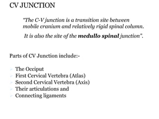
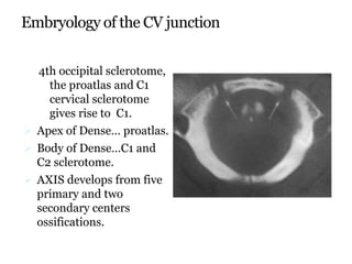
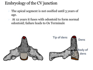
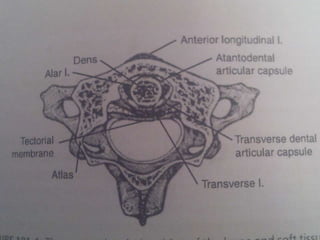
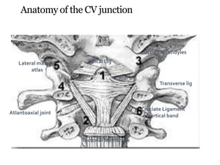
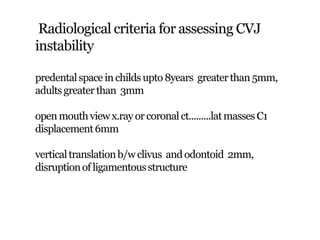
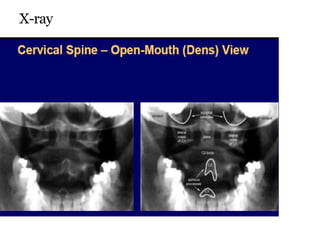
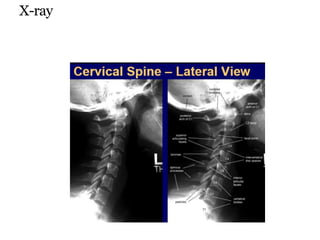
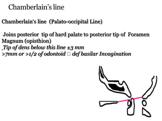
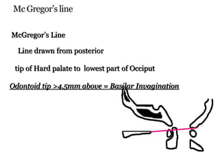
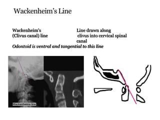
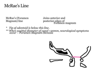
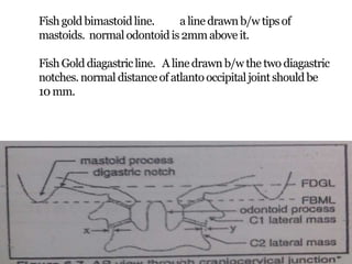
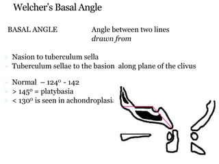
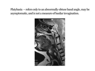
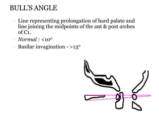
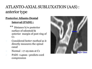
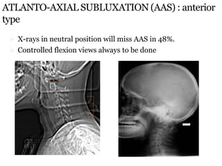
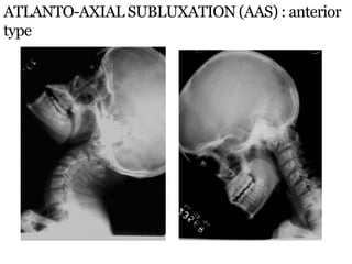
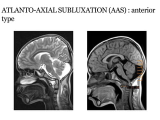
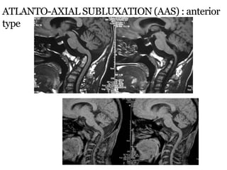
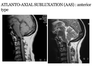
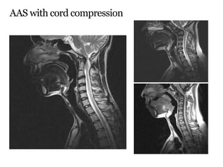
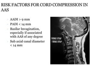
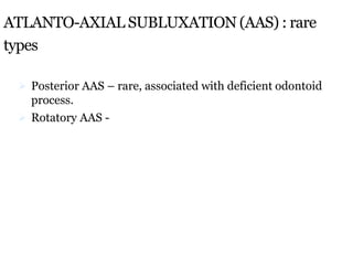
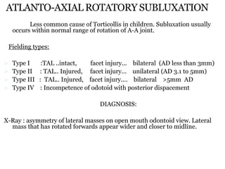
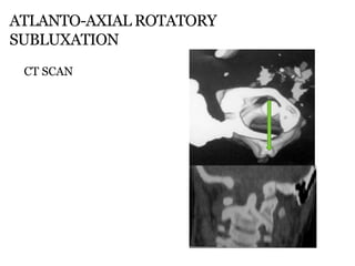
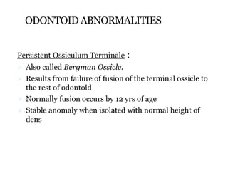
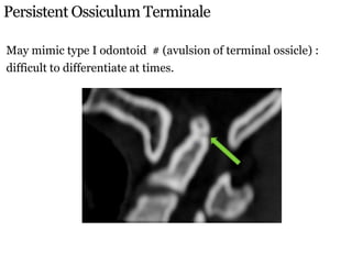
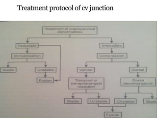
This document discusses abnormalities of the craniovertebral junction (CVJ). It begins by defining the anatomical structures that make up the CVJ. It then covers the embryology and normal anatomy, including range of motion measurements. Radiological assessment criteria are outlined. Various congenital and acquired disorders are described such as basilar invagination and atlantoaxial subluxation. Specific conditions involving the occiput, atlas, and odontoid are also detailed.
Foramen magnum meningiomas are challenging tumors, requiring special considerations because of the vicinity of the medulla oblongata, the lower cranial nerves, and the vertebral artery. It accounts for 1-3% of all intracranial Meningioma.
Applied surgical anatomy of the craniovertebral spineKshitij Chaudhary
This presentation was made at the Advanced Cervical Spine Course conducted by Dr. Sandeep Sonone and Dr. Kshitij Chaudhary for the Bombay Orthopaedic Society. http://bombayorth.org/academics/instructional-courses/
Applied surgical anatomy of the craniovertebral spineKshitij Chaudhary
This presentation was made at the Advanced Cervical Spine Course conducted by Dr. Sandeep Sonone and Dr. Kshitij Chaudhary for the Bombay Orthopaedic Society. http://bombayorth.org/academics/instructional-courses/
Apresentação sobre deformidades no pulso devido a Exostoses multiplas hereditárias.
Apresentação feita pelo Dr. Jeff Auyeung, cirurgião consultor do Hospital Universitário de North Dunham.
MADELUNG
AND MULTIPLE EXOSTOSES
Jeff Auyeung
Consultant Hand Surgeon
University Hospital of North Durham
NVBDCP.pptx Nation vector borne disease control programSapna Thakur
NVBDCP was launched in 2003-2004 . Vector-Borne Disease: Disease that results from an infection transmitted to humans and other animals by blood-feeding arthropods, such as mosquitoes, ticks, and fleas. Examples of vector-borne diseases include Dengue fever, West Nile Virus, Lyme disease, and malaria.
micro teaching on communication m.sc nursing.pdfAnurag Sharma
Microteaching is a unique model of practice teaching. It is a viable instrument for the. desired change in the teaching behavior or the behavior potential which, in specified types of real. classroom situations, tends to facilitate the achievement of specified types of objectives.
Antimicrobial stewardship to prevent antimicrobial resistanceGovindRankawat1
India is among the nations with the highest burden of bacterial infections.
India is one of the largest consumers of antibiotics worldwide.
India carries one of the largest burdens of drug‑resistant pathogens worldwide.
Highest burden of multidrug‑resistant tuberculosis,
Alarmingly high resistance among Gram‑negative and Gram‑positive bacteria even to newer antimicrobials such as carbapenems.
NDM‑1 ( New Delhi Metallo Beta lactamase 1, an enzyme which inactivates majority of Beta lactam antibiotics including carbapenems) was reported in 2008
ABDOMINAL TRAUMA in pediatrics part one.drhasanrajab
Abdominal trauma in pediatrics refers to injuries or damage to the abdominal organs in children. It can occur due to various causes such as falls, motor vehicle accidents, sports-related injuries, and physical abuse. Children are more vulnerable to abdominal trauma due to their unique anatomical and physiological characteristics. Signs and symptoms include abdominal pain, tenderness, distension, vomiting, and signs of shock. Diagnosis involves physical examination, imaging studies, and laboratory tests. Management depends on the severity and may involve conservative treatment or surgical intervention. Prevention is crucial in reducing the incidence of abdominal trauma in children.
Recomendações da OMS sobre cuidados maternos e neonatais para uma experiência pós-natal positiva.
Em consonância com os ODS – Objetivos do Desenvolvimento Sustentável e a Estratégia Global para a Saúde das Mulheres, Crianças e Adolescentes, e aplicando uma abordagem baseada nos direitos humanos, os esforços de cuidados pós-natais devem expandir-se para além da cobertura e da simples sobrevivência, de modo a incluir cuidados de qualidade.
Estas diretrizes visam melhorar a qualidade dos cuidados pós-natais essenciais e de rotina prestados às mulheres e aos recém-nascidos, com o objetivo final de melhorar a saúde e o bem-estar materno e neonatal.
Uma “experiência pós-natal positiva” é um resultado importante para todas as mulheres que dão à luz e para os seus recém-nascidos, estabelecendo as bases para a melhoria da saúde e do bem-estar a curto e longo prazo. Uma experiência pós-natal positiva é definida como aquela em que as mulheres, pessoas que gestam, os recém-nascidos, os casais, os pais, os cuidadores e as famílias recebem informação consistente, garantia e apoio de profissionais de saúde motivados; e onde um sistema de saúde flexível e com recursos reconheça as necessidades das mulheres e dos bebês e respeite o seu contexto cultural.
Estas diretrizes consolidadas apresentam algumas recomendações novas e já bem fundamentadas sobre cuidados pós-natais de rotina para mulheres e neonatos que recebem cuidados no pós-parto em unidades de saúde ou na comunidade, independentemente dos recursos disponíveis.
É fornecido um conjunto abrangente de recomendações para cuidados durante o período puerperal, com ênfase nos cuidados essenciais que todas as mulheres e recém-nascidos devem receber, e com a devida atenção à qualidade dos cuidados; isto é, a entrega e a experiência do cuidado recebido. Estas diretrizes atualizam e ampliam as recomendações da OMS de 2014 sobre cuidados pós-natais da mãe e do recém-nascido e complementam as atuais diretrizes da OMS sobre a gestão de complicações pós-natais.
O estabelecimento da amamentação e o manejo das principais intercorrências é contemplada.
Recomendamos muito.
Vamos discutir essas recomendações no nosso curso de pós-graduação em Aleitamento no Instituto Ciclos.
Esta publicação só está disponível em inglês até o momento.
Prof. Marcus Renato de Carvalho
www.agostodourado.com
Knee anatomy and clinical tests 2024.pdfvimalpl1234
This includes all relevant anatomy and clinical tests compiled from standard textbooks, Campbell,netter etc..It is comprehensive and best suited for orthopaedicians and orthopaedic residents.
Basavarajeeyam is a Sreshta Sangraha grantha (Compiled book ), written by Neelkanta kotturu Basavaraja Virachita. It contains 25 Prakaranas, First 24 Chapters related to Rogas& 25th to Rasadravyas.
Adv. biopharm. APPLICATION OF PHARMACOKINETICS : TARGETED DRUG DELIVERY SYSTEMSAkankshaAshtankar
MIP 201T & MPH 202T
ADVANCED BIOPHARMACEUTICS & PHARMACOKINETICS : UNIT 5
APPLICATION OF PHARMACOKINETICS : TARGETED DRUG DELIVERY SYSTEMS By - AKANKSHA ASHTANKAR
Here is the updated list of Top Best Ayurvedic medicine for Gas and Indigestion and those are Gas-O-Go Syp for Dyspepsia | Lavizyme Syrup for Acidity | Yumzyme Hepatoprotective Capsules etc
Rasamanikya is a excellent preparation in the field of Rasashastra, it is used in various Kushtha Roga, Shwasa, Vicharchika, Bhagandara, Vatarakta, and Phiranga Roga. In this article Preparation& Comparative analytical profile for both Formulationon i.e Rasamanikya prepared by Kushmanda swarasa & Churnodhaka Shodita Haratala. The study aims to provide insights into the comparative efficacy and analytical aspects of these formulations for enhanced therapeutic outcomes.
2. CV JUNCTION
Parts of CV Junction include:-
The Occiput
First Cervical Vertebra (Atlas)
Second Cervical Vertebra (Axis)
Their articulations and
Connecting ligaments
“The C-V junction is a transition site between
mobile cranium and relatively rigid spinal column.
It is also the site of the medullo spinal junction”.
3. Embryology of the CV junction
4th occipital sclerotome,
the proatlas and C1
cervical sclerotome
gives rise to C1.
Apex of Dense… proatlas.
Body of Dense…C1 and
C2 sclerotome.
AXIS develops from five
primary and two
secondary centers
ossifications.
4. Embryology of the CV junction
The apical segment is not ossified until 3 years of
age.
At 12 years it fuses with odontoid to form normal
odontoid; failure leads to Os Terminale
Tip of dens
Body of
dens
Dens
5. Anatomy of the CV junction
ATLANTO-AXIAL JOINT:
Normal range of cervical motion is 900 on each side,
range of rotation of atlas on axis being 25-530
Rotation of >560 on one side or a R-L diff >80
implies hyper mobility
Rotation of <280 implies hypo mobility
9. Anatomy of the CV junction
Occipital condyles
Atlantoaxial joint
Tectorial Membrane
Lateral mass of
atlas
Transverse lig
Cruciate Ligament
vertical band
Apical Lig
Alar Lig
10. Radiological criteria for assessing CVJ
instability
predentalspace inchildsupto 8years greaterthan 5mm,
adultsgreaterthan 3mm
open mouthviewx.ray or coronalct.........latmasses C1
displacement6mm
verticaltranslationb/w clivus and odontoid 2mm,
disruptionof ligamentousstructure
13. Lines and Angles
The important lines are
Chamberlain’s line
Wackenheim’s clivus canal line
Mc Gregor’s line (basal line)
McRae,s line
Basal angle
Bull,s angle
14. Chamberlain’s line
Chamberlain‘s line (Palato-occipital Line)
Joins posterior tip of hard palate to posterior tip of Foramen
Magnum (opisthion)
Tip of dens below this line ±3 mm
>7mm or >1/2 of odontoid def basilar Invagination
15. Mc Gregor’s line
McGregor’s Line
Line drawn from posterior
tip of Hard palate to lowest part of Occiput
Odontoid tip >4.5mm above = Basilar Invagination
16. Wackenheim’s Line
Wackenheim's Line drawn along
(Clivus canal) line clivus into cervical spinal
canal
Odontoid is ventral and tangential to this line
17. McRae’s Line
McRae's (Foramen Joins anterior and
Magnum) line posterior edges of
Foramen magnum
* Tip of odontoid is below this line.
** When sagittal diameter of canal <20mm, neurological symptoms
occur – Foramen Magnum Stenosis
18. Fishgold bimastoidline. a line drawnb/w tipsof
mastoids. normalodontoidis2mmabove it.
FishGold diagastricline. A linedrawn b/w the two diagastric
notches.normal distanceof atlantooccipitaljointshouldbe
10 mm.
19. Welcher’s Basal Angle
BASAL ANGLE Angle between two lines
drawn from
Nasion to tuberculum sella
Tuberculum sellae to the basion along plane of the clivus
Normal – 1240 - 142
> 1450 = platybasia
< 1300 is seen in achondroplasiaaaasdaaaaaaaaa
21. BULL’S ANGLE
Line representing prolongation of hard palate and
line joining the midpoints of the ant & post arches
of C1.
Normal : <100
Basilar invagination - >130
22. Lymphatic drainage
Occipitoatlantoaxial joint drain through
retropharyngeal gland to deep cervical lymph
channels.
Paeds. nasopharyngeal infections cause
inflammatory reaction of CVJ.
C1-2 sublaxation
Refferd as GRISEL SYNDROME
23. Signs and sympyoms
Myelopathy different degrees of extremities
weakness
Brainstem symptoms
Cranial nerves deficit loss of gag
reflex,nystagmus,hearing loss
Vascular compromise syncope,vertigo,episodic
hemiparesis,transient loss of vision,altered conscious
level
Restricted neck movement
Neck and occipital pain
24. Disorders of the CV junction
Congenital bony malformations
Basilar invagination
Anomalies of atlas
Odontoid abnormality
Atlanto-axial instability
Others
25. Disorders of the CV junction
ACQUIRED MALFORMATIONS
Trauma
Arthritides
Infection
Degeneration
Tumours
26. Basilar invigination….
The upward displacement of upper cervical spine
including odontoid through the foramen magnum
into posterior fossa.
27. Pathogenesis
1. Emberyological dysgenesis,genetics,
maldevelopment of craniovertebral transition
region.
2. Secondary abnormally alignment of fascet joints of
atlas and axis leading to progressive slippage of atlas
over axis which results in odondoid tip invigination
superoir and posterior into cranio cervical cord.
29. Classification
Type1. the odontoid tip inviginates into foramen magnum
indented into brainstem. atlanto odontoid distance
increases. odentoid tip is above CL,McR,wccl.
volume of posterior fossa and Cl angle z normal.
Type2. odontoid tip,ant arch of C1 and clivus migrate
superiorly in unison, results in small post fossa causing
ACM. odontoid tip is only above CL not wccl,McR.
30. Type A.
Based on mechanical instability.just like type1 but
normal horizontal poition of fascet joint changes into
oblique position.which leads progressive slippage.
Type B.
there is congenital dysgenesis , and atlantoaxial
joints were normal or entirely fused.
31. Treatment
Type1.
85% can be reduced with traction
Transoral decompression and posterior fusion
Its superior to include craniovertebral realignment
procedure.
Type2.
only 15% reduced with traction.foramen magnum
decompression is appropriate
36. ATLANTO-AXIAL SUBLUXATION (AAS) : anterior
type
Anterior Atlanto-Dental Interval (AADI) :
AAS is present when it is >3mm in adults & >5mm in
children
Measured from posteroinferior margin of ant arch of C1
to the ant surface of odontoid
AADI 3-6 mm trans lig. damage
AADI >6mm alar lig. damage also
AADI >9mm surgical stabilization
37. ATLANTO-AXIAL SUBLUXATION (AAS) :
anterior type
Posterior Atlanto-Dental
Interval (PADI) :
** Distance b/w posterior
surface of odontoid &
anterior margin of post ring of
C1
Considered better method as it
directly measures the spinal
canal
Normal : 17-29 mm at C1
PADI <14mm : predicts cord
compression
38. ATLANTO-AXIAL SUBLUXATION (AAS) : anterior
type
X-rays in neutral position will miss AAS in 48%.
Controlled flexion views always to be done
45. RISK FACTORS FOR CORD COMPRESSION IN
AAS
AADI > 9 mm
PADI < 14 mm
Basilar Invagination,
especially if associated
with AAS of any degree
Sub axial canal diameter
< 14 mm
47. ATLANTO-AXIAL ROTATORY SUBLUXATION
Less common cause of Torticollis in children. Subluxation usually
occurs within normal range of rotation of A-A joint.
Fielding types:
Type I :TAL ..intact, facet injury... bilateral (AD less than 3mm)
Type II : TAL.. Injured, facet injury... unilateral (AD 3.1 to 5mm)
Type III : TAL.. Injured, facet injury.... bilateral >5mm AD
Type IV : Incompetence of odotoid with posterior dispacement
DIAGNOSIS:
X-Ray : asymmetry of lateral masses on open mouth odontoid view. Lateral
mass that has rotated forwards appear wider and closer to midline.
50. Specific Anomalies – Occiput anomalies
Condylus Tertius (IIIrd
occipital condyle) :
when proatlas persists or fails to
migrate, an ossified remnant is
seen at distal end of clivus
May form pseudo joint with
odontoid or ant arch of C1 and
limit mobility of CVJ
Increased prevalence of Os
Odontoideum seen
51. ATLAS ASSIMILATION
Represents most cephalic
‘blocked vertebra’
0.25% of population
Usually occurs in
association with other
anomalies such as BI and
Klippel Feil syndrome.
Associated with
segmentation failures
b/w C2-3 : atlanto-axial
subluxation in 50%.
57. ODONTOID ABNORMALITIES
Persistent Ossiculum Terminale :
Also called Bergman Ossicle.
Results from failure of fusion of the terminal ossicle to
the rest of odontoid
Normally fusion occurs by 12 yrs of age
Stable anomaly when isolated with normal height of
dens