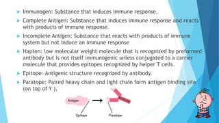
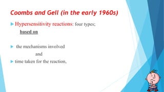
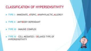
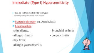
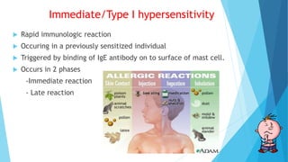
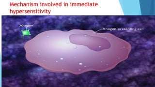
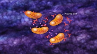
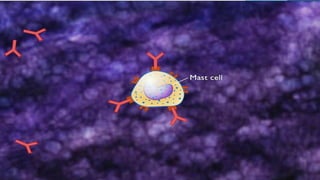
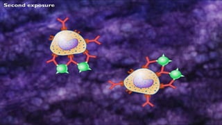
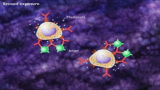
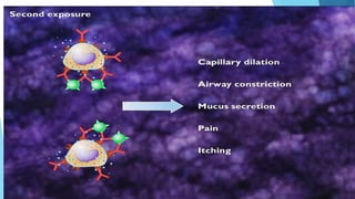
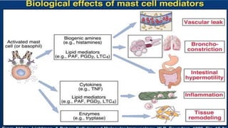
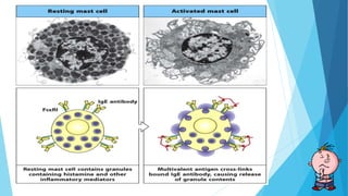
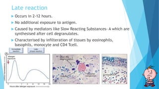
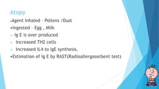
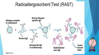
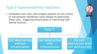
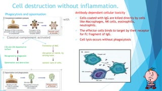
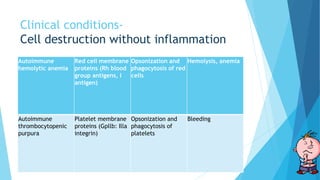
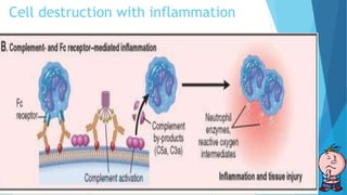
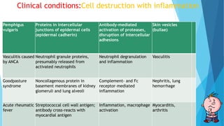
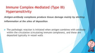
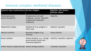
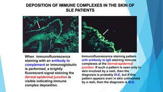
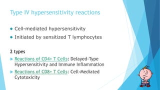
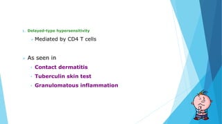
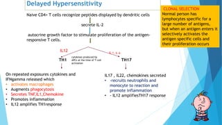
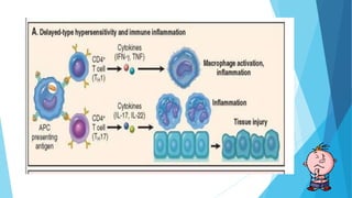
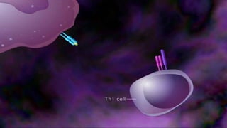
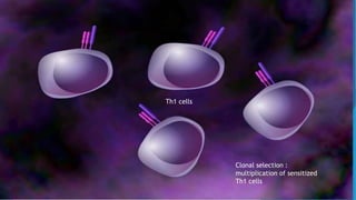
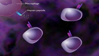
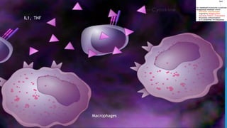
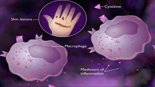
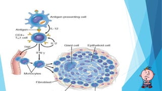
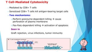
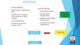
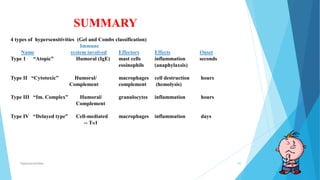
Hypersensitivity
Speaker : Dr Joyshree Panda
Moderator : Dr Atanu kumar Bal
Headings I will be covering:
 Definitions
 Class switching
 Complement System
 Classification of hypersensitivity
 Type I hypersensitivity
 Type II hypersensitivity
 Type III hypersensitivity
 Type IV hypersensitivity
Definitions
 Hypersensitivity - Immune & inflammatory responses that are
harmful to the host (von Pirquet, 1906)
 Hypersensitivity reactions - Harmful antigen-specific immune
responses, occur when an individual who has been primed by an
innocuous antigen subsequently encounters the same antigen,
produce tissue injury and dysfunction.
 Atopy: Genetic tendency to develop classic allergic diseases.
 Allergy: Damaging immune response by the body to a substance.
 Allergen: Substance causing allergic reaction.
 Immunogen: Substance that induces immune response.
 Complete Antigen: Substance that induces immune response and reacts
with products of immune response.
 Incomplete Antigen: Substance that reacts with products of immune
system but not induce an immune response
 Hapten: low molecular weight molecule that is recognized by preformed
antibody but is not itself immunogenic unless conjugated to a carrier
molecule that provides epitopes recognized by helper T cells.
 Epitope: Antigenic structure recognized by antibody.
 Paratope: Paired heavy chain and light chain form antigen binding site
(on top of Y ).
Class switching
 Biological mechanism that changes a B cell’s production
of immunoglobulin (antibodies) from one type to another, such as from
the isotype IgM to the isotype IgG.
 During this process, the constant-region portion of the antibody heavy
chain is changed, but the variable region of the heavy chain stays the
same
 Since the variable region does not change, class switching does not
affect antigen specificity. Instead, the antibody retains affinity for the
same antigens, but can interact with different effector molecules.
Structure of Immunoglobulin
Complement system
 The complement system is activated when
antigen combines with antibody
 It consists of more than 20 proteins, some of
which are numbered C1 through C9.
 C1 is a helical structure made of C1q, 2 C1r,
2C1s.
 C1q binds to antibody and fixes the
complement pathway.
 Complements C2-C5 are cleaved into two parts.
One big (b) one small (a).
 a ones are anaphylatoxins and b are opsonins
except in case of C2. (C3a, C4a, C5a and in C2 it
is C2b all are anaphylatoxins)
 C5a is chemotactic too.
 C5-C9 make membrane attack complex by joining
of multiple C9. and cell lysis occurs.
Coombs and Gell (in the early 1960s)
 Hypersensitivity reactions: four types;
based on
 the mechanisms involved
and
 time taken for the reaction,
CLASSIFICATION OF HYPERSENSITIVITY
 TYPE I – IMMEDIATE, ATOPIC, ANAPHYLACTIC,ALLERGY
 TYPE II – ANTIBODY DEPENDANT
 TYPE III – IMMUNE COMPLEX
 TYPE IV – CELL MEDIATED / DELAYED TYPE OF
HYPERSENSITIVITY
Immediate (Type I) Hypersensitivity
 Can be further divided into two types
( depending on the portal of entry of the allergen)
Systemic disorder- eg. Anaphylaxis
Local reaction
-skin allergy, - bronchial asthma
-allergic rhinitis - conjunctivitis
-hay fever,
-allergic gastroenteritis
Immediate/Type I hypersensitivity
 Rapid immunologic reaction
 Occuring in a previously sensitized individual
 Triggered by binding of IgE antibody on to surface of mast cell.
 Occurs in 2 phases
-Immediate reaction
- Late reaction
Mechanism involved in immediate
hypersensitivity
CD 4 T Cell
TCR
Activation of TH2 cells and production of
IgE antibody
 Presentation of antigen to naïve CD4 helper T cells,Produce cytokines and
interleukins IL-4, IL-5, and IL-13
TH2 cells
Acts on B
cells IL13
Enhance IgE
production
Acts on epithelial
cells to stimulate
mucus secretion
IL-5
Eosinophil
development
and
activation.
IL-4
Class switching
to IgE
T cell
B cell
B cell
Antigen
Mast cells can also be triggered by other
stimuli- Anaphylactoid reactions.
 C3a, C5a- Act by binding to receptor on mast cell
membrane
 Chemokines – IL8
 Drugs like Codeine, Morphine, Adenosine etc
 Mellitin(present in bee venom), physical stimulus
like heat , cold, sunlight.
Late reaction
 Occurs in 2-12 hours.
 No additional exposure to antigen.
 Caused by mediators like Slow Reacting Substances- A which are
synthesised after cell degranulates.
 Characterised by infilteration of tissues by eosinophils,
basophils, monocyte and CD4 Tcell.
Atopy
•Agent Inhaled – Pollens /Dust
•Ingested – Egg , Milk
 Ig E is over produced
 Increased TH2 cells
 Increased IL4 to IgE synthesis.
•Estimation of Ig E by RAST(Radioallergosorbent test)
Type II hypersensitivity reactions
 Antibodies that react with antigens present on cell surface
or extracellular membrane cause disease by destroying
these cells , triggering inflammation or interfering with
normal function.
Type-II.
Cell destruction
without
inflammation
Cell destruction
with
inflammation
No cell
destruction only
cell dysfunction
Cell destruction without inflammation.
Phagocytosis and opsonisation
 Responsible for depletion of cells coated with
antibody
 Cells opsonized by IgG are recognized by
Phagocyte Fc receptors
 Classical complement activated
Antibody dependant cellular toxicity
 Cells coated with IgG are killed directly by cells
like Macrophages, NK cells, eosinophills,
neutrophils.
 The effector cells binds to target by their receptor
for Fc fragment of IgG.
 Cell lysis occurs without phagocytosis
C3b and C4b deposited on
surface
Recognised by phagocytes
Opsonisation and destruction
Formation of MAC
disruption of memb. by
drilling holes
osmotic lysis of cells.
Clinical conditions-
Cell destruction without inflammation
Autoimmune
hemolytic anemia
Red cell membrane
proteins (Rh blood
group antigens, I
antigen)
Opsonization and
phagocytosis of red
cells
Hemolysis, anemia
Autoimmune
thrombocytopenic
purpura
Platelet membrane
proteins (Gpllb: Illa
integrin)
Opsonization and
phagocytosis of
platelets
Bleeding
Cell destruction with inflammation
When antibodies deposit in fixed tissues, such as basement membranes and
extracellular matrix, the resultant injury is due to inflammation.
The deposited antibodies activate complement, generating by-products,
including chemotactic agents (mainly C5a), which direct the migration of
polymorphonuclear leukocytes and monocytes and anaphylatoxins (C3a and
C5a), which increase vascular permeability
release or generation of a variety of pro-inflammatory substances, including
prostaglandins, vasodilator peptides, and chemotactic substances
Damage of tissues by lysosomal enzymes, including proteases capable of
digesting basement membrane, collagen, elastin, and cartilage, and reactive
oxygen species.
Clinical conditions:Cell destruction with inflammation
Pemphigus
vulgaris
Proteins in intercellular
junctions of epidermal cells
(epidermal cadherin)
Antibody-mediated
activation of proteases,
disruption of intercellular
adhesions
Skin vesicles
(bullae)
Vasculitis caused
by ANCA
Neutrophil granule proteins,
presumably released from
activated neutrophils
Neutrophil degranulation
and inflammation
Vasculitis
Goodpasture
syndrome
Noncollagenous protein in
basement membranes of kidney
glomeruli and lung alveoli
Complement- and Fc
receptor–mediated
inflammation
Nephritis, lung
hemorrhage
Acute rheumatic
fever
Streptococcal cell wall antigen;
antibody cross-reacts with
myocardial antigen
Inflammation, macrophage
activation
Myocarditis,
arthritis
No cell destruction only cell dysfunction
 Antibody directed against cell surface receptors impair or dysregulate
function without causing cell injury or inflammation
 Myasthenia gravis, antibodies reactive with acetylcholine receptors in
the motor end plates of skeletal muscles block neuromuscular
transmission and therefore cause muscle weakness.
 In Graves disease antibodies against the thyroid-stimulating hormone
receptor on thyroid epithelial cells stimulate the cells, resulting in
hyperthyroidism
No cell destruction only cell dysfunction
Myasthenia gravis Acetylcholine
receptor
Antibody inhibits
acetylcholine binding,
down-modulates receptors
Muscle weakness,
paralysis
Graves disease
(hyperthyroidism)
TSH receptor Antibody-mediated
stimulation of TSH
receptors
Hyperthyroidism
Insulin-resistant diabetes Insulin receptor Antibody inhibits binding of
insulin
Hyperglycemia,
ketoacidosis
Pernicious anemia Intrinsic factor of
gastric parietal cells
Neutralization of intrinsic
factor, decreased
absorption of vitamin B12
Abnormal erythropoiesis,
anemia
Immune Complex–Mediated (Type III)
Hypersensitivity
Antigen-antibody complexes produce tissue damage mainly by eliciting
inflammation at the sites of deposition.
 The pathologic reaction is initiated when antigen combines with antibody
within the circulation (circulating immune complexes), and these are
deposited typically in vessel walls
Antigens that form immune complexes
 Exogenous- foreign protein that is injected or
produced by an infectious microbe
 Endogenous - individual produces antibody against
self-components (autoimmunity).
Formation of Immune Complexes.
Introduction of a protein antigen
triggers an immune response
formation of antibodies, a week after the injection of the
protein.
These antibodies are secreted into the blood,
react with the antigen still present in the circulation and
form antigen-antibody complexes.
Deposition of immune complex depends on
Size
 Large immune complexes are deposited in tissues or phagocytosed.
 Tiny immune complexes are easily cleared by phagocytosis
 Medium sized immune complexes most pathogenic
Ratio of Antigen: antibody
 Slight antigen excess more pathogenic
Blood Flow
 Places where blood is filtered at high pressure are more prone.
Eg glomeruli, joints
Systemic Immune Complex Disease
 Acute serum sickness is the prototype of a systemic
immune complex disease; occurs due to administration
of large amounts of foreign serum (e.g., serum from
immunized horses used for protection against
diphtheria).
 The pathogenesis can be divided into three phases:
 (1) formation of antigen-antibody complexes in the
circulation;
 (2) deposition of the immune complexes in various
tissues
 (3) an inflammatory reaction at the sites of immune
complex deposition
Pathogenesis of immune complexed mediated
Local Immune Complex Disease (Arthus Reaction)
 The Arthus reaction is a localized area of tissue
necrosis resulting from acute immune complex
vasculitis, usually elicited in the skin.
 experimental intracutaneous injection of antigen
in a previously immunized person.
 antigen diffuses into the vascular wall, it binds the
preformed antibody, and large immune complexes
are formed locally.
 These complexes precipitate in the vessel walls
and cause fibrinoid necrosis, and superimposed
thrombosis worsens the ischemic injury.
Immune complex mediated diseases
Systemic lupus erythematosus Nuclear antigens Nephritis, skin lesions,
arthritis, others
Poststreptococcal
glomerulonephritis
Streptococcal cell wall
antigen(s); may be “planted”
in glomerular basement
membrane
Nephritis
Polyarteritis nodosa Hepatitis B virus antigens in
some cases
Systemic vasculitis
Reactive arthritis Bacterial antigens (e.g.,
Yersinia)
Acute arthritis
Serum sickness Various proteins, e.g., foreign
serum protein (horse anti-
thymocyte globulin)
Arthritis, vasculitis, nephritis
Arthus reaction (experimental) Various foreign proteins Cutaneous vasculitis
When immunofluorescence
staining with an antibody to
complement or immunoglobulin
is performed, a brightly
fluorescent signal staining the
dermal epidermal junction is
visible indicating immune
complex deposition.
Immunofluorescence staining pattern
with antibody to IgG staining immune
complexes at the dermal-epidermal
junction. If such a pattern is seen only in
skin involved by a rash, then the
diagnosis is probably DLE, but if this
pattern appears even in skin uninvolved
by a rash, then the diagnosis is SLE.
DEPOSITION OF IMMUNE COMPLEXES IN THE SKIN OF
SLE PATIENTS
Type IV hypersensitivity reactions
Cell-mediated hypersensitivity
Initiated by sensitized T lymphocytes
2 types
 Reactions of CD4+ T Cells: Delayed-Type
Hypersensitivity and Immune Inflammation
 Reactions of CD8+ T Cells: Cell-Mediated
Cytotoxicity
1. Delayed-type hypersensitivity
 Mediated by CD4 T cells
 As seen in
• Contact dermatitis
• Tuberculin skin test
• Granulomatous inflammation
Delayed Hypersensitivity
Naive CD4+ T cells recognize peptides displayed by dendritic cells
secrete IL-2
autocrine growth factor to stimulate proliferation of the antigen-
responsive T cells.
TH1 TH17
cytokines produced by
APCs at the time of T-cell
activation
IL12
On repeated exposures cytokines and
IFNgamma released which
• activates macrophages
• Augments phagocytosis
• Secretes TNF,IL1,Chemokine
• Promotes inflammation
• IL12 amplifies TH1response
IL17 , IL22, chemokines secreted
• -recruits neutrophils and
monocyte to reaction and
promote inflammation
• - IL12 amplifiesTH17 response
CLONAL SELECTION
Normal person has
lymphocytes specific for a
large number of antigens,
but when an antigen enters it
selectively activates the
antigen specific cells and
their proliferation occurs
Mechanism of T-cell mediated contact dermatitis
MHC 2
Antigen
T-Cell
IL2
released
Th1 cells
Clonal selection :
multiplication of sensitized
Th1 cells
Macrophages
IL1, TNF
Pathogenesis of tuberculosis and tuberculin test
Mtb activate TLR2 which
makes apc produce IL12
T Cell-Mediated Cytotoxicity
 Mediated by CD8+ T cells
 Sensitized CD8+ T cells kill antigen-bearing target cells
 Two mechanisms:
a)Perforin-granzyme-dependent killing  cause
perforation of plasma membrane
b)Fas-FasL-dependent killing  activation of apoptosis
Seen in
Graft rejection, virus infections, tumor immunity
APOPTOSIS
o Intrinsic Pathway
o Mitochondria , regulated by
Bcl2
o Cytochrome C released
which attaches to APAF
complex
o Forms Apoptosome
o Activation of initiator
caspase (8/9)
Extrinsic Pathway
 Extracellular signals
 Surface death
receptors(fas)
 Adaptor protein(FADD)
binds to Fasl
 Activation of initiator
caspase(8/9)
Executional
cascpase 3/6
Apoptotic
fragmentation
GRANZYME
Fas-FasL-
dependent
killing
Fas-FasL-
dependent
killing
Perforin-granzyme-dependent killing
Fas-FasL-dependent killing
Hypersensitivities 68
SUMMARY
4 types of hypersensitivities (Gel and Combs classification)
Immune
Name system involved Effectors Effects Onset
Type 1 “Atopic” Humoral (IgE) mast cells inflammation seconds
eosinophils (anaphylaxsis)
Type II “Cytotoxic” Humoral/ macrophages cell destruction hours
Complement complement (hemolysis)
Type III “Im. Complex” Humoral/ granulocytes inflammation hours
Complement
Type IV “Delayed type” Cell-mediated macrophages inflammation days
-- TH1
Hypersensitivity Reactions

Hypersensitivity Reactions

  • 1.
    Hypersensitivity Speaker : DrJoyshree Panda Moderator : Dr Atanu kumar Bal
  • 2.
    Headings I willbe covering:  Definitions  Class switching  Complement System  Classification of hypersensitivity  Type I hypersensitivity  Type II hypersensitivity  Type III hypersensitivity  Type IV hypersensitivity
  • 3.
    Definitions  Hypersensitivity -Immune & inflammatory responses that are harmful to the host (von Pirquet, 1906)  Hypersensitivity reactions - Harmful antigen-specific immune responses, occur when an individual who has been primed by an innocuous antigen subsequently encounters the same antigen, produce tissue injury and dysfunction.  Atopy: Genetic tendency to develop classic allergic diseases.  Allergy: Damaging immune response by the body to a substance.  Allergen: Substance causing allergic reaction.
  • 4.
     Immunogen: Substancethat induces immune response.  Complete Antigen: Substance that induces immune response and reacts with products of immune response.  Incomplete Antigen: Substance that reacts with products of immune system but not induce an immune response  Hapten: low molecular weight molecule that is recognized by preformed antibody but is not itself immunogenic unless conjugated to a carrier molecule that provides epitopes recognized by helper T cells.  Epitope: Antigenic structure recognized by antibody.  Paratope: Paired heavy chain and light chain form antigen binding site (on top of Y ).
  • 5.
    Class switching  Biologicalmechanism that changes a B cell’s production of immunoglobulin (antibodies) from one type to another, such as from the isotype IgM to the isotype IgG.  During this process, the constant-region portion of the antibody heavy chain is changed, but the variable region of the heavy chain stays the same  Since the variable region does not change, class switching does not affect antigen specificity. Instead, the antibody retains affinity for the same antigens, but can interact with different effector molecules.
  • 6.
  • 7.
    Complement system  Thecomplement system is activated when antigen combines with antibody  It consists of more than 20 proteins, some of which are numbered C1 through C9.  C1 is a helical structure made of C1q, 2 C1r, 2C1s.  C1q binds to antibody and fixes the complement pathway.  Complements C2-C5 are cleaved into two parts. One big (b) one small (a).  a ones are anaphylatoxins and b are opsonins except in case of C2. (C3a, C4a, C5a and in C2 it is C2b all are anaphylatoxins)  C5a is chemotactic too.  C5-C9 make membrane attack complex by joining of multiple C9. and cell lysis occurs.
  • 8.
    Coombs and Gell(in the early 1960s)  Hypersensitivity reactions: four types; based on  the mechanisms involved and  time taken for the reaction,
  • 9.
    CLASSIFICATION OF HYPERSENSITIVITY TYPE I – IMMEDIATE, ATOPIC, ANAPHYLACTIC,ALLERGY  TYPE II – ANTIBODY DEPENDANT  TYPE III – IMMUNE COMPLEX  TYPE IV – CELL MEDIATED / DELAYED TYPE OF HYPERSENSITIVITY
  • 10.
    Immediate (Type I)Hypersensitivity  Can be further divided into two types ( depending on the portal of entry of the allergen) Systemic disorder- eg. Anaphylaxis Local reaction -skin allergy, - bronchial asthma -allergic rhinitis - conjunctivitis -hay fever, -allergic gastroenteritis
  • 11.
    Immediate/Type I hypersensitivity Rapid immunologic reaction  Occuring in a previously sensitized individual  Triggered by binding of IgE antibody on to surface of mast cell.  Occurs in 2 phases -Immediate reaction - Late reaction
  • 12.
    Mechanism involved inimmediate hypersensitivity
  • 14.
    CD 4 TCell TCR
  • 15.
    Activation of TH2cells and production of IgE antibody  Presentation of antigen to naïve CD4 helper T cells,Produce cytokines and interleukins IL-4, IL-5, and IL-13 TH2 cells Acts on B cells IL13 Enhance IgE production Acts on epithelial cells to stimulate mucus secretion IL-5 Eosinophil development and activation. IL-4 Class switching to IgE
  • 16.
  • 17.
  • 21.
  • 25.
    Mast cells canalso be triggered by other stimuli- Anaphylactoid reactions.  C3a, C5a- Act by binding to receptor on mast cell membrane  Chemokines – IL8  Drugs like Codeine, Morphine, Adenosine etc  Mellitin(present in bee venom), physical stimulus like heat , cold, sunlight.
  • 26.
    Late reaction  Occursin 2-12 hours.  No additional exposure to antigen.  Caused by mediators like Slow Reacting Substances- A which are synthesised after cell degranulates.  Characterised by infilteration of tissues by eosinophils, basophils, monocyte and CD4 Tcell.
  • 27.
    Atopy •Agent Inhaled –Pollens /Dust •Ingested – Egg , Milk  Ig E is over produced  Increased TH2 cells  Increased IL4 to IgE synthesis. •Estimation of Ig E by RAST(Radioallergosorbent test)
  • 29.
    Type II hypersensitivityreactions  Antibodies that react with antigens present on cell surface or extracellular membrane cause disease by destroying these cells , triggering inflammation or interfering with normal function. Type-II. Cell destruction without inflammation Cell destruction with inflammation No cell destruction only cell dysfunction
  • 30.
    Cell destruction withoutinflammation. Phagocytosis and opsonisation  Responsible for depletion of cells coated with antibody  Cells opsonized by IgG are recognized by Phagocyte Fc receptors  Classical complement activated Antibody dependant cellular toxicity  Cells coated with IgG are killed directly by cells like Macrophages, NK cells, eosinophills, neutrophils.  The effector cells binds to target by their receptor for Fc fragment of IgG.  Cell lysis occurs without phagocytosis C3b and C4b deposited on surface Recognised by phagocytes Opsonisation and destruction Formation of MAC disruption of memb. by drilling holes osmotic lysis of cells.
  • 31.
    Clinical conditions- Cell destructionwithout inflammation Autoimmune hemolytic anemia Red cell membrane proteins (Rh blood group antigens, I antigen) Opsonization and phagocytosis of red cells Hemolysis, anemia Autoimmune thrombocytopenic purpura Platelet membrane proteins (Gpllb: Illa integrin) Opsonization and phagocytosis of platelets Bleeding
  • 32.
    Cell destruction withinflammation When antibodies deposit in fixed tissues, such as basement membranes and extracellular matrix, the resultant injury is due to inflammation. The deposited antibodies activate complement, generating by-products, including chemotactic agents (mainly C5a), which direct the migration of polymorphonuclear leukocytes and monocytes and anaphylatoxins (C3a and C5a), which increase vascular permeability release or generation of a variety of pro-inflammatory substances, including prostaglandins, vasodilator peptides, and chemotactic substances Damage of tissues by lysosomal enzymes, including proteases capable of digesting basement membrane, collagen, elastin, and cartilage, and reactive oxygen species.
  • 33.
    Clinical conditions:Cell destructionwith inflammation Pemphigus vulgaris Proteins in intercellular junctions of epidermal cells (epidermal cadherin) Antibody-mediated activation of proteases, disruption of intercellular adhesions Skin vesicles (bullae) Vasculitis caused by ANCA Neutrophil granule proteins, presumably released from activated neutrophils Neutrophil degranulation and inflammation Vasculitis Goodpasture syndrome Noncollagenous protein in basement membranes of kidney glomeruli and lung alveoli Complement- and Fc receptor–mediated inflammation Nephritis, lung hemorrhage Acute rheumatic fever Streptococcal cell wall antigen; antibody cross-reacts with myocardial antigen Inflammation, macrophage activation Myocarditis, arthritis
  • 34.
    No cell destructiononly cell dysfunction  Antibody directed against cell surface receptors impair or dysregulate function without causing cell injury or inflammation  Myasthenia gravis, antibodies reactive with acetylcholine receptors in the motor end plates of skeletal muscles block neuromuscular transmission and therefore cause muscle weakness.  In Graves disease antibodies against the thyroid-stimulating hormone receptor on thyroid epithelial cells stimulate the cells, resulting in hyperthyroidism
  • 35.
    No cell destructiononly cell dysfunction Myasthenia gravis Acetylcholine receptor Antibody inhibits acetylcholine binding, down-modulates receptors Muscle weakness, paralysis Graves disease (hyperthyroidism) TSH receptor Antibody-mediated stimulation of TSH receptors Hyperthyroidism Insulin-resistant diabetes Insulin receptor Antibody inhibits binding of insulin Hyperglycemia, ketoacidosis Pernicious anemia Intrinsic factor of gastric parietal cells Neutralization of intrinsic factor, decreased absorption of vitamin B12 Abnormal erythropoiesis, anemia
  • 36.
    Immune Complex–Mediated (TypeIII) Hypersensitivity Antigen-antibody complexes produce tissue damage mainly by eliciting inflammation at the sites of deposition.  The pathologic reaction is initiated when antigen combines with antibody within the circulation (circulating immune complexes), and these are deposited typically in vessel walls
  • 37.
    Antigens that formimmune complexes  Exogenous- foreign protein that is injected or produced by an infectious microbe  Endogenous - individual produces antibody against self-components (autoimmunity).
  • 38.
    Formation of ImmuneComplexes. Introduction of a protein antigen triggers an immune response formation of antibodies, a week after the injection of the protein. These antibodies are secreted into the blood, react with the antigen still present in the circulation and form antigen-antibody complexes.
  • 39.
    Deposition of immunecomplex depends on Size  Large immune complexes are deposited in tissues or phagocytosed.  Tiny immune complexes are easily cleared by phagocytosis  Medium sized immune complexes most pathogenic Ratio of Antigen: antibody  Slight antigen excess more pathogenic Blood Flow  Places where blood is filtered at high pressure are more prone. Eg glomeruli, joints
  • 40.
    Systemic Immune ComplexDisease  Acute serum sickness is the prototype of a systemic immune complex disease; occurs due to administration of large amounts of foreign serum (e.g., serum from immunized horses used for protection against diphtheria).  The pathogenesis can be divided into three phases:  (1) formation of antigen-antibody complexes in the circulation;  (2) deposition of the immune complexes in various tissues  (3) an inflammatory reaction at the sites of immune complex deposition
  • 41.
    Pathogenesis of immunecomplexed mediated
  • 42.
    Local Immune ComplexDisease (Arthus Reaction)  The Arthus reaction is a localized area of tissue necrosis resulting from acute immune complex vasculitis, usually elicited in the skin.  experimental intracutaneous injection of antigen in a previously immunized person.  antigen diffuses into the vascular wall, it binds the preformed antibody, and large immune complexes are formed locally.  These complexes precipitate in the vessel walls and cause fibrinoid necrosis, and superimposed thrombosis worsens the ischemic injury.
  • 44.
    Immune complex mediateddiseases Systemic lupus erythematosus Nuclear antigens Nephritis, skin lesions, arthritis, others Poststreptococcal glomerulonephritis Streptococcal cell wall antigen(s); may be “planted” in glomerular basement membrane Nephritis Polyarteritis nodosa Hepatitis B virus antigens in some cases Systemic vasculitis Reactive arthritis Bacterial antigens (e.g., Yersinia) Acute arthritis Serum sickness Various proteins, e.g., foreign serum protein (horse anti- thymocyte globulin) Arthritis, vasculitis, nephritis Arthus reaction (experimental) Various foreign proteins Cutaneous vasculitis
  • 45.
    When immunofluorescence staining withan antibody to complement or immunoglobulin is performed, a brightly fluorescent signal staining the dermal epidermal junction is visible indicating immune complex deposition. Immunofluorescence staining pattern with antibody to IgG staining immune complexes at the dermal-epidermal junction. If such a pattern is seen only in skin involved by a rash, then the diagnosis is probably DLE, but if this pattern appears even in skin uninvolved by a rash, then the diagnosis is SLE. DEPOSITION OF IMMUNE COMPLEXES IN THE SKIN OF SLE PATIENTS
  • 46.
    Type IV hypersensitivityreactions Cell-mediated hypersensitivity Initiated by sensitized T lymphocytes 2 types  Reactions of CD4+ T Cells: Delayed-Type Hypersensitivity and Immune Inflammation  Reactions of CD8+ T Cells: Cell-Mediated Cytotoxicity
  • 47.
    1. Delayed-type hypersensitivity Mediated by CD4 T cells  As seen in • Contact dermatitis • Tuberculin skin test • Granulomatous inflammation
  • 48.
    Delayed Hypersensitivity Naive CD4+T cells recognize peptides displayed by dendritic cells secrete IL-2 autocrine growth factor to stimulate proliferation of the antigen- responsive T cells. TH1 TH17 cytokines produced by APCs at the time of T-cell activation IL12 On repeated exposures cytokines and IFNgamma released which • activates macrophages • Augments phagocytosis • Secretes TNF,IL1,Chemokine • Promotes inflammation • IL12 amplifies TH1response IL17 , IL22, chemokines secreted • -recruits neutrophils and monocyte to reaction and promote inflammation • - IL12 amplifiesTH17 response CLONAL SELECTION Normal person has lymphocytes specific for a large number of antigens, but when an antigen enters it selectively activates the antigen specific cells and their proliferation occurs
  • 50.
    Mechanism of T-cellmediated contact dermatitis
  • 53.
  • 55.
  • 56.
    Th1 cells Clonal selection: multiplication of sensitized Th1 cells
  • 59.
  • 61.
    Pathogenesis of tuberculosisand tuberculin test Mtb activate TLR2 which makes apc produce IL12
  • 63.
    T Cell-Mediated Cytotoxicity Mediated by CD8+ T cells  Sensitized CD8+ T cells kill antigen-bearing target cells  Two mechanisms: a)Perforin-granzyme-dependent killing  cause perforation of plasma membrane b)Fas-FasL-dependent killing  activation of apoptosis Seen in Graft rejection, virus infections, tumor immunity
  • 64.
    APOPTOSIS o Intrinsic Pathway oMitochondria , regulated by Bcl2 o Cytochrome C released which attaches to APAF complex o Forms Apoptosome o Activation of initiator caspase (8/9) Extrinsic Pathway  Extracellular signals  Surface death receptors(fas)  Adaptor protein(FADD) binds to Fasl  Activation of initiator caspase(8/9) Executional cascpase 3/6 Apoptotic fragmentation GRANZYME Fas-FasL- dependent killing Fas-FasL- dependent killing
  • 65.
  • 66.
  • 68.
    Hypersensitivities 68 SUMMARY 4 typesof hypersensitivities (Gel and Combs classification) Immune Name system involved Effectors Effects Onset Type 1 “Atopic” Humoral (IgE) mast cells inflammation seconds eosinophils (anaphylaxsis) Type II “Cytotoxic” Humoral/ macrophages cell destruction hours Complement complement (hemolysis) Type III “Im. Complex” Humoral/ granulocytes inflammation hours Complement Type IV “Delayed type” Cell-mediated macrophages inflammation days -- TH1