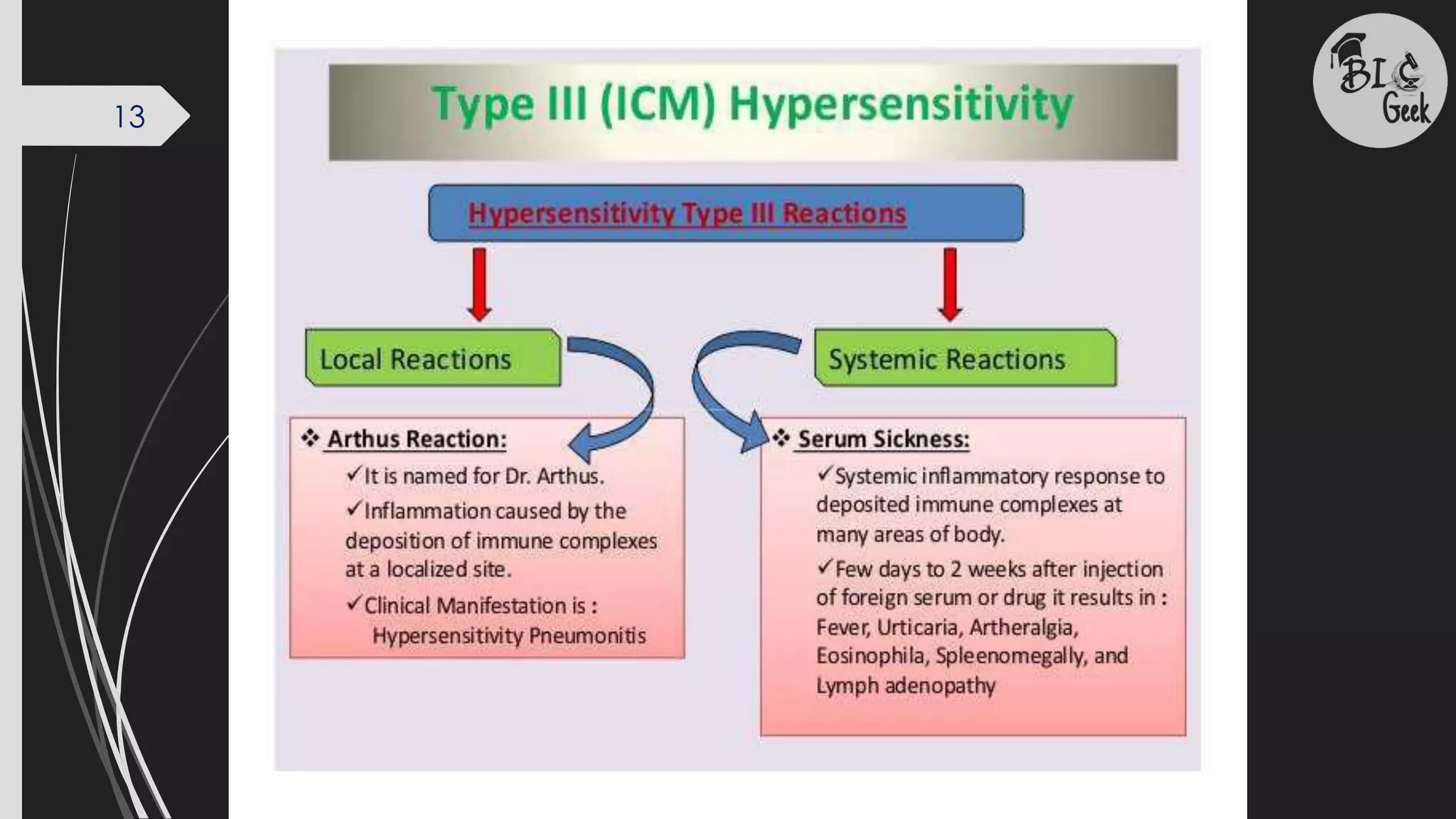
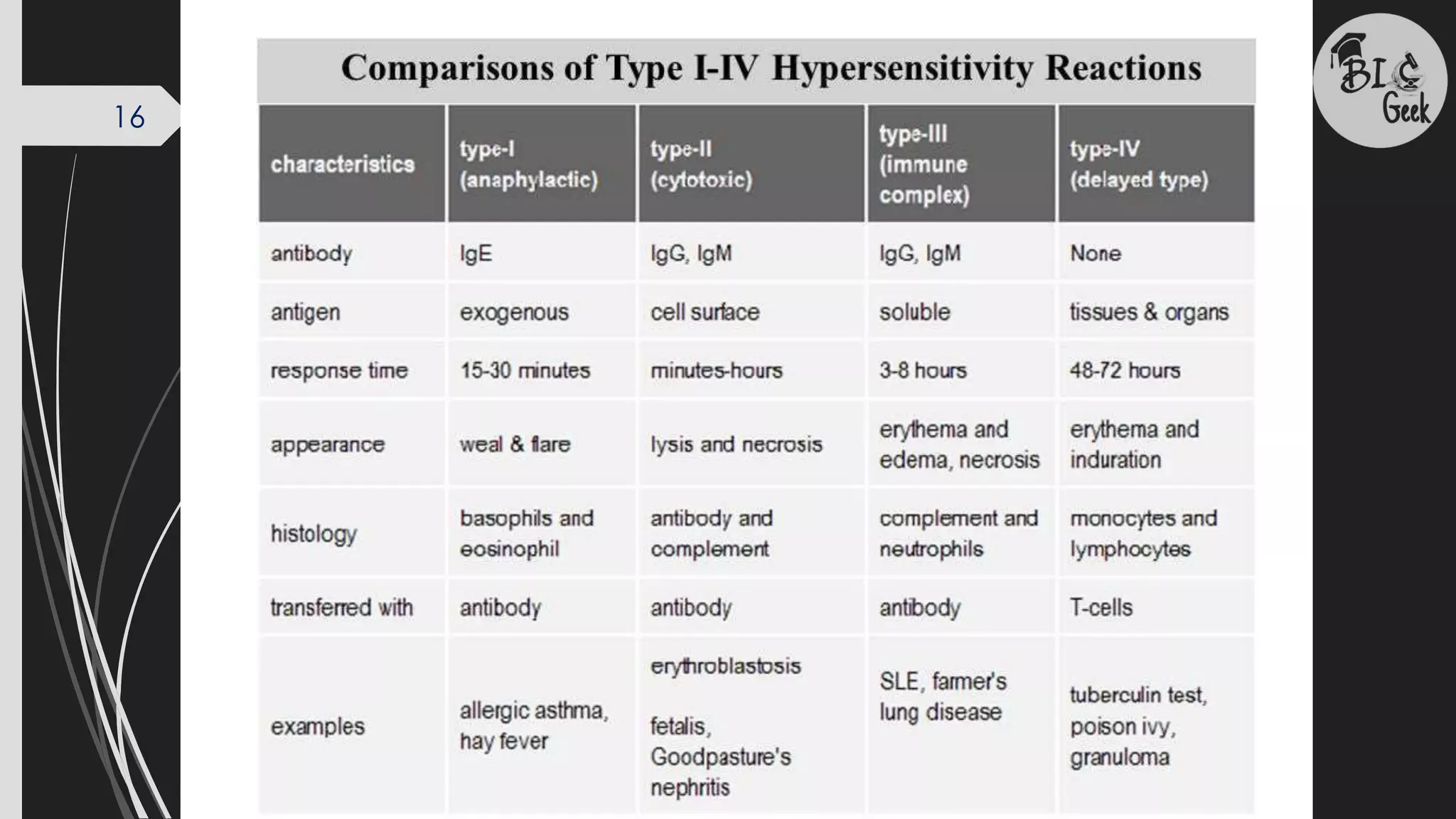
8/11/2023
1
Immunology
Mr. Akash Bagade
M.Sc. Zoology, CSIR-UGC NET, MH-SET and GATE
Senior Research Fellow
Dr. Babasaheb Ambedkar Research and Training
HYPERSENSITIVITY
SAVITRIBAI PHULE PUNE UNIVERSITY
B.Sc. And M.Sc.-
Zoology/Life Science
2
INTRODUCTION
 Hypersensitivity can be defined as a state of altered
immune response against an antigen characterized by
hyper reactivity leading to immunopathology
Hypersensitivity reactions require a pre-sensitized (immune)
state of the host.
There are two categories of adaptive hypersensitivities:
–Immediate hypersensitivities refer to humoral immunity
(antigen/antibody reactions)
–Delayed hypersensitivities refer to cell-mediated
immunity(cytotoxic T-lymphocytes, macrophages, and
cytokines)
3
CLASSIFICATION OF HYPERSENSITIVITY
Coomb and Gell in 1963 classified hypersensitivity reactions
into the following four types based on the mechanism involved
and time taken for the reaction.
4
Type I Hypersensitivity
(IgE Mediated Anaphylactic Hypersensitivity)
Characteristics of Type-I hypersensitivity
It is also known as immediate or anaphylactic hypersensitivity and is mediated by IgE.
 The reaction occurs on exposure to allergen second time. The first exposure
(sensitizing dose) results in sensitization of host to allergen and subsequent
exposure (s) (shocking dose) will cause reaction
 Anaphylactic shock occurs in sensitized animals within seconds to minutes (15-
30after exposure to the allergen) after exposure to the antigen, now called as an
allergen.
 Sometimes the reaction may have a delayed onset (10-12 hours).
 In type I hypersensitivity reactions, the allergens are proteins with a molecular
weight ranging from 10 to 40 kDa.
 Diagnostic tests for immediate hypersensitivity include skin (prick and intradermal)
tests resulting in wheal and flare reaction, measurement of total IgE and specific
IgE antibodies against the suspected allergens by ELISA, Radioallergosorbent test
(RAST)
5
Mechanism of Type-I hypersensitivity
The mechanism of reaction involves preferential production of IgE, in response to
certain antigens (allergens).
 Individuals prone to type-I hypersensitivity preferentially produce IL-4 and IL-13
that favor IgE class switch.
 IgE has very high affinity for its receptor (FcεIII; CD23) expressed on surface of
mast cells and basophils; the Fc portion of IgE binds to these receptors.
 A subsequent exposure to the same allergen cross links the cell bound IgE and
triggers the release of various pharmacologically active substances by a process
called as ‘degranulation’; mast cell degranulation is preceded by increased
Ca++ influx, which is a crucial process.
 These agents cause the early phase of allergic reactions that appears within
minutes after exposure to the antigen.
Cross-linking of IgE Fc-receptor is important in mast cell triggering
6
Mechanism of Type-I hypersensitivity
Degranulation of cells result in the synthesis and secretion of inflammatory
mediators such as platelet-activating factor, leukotreins,bradykinins, prostaglandins,
and cytokines that contribute to inflammation.
 The reaction is amplified by PAF (platelet activation factor) that causes platelet
aggregation and release of histamine, heparin and vasoactive amines.
 Eosinophil chemotactic factor of anaphylaxis (ECF-A) and neutrophil chemotactic
factors attract eosinophils and neutrophils, respectively, which release various
hydrolytic enzymes that cause necrosis
 These agents cause the early phase of allergic reactions that appears within
minutes after exposure to the antigen.
 Late phase allergic reactions may begin several hours after exposure to antigen.
 Cell-bound IgE on the surface of basophils of sensitive individuals binds a
substance called histamine releasing factor (possibly produced by macrophages
and B-lymphocytes) causing further histamine release
7
The inflammatory agents released or produced cause the following:
 dilation of blood vessels, which causes local redness (erythema)
at the site of allergen delivery. If dilation is widespread, this can
contribute to decreased vascular resistance, a drop in blood
pressure, and shock.
 increased capillary permeability, which causes swelling of local
tissues (edema). If widespread, it can contribute to decreased
blood volume and shock.
 constriction of bronchial airways, which leads to wheezing and
difficulty in breathing.
 stimulation of mucous secretion, which leads to congestion of
airways.
 stimulation of nerve endings, which leads to itching and pain in
the skin.
Effects of Type-I Hypersensitivity
8
Type II Hypersensitivity
(Antibody-mediated cytotoxic hypersensitivity)
Characteristics of Type II Hypersensitivity
Type II hypersensitivity are antibody mediated cytotoxic reactions occurring when
an antibody binds to antigens located on the surface of cells (usually RBCs). The
reaction time is minutes to hours.
 It is mediated, primarily, by antibodies of IgM or IgG class.
 The bound antibody can cause cell lysis by activating the classical complement
pathway, promoting phagocytosis (opsonization) or through ADCC.
 Many different antigens may trigger this cell destruction, but an infection in a
genetically predisposed animal appears to be a major triggering pathway.
 Antigens are normally endogenous, although exogenous chemicals (haptens, such
as ivy or drugs) that can attach to cell membranes can also induce type II reactions.
 Autoimmune haemolytic anaemia, Blood transfusion reactions, Erythroblastosis
fetalis, drug-induced hemolytic anemia, granulocytopenia and thrombocytopenia
are examples.
9
• IgM or IgG antibodies are made against self antigens
either as a result of failure of immune tolerance or
cross-reactive antibodies can develop during
infections, which may bind to normal tissue antigens
and trigger antibody-mediated cytotoxicity.
• Subsequent binding of these antibodies to the
surface of host cells leads to:
 Opsonization of the host cells whereby
phagocytes stick to host cells by way of IgG, C3b,
or C4b and discharge their lysosomes.
 Activation of the classical complement pathway
causing MAC induced lysis.
 ADCC mediated destruction of the host cells
whereby NK cells attach to the Fc portion of the
antibodies.
Mechanism of Type II Hypersensitivity
10
Examples of Type II Hypersensitivity
AB and Rh blood group reactions (blood transfusion reactions,erythroblastosis
foetalis)
Autoimmune diseases:
Rheumatic fever where antibodies result in joint and heart valvedamage;
Idiopathic thrombocytopenia purpura where antibodies result in the destruction of
platelets;
myasthenia gravis where antibodies bind to the acetylcholine receptors on muscle
cells causing faulty enervation of muscles;
Goodpasture's syndrome where antibodies lead to destruction of cells in the
kidney.
11
TYPE – III HYPERSENSITIVITY
(Immune complex mediated hypersensitivity)
Characteristics of Type III Hypersensitivity
• In type III hypersensitivity, soluble immune complexes are formed in blood and are
deposited in various tissues (typically the skin, kidney and joints), activate classical
complement pathway and cause inflammatory damage.
• It is mediated by soluble immune complexes. They are mostly of IgG class,
although IgM may also be involved.
• The reaction takes hours to days (3-10 hours) to develop.
• The antigen may be exogenous (chronic bacterial, viral or parasitic infections), or
endogenous (non-organ specific autoimmunity: e.g., Systemic Lupus Erythematosus-
SLE).
• The antigen is soluble and not attached to the organ involved.
• The prerequisite for the development of immune-complex disease is the persistent
presence of soluble antigen and antibody.
Soluble antigen-antibody (IgG or IgM) complexes are normally removed by
macrophages in the spleen and liver.
On formation of excessive amount or large immune complexes these gets lodged
in capillaries, pass between endothelial cells of blood vessels – specially those in the
skin, joints, and kidneys - and become trapped on the surrounding basement
membrane beneath these cells.
The Ag-Ab complexes then activate the classical complement pathway; the
damage being caused by platelets and neutrophils :
massive inflammation, due to complement protein C5a;
influx of neutrophils, due to complement protein C5a , resulting in neutrophils discharging their
lysosomes and causing tissue destruction and further inflammation;
MAC induced lysis of surrounding tissue cells; and
aggregation of platelets, resulting in more inflammation and the formation of microthrombi that
block capillaries.
Mechanism of Type III Hypersensitivity
12
13
TYPE – IV HYPERSENSITIVITY
(Cell mediated hypersensitivity)
(Delayed type hypersensitivity)
Characteristics of Type IV Hypersensitivity
• Type IV hypersensitivity is often called delayed type hypersensitivity as the reaction
takes more than 12 hours to develop. Typically the maximal reaction time occurs
between 48 to 72 hours.
• It is mediated by cells that cause an inflammatory reaction to either exogenous or
autoantigens.
• The major cells involved are T lymphocytes and monocytes/macrophages.
• This reaction to exogenous antigens involves T cells and also antigen-presenting cells
(APC), all produce cytokines that stimulate a local inflammatory response in a
sensitized individual.
• DHR cannot be transferred from an animal to another by means of antibodies or
serum. However, it can be transferred by T cells, particularly CD4 Th1 cells.
14
Mechanism of Type IV Hypersensitivity
CD8 cytotoxic T cells and CD4 helper T cells recognize antigen in a complex with
either type I or II MHC antigens
• The antigen-presenting cells in this case are macrophages and they release
interleukin 1, which further stimulates the proliferation of CD4 cells.
• These cells release IL-2 and IFN-λ, which together regulate the immune reaction;
other lymphokines involved in delayed hypersensitivity reaction include monocyte
chemotactic factor, TNF α etc.
• Cytokines produced by keratinocytes, APC, and T cells recruit antigen-
nonspecific T cells and macrophages to participate in a local inflammatory
reaction.
• Activated CD8 cells destroy target cells on contact, while activated macrophages
produce hydrolytic enzymes and, transform into multinucleated giant cells.
15
16
17
Thank you

Hypersensitivity.pdf

  • 1.
    8/11/2023 1 Immunology Mr. Akash Bagade M.Sc.Zoology, CSIR-UGC NET, MH-SET and GATE Senior Research Fellow Dr. Babasaheb Ambedkar Research and Training HYPERSENSITIVITY SAVITRIBAI PHULE PUNE UNIVERSITY B.Sc. And M.Sc.- Zoology/Life Science
  • 2.
    2 INTRODUCTION  Hypersensitivity canbe defined as a state of altered immune response against an antigen characterized by hyper reactivity leading to immunopathology Hypersensitivity reactions require a pre-sensitized (immune) state of the host. There are two categories of adaptive hypersensitivities: –Immediate hypersensitivities refer to humoral immunity (antigen/antibody reactions) –Delayed hypersensitivities refer to cell-mediated immunity(cytotoxic T-lymphocytes, macrophages, and cytokines)
  • 3.
    3 CLASSIFICATION OF HYPERSENSITIVITY Coomband Gell in 1963 classified hypersensitivity reactions into the following four types based on the mechanism involved and time taken for the reaction.
  • 4.
    4 Type I Hypersensitivity (IgEMediated Anaphylactic Hypersensitivity) Characteristics of Type-I hypersensitivity It is also known as immediate or anaphylactic hypersensitivity and is mediated by IgE.  The reaction occurs on exposure to allergen second time. The first exposure (sensitizing dose) results in sensitization of host to allergen and subsequent exposure (s) (shocking dose) will cause reaction  Anaphylactic shock occurs in sensitized animals within seconds to minutes (15- 30after exposure to the allergen) after exposure to the antigen, now called as an allergen.  Sometimes the reaction may have a delayed onset (10-12 hours).  In type I hypersensitivity reactions, the allergens are proteins with a molecular weight ranging from 10 to 40 kDa.  Diagnostic tests for immediate hypersensitivity include skin (prick and intradermal) tests resulting in wheal and flare reaction, measurement of total IgE and specific IgE antibodies against the suspected allergens by ELISA, Radioallergosorbent test (RAST)
  • 5.
    5 Mechanism of Type-Ihypersensitivity The mechanism of reaction involves preferential production of IgE, in response to certain antigens (allergens).  Individuals prone to type-I hypersensitivity preferentially produce IL-4 and IL-13 that favor IgE class switch.  IgE has very high affinity for its receptor (FcεIII; CD23) expressed on surface of mast cells and basophils; the Fc portion of IgE binds to these receptors.  A subsequent exposure to the same allergen cross links the cell bound IgE and triggers the release of various pharmacologically active substances by a process called as ‘degranulation’; mast cell degranulation is preceded by increased Ca++ influx, which is a crucial process.  These agents cause the early phase of allergic reactions that appears within minutes after exposure to the antigen. Cross-linking of IgE Fc-receptor is important in mast cell triggering
  • 6.
    6 Mechanism of Type-Ihypersensitivity Degranulation of cells result in the synthesis and secretion of inflammatory mediators such as platelet-activating factor, leukotreins,bradykinins, prostaglandins, and cytokines that contribute to inflammation.  The reaction is amplified by PAF (platelet activation factor) that causes platelet aggregation and release of histamine, heparin and vasoactive amines.  Eosinophil chemotactic factor of anaphylaxis (ECF-A) and neutrophil chemotactic factors attract eosinophils and neutrophils, respectively, which release various hydrolytic enzymes that cause necrosis  These agents cause the early phase of allergic reactions that appears within minutes after exposure to the antigen.  Late phase allergic reactions may begin several hours after exposure to antigen.  Cell-bound IgE on the surface of basophils of sensitive individuals binds a substance called histamine releasing factor (possibly produced by macrophages and B-lymphocytes) causing further histamine release
  • 7.
    7 The inflammatory agentsreleased or produced cause the following:  dilation of blood vessels, which causes local redness (erythema) at the site of allergen delivery. If dilation is widespread, this can contribute to decreased vascular resistance, a drop in blood pressure, and shock.  increased capillary permeability, which causes swelling of local tissues (edema). If widespread, it can contribute to decreased blood volume and shock.  constriction of bronchial airways, which leads to wheezing and difficulty in breathing.  stimulation of mucous secretion, which leads to congestion of airways.  stimulation of nerve endings, which leads to itching and pain in the skin. Effects of Type-I Hypersensitivity
  • 8.
    8 Type II Hypersensitivity (Antibody-mediatedcytotoxic hypersensitivity) Characteristics of Type II Hypersensitivity Type II hypersensitivity are antibody mediated cytotoxic reactions occurring when an antibody binds to antigens located on the surface of cells (usually RBCs). The reaction time is minutes to hours.  It is mediated, primarily, by antibodies of IgM or IgG class.  The bound antibody can cause cell lysis by activating the classical complement pathway, promoting phagocytosis (opsonization) or through ADCC.  Many different antigens may trigger this cell destruction, but an infection in a genetically predisposed animal appears to be a major triggering pathway.  Antigens are normally endogenous, although exogenous chemicals (haptens, such as ivy or drugs) that can attach to cell membranes can also induce type II reactions.  Autoimmune haemolytic anaemia, Blood transfusion reactions, Erythroblastosis fetalis, drug-induced hemolytic anemia, granulocytopenia and thrombocytopenia are examples.
  • 9.
    9 • IgM orIgG antibodies are made against self antigens either as a result of failure of immune tolerance or cross-reactive antibodies can develop during infections, which may bind to normal tissue antigens and trigger antibody-mediated cytotoxicity. • Subsequent binding of these antibodies to the surface of host cells leads to:  Opsonization of the host cells whereby phagocytes stick to host cells by way of IgG, C3b, or C4b and discharge their lysosomes.  Activation of the classical complement pathway causing MAC induced lysis.  ADCC mediated destruction of the host cells whereby NK cells attach to the Fc portion of the antibodies. Mechanism of Type II Hypersensitivity
  • 10.
    10 Examples of TypeII Hypersensitivity AB and Rh blood group reactions (blood transfusion reactions,erythroblastosis foetalis) Autoimmune diseases: Rheumatic fever where antibodies result in joint and heart valvedamage; Idiopathic thrombocytopenia purpura where antibodies result in the destruction of platelets; myasthenia gravis where antibodies bind to the acetylcholine receptors on muscle cells causing faulty enervation of muscles; Goodpasture's syndrome where antibodies lead to destruction of cells in the kidney.
  • 11.
    11 TYPE – IIIHYPERSENSITIVITY (Immune complex mediated hypersensitivity) Characteristics of Type III Hypersensitivity • In type III hypersensitivity, soluble immune complexes are formed in blood and are deposited in various tissues (typically the skin, kidney and joints), activate classical complement pathway and cause inflammatory damage. • It is mediated by soluble immune complexes. They are mostly of IgG class, although IgM may also be involved. • The reaction takes hours to days (3-10 hours) to develop. • The antigen may be exogenous (chronic bacterial, viral or parasitic infections), or endogenous (non-organ specific autoimmunity: e.g., Systemic Lupus Erythematosus- SLE). • The antigen is soluble and not attached to the organ involved. • The prerequisite for the development of immune-complex disease is the persistent presence of soluble antigen and antibody.
  • 12.
    Soluble antigen-antibody (IgGor IgM) complexes are normally removed by macrophages in the spleen and liver. On formation of excessive amount or large immune complexes these gets lodged in capillaries, pass between endothelial cells of blood vessels – specially those in the skin, joints, and kidneys - and become trapped on the surrounding basement membrane beneath these cells. The Ag-Ab complexes then activate the classical complement pathway; the damage being caused by platelets and neutrophils : massive inflammation, due to complement protein C5a; influx of neutrophils, due to complement protein C5a , resulting in neutrophils discharging their lysosomes and causing tissue destruction and further inflammation; MAC induced lysis of surrounding tissue cells; and aggregation of platelets, resulting in more inflammation and the formation of microthrombi that block capillaries. Mechanism of Type III Hypersensitivity 12
  • 13.
  • 14.
    TYPE – IVHYPERSENSITIVITY (Cell mediated hypersensitivity) (Delayed type hypersensitivity) Characteristics of Type IV Hypersensitivity • Type IV hypersensitivity is often called delayed type hypersensitivity as the reaction takes more than 12 hours to develop. Typically the maximal reaction time occurs between 48 to 72 hours. • It is mediated by cells that cause an inflammatory reaction to either exogenous or autoantigens. • The major cells involved are T lymphocytes and monocytes/macrophages. • This reaction to exogenous antigens involves T cells and also antigen-presenting cells (APC), all produce cytokines that stimulate a local inflammatory response in a sensitized individual. • DHR cannot be transferred from an animal to another by means of antibodies or serum. However, it can be transferred by T cells, particularly CD4 Th1 cells. 14
  • 15.
    Mechanism of TypeIV Hypersensitivity CD8 cytotoxic T cells and CD4 helper T cells recognize antigen in a complex with either type I or II MHC antigens • The antigen-presenting cells in this case are macrophages and they release interleukin 1, which further stimulates the proliferation of CD4 cells. • These cells release IL-2 and IFN-λ, which together regulate the immune reaction; other lymphokines involved in delayed hypersensitivity reaction include monocyte chemotactic factor, TNF α etc. • Cytokines produced by keratinocytes, APC, and T cells recruit antigen- nonspecific T cells and macrophages to participate in a local inflammatory reaction. • Activated CD8 cells destroy target cells on contact, while activated macrophages produce hydrolytic enzymes and, transform into multinucleated giant cells. 15
  • 16.
  • 17.