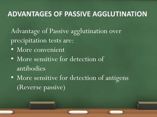
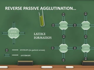
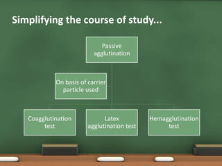
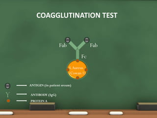
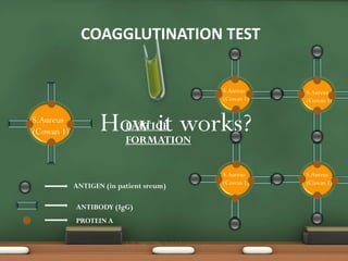
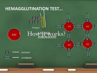
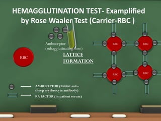
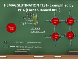
Direct
Passive
Reverse Passive
Agglutination Inhibition
Coagglutination
Agglutination tests can be done :
On slides
In tubes
In microtritation plates
-Difference between precipitation and agglutination reaction.
Antigen-antibody interactions can be quantified using various serological tests. Common types include precipitation tests like immunodiffusion that form visible precipitate lines, agglutination tests where antigens clump together, neutralization tests using viruses and complement fixation assays. Enzyme-linked immunosorbent assays (ELISAs) are now widely used as they are sensitive, specific and can be quantitative or qualitative. Fluorescent antibody techniques use fluorescent dyes to label antibodies or cells for detection under a microscope.
This document provides an overview of antigen-antibody interactions and their applications in infectious disease diagnosis. It discusses how immunochemical methods can detect microorganisms in patient specimens using antigens and antibodies. Various serological tests that utilize antigen-antibody reactions are described, including precipitation reactions, agglutination reactions, complement fixation tests, neutralization tests, and immunoassays. Specific techniques like immunodiffusion, immunoelectrophoresis, and slide and tube agglutination are also summarized. The document aims to explain the basic principles of antigen-antibody reactions and their uses in clinical diagnosis and epidemiology.
Reverse passive agglutination test coats antibodies onto carrier molecules that detect antigens in a patient's serum. For example, this test can be used to detect cholera toxin by coating cholera toxin antibodies onto carrier molecules which will agglutinate or clump if the cholera toxin antigen is present in a serum sample. This provides a detection method for certain antigens by reversing the roles of the antibody and antigen compared to a standard agglutination test.
The lecture was presented to the students of Saudi board of Community Medicine to help them know about the various serological methods applicable in the diagnosis of infectious diseases in general with attention upon the specificity and sensitivity of various diagnostic modalities. The lecture covers the basic principles of each test and the clinical applications with the advantages and disadvantages of each.
Immunologic methods are used in the laboratory diagnosis of infections by detecting the interaction between antigens and antibodies. Common techniques include agglutination, immunofluorescence, ELISA, and immunoblotting. Agglutination involves clumping of antigens by antibodies that can be seen visually. Immunofluorescence uses antibodies coupled to fluorescent dyes to identify antigens under UV light. ELISA detects antigens or antibodies through an enzymatic reaction, while immunoblotting confirms antibody presence by blotting proteins and detecting binding. These methods exploit the high specificity of the antigen-antibody reaction for diagnostic purposes.
This document summarizes various serological tests used to detect antigens and antibodies, including:
- Primary tests like ELISA, IFAT, RIA that detect markers
- Secondary tests like agglutination, complement fixation, precipitation that detect interactions
- Tertiary tests that assess protective value of antiserum in animals
It then provides details on specific tests like agglutination, Coombs test, hemagglutination inhibition, precipitation, complement fixation, ELISA and their applications in medicine, food/plant pathology, and quality control.
Complement fixation tests (CFT) detect antibodies that do not agglutinate or precipitate by measuring their ability to fix complement. CFT involves incubating patient serum with antigen and complement, then determining if complement is still available to lyse indicator cells. If complement is fixed in the antigen-antibody complex, it cannot lyse the indicator cells, indicating antibody presence. CFT can detect antibody levels below 1 microgram/mL, but it is time-consuming and not sensitive enough for immunity screening due to occasional nonspecific reactions. Interpretation involves whether indicator cell lysis occurs, indicating the absence or presence of antibodies in the patient serum.
Agglutination is the clumping together of antigens and antibodies. It occurs when the antibodies bind to particulate antigens. This causes the antigens to crosslink and form visible aggregates. Common applications of agglutination tests include blood typing (ABO and Rh), diagnosis of typhoid (Widal test), and identification of antibodies against Rh antigens (Coombs test). The titer or end point of an agglutination test refers to the highest dilution at which antigen-antibody clumping is still visible.
1) Agglutination tests detect antigens or antibodies by exploiting the ability of antibodies to cross-link antigen-coated particles, forming visible clumps or lattices.
2) There are several types of agglutination tests including direct, passive, and reverse passive agglutination as well as hemagglutination and hemagglutination inhibition.
3) Agglutination tests are useful, rapid techniques for detecting various infectious diseases and other analytes but can be limited by prozone effects at high antibody concentrations.
The document provides an overview of the complement system. It discusses the history and components of the three complement pathways: the classical pathway, lectin pathway, and alternative pathway. It also describes the roles of complement components in opsonization, chemotaxis, and formation of the membrane attack complex to lyse cells. The complement system is regulated to prevent damage to host cells. Deficiencies in complement proteins can increase susceptibility to certain infections.
Comparison of hormonal assay by ELISA , ELFA and ECLrijaa
The document discusses various methods for hormone measurement and immunoassay techniques. It explains that hormone assays are important for clinical diagnosis and treatment monitoring. While early methods like bioassay and chemical analysis had low sensitivity, radioimmunoassay (RIA) introduced in 1959 improved detection. However, due to health risks, safer alternatives were sought. Enzyme-linked immunosorbent assay (ELISA) was developed in the 1970s as a replacement for RIA using enzymes rather than radioisotopes. More recent techniques like chemiluminescence, fluorescence, and electrochemiluminescence provide adequate sensitivity without radioactivity. The document compares the principles and properties of various immunoassay methods.
This document describes single radial immunodiffusion (RID), a quantitative technique used to determine the concentration of an antigen in a sample. RID involves incorporating specific antibody into an agarose medium with a central well for the antigen sample. The antigen diffuses radially and reacts with the antibody, forming a visible precipitate ring whose diameter relates to the antigen concentration. The technique is simple, cost-effective, and can quantify immunoglobulins, complement components, or other antigens from biological fluids by measuring ring diameters against a standard curve.
This document provides an overview of the ELISA (Enzyme Linked Immuno Sorbent Assay) technique. It was developed in 1971 as a method to detect antigens or antibodies. The principle involves forming an antigen-antibody complex that is detected using an enzyme-conjugated secondary antibody. There are four main types of ELISA: direct, indirect, sandwich, and competitive. ELISA has various applications in diagnostics, food testing, and more due to its sensitivity, availability of equipment, and low cost of reagents.
Immunological techniques use antigens and antibodies to detect pathogens or their components in patient specimens. Agglutination tests couple antigens or antibodies to particles and look for cross-linking and agglutination. Complement fixation tests measure complement-consuming antibodies by incubating specimens with complement and antigens. Enzyme immunoassays like ELISA use enzyme-linked antibodies to detect antigens and quantify antibodies. Precipitation tests look for visible precipitation of antigen-antibody complexes to detect antigens or antibodies.
The document discusses antigen-antibody reactions. It begins by introducing antigens and antibodies and how they specifically combine in antigen-antibody reactions. The reactions occur in three stages: formation of an antigen-antibody complex, leading to visible events like precipitation or agglutination, and destruction or neutralization of the antigen. Key features of antigen-antibody reactions are their specificity, the formation of immune complexes, antigen binding sites called epitopes, and the binding force between antigens and antibodies. Common types of antigen-antibody reactions include precipitation, agglutination, complement fixation, ELISA, and immunofluorescence.
The document discusses various serological assays used to detect antibodies and antigens. It describes the basic principles of agglutination tests, precipitation tests, immunofluorescence, radioimmunoassay, ELISA, complement fixation tests, and serum neutralization tests. It provides examples of the application of these assays for diagnosing diseases like brucellosis, influenza, and foot-and-mouth disease. The document emphasizes the importance of assay sensitivity, specificity, reproducibility, and using appropriate antigen and antibody concentrations.
This document provides an overview of basic principles of immunohematology. It defines key terms like antigen and antibody. It describes the characteristics of antigens and factors that contribute to antigen immunogenicity. It also discusses the different types of immunoglobulins involved in blood group antibodies, and the differences between naturally occurring versus immune antibodies. Finally, it explains the stages of antigen-antibody reactions including sensitization and agglutination, and factors that can influence these reactions.
This document provides an overview of ELISA (Enzyme-Linked Immunosorbent Assay). It describes ELISA as a method for detecting antibodies or antigens using enzyme-linked antibodies and a chromogenic substrate. The document outlines the basic ELISA procedure and discusses different ELISA types including direct, indirect, sandwich, and competitive ELISA. It also notes applications of ELISA for detecting antigens and antibodies and compares ELISA to other detection methods.
This document discusses electrophoresis and immunofixation tests used to analyze serum proteins. Electrophoresis separates plasma proteins based on their charge, but cannot identify specific protein subtypes. Immunofixation adds antibodies that bind to specific protein antigens like IgG, IgM, IgA, kappa and lambda to identify the subtype of any abnormal monoclonal proteins present. Together, electrophoresis and immunofixation can detect monoclonal gammopathies and determine the exact monoclonal protein, aiding diagnosis of conditions like multiple myeloma. The case example describes using these tests to identify a monoclonal IgG lambda protein in a patient with bone pain, leading to a diagnosis of multiple myeloma.
ELISA is a biochemical technique used in immunology to detect the presence of an antibody or antigen in a sample. It involves coating microtiter plate wells with an antigen or antibody and using conjugated enzymes and substrates to produce a colored product to indicate a positive result. There are different types of ELISA including direct, indirect, sandwich, and competitive ELISA which are used to test for various antigens or antibodies. ELISA has many applications such as measuring serum antibody concentrations, detecting food allergens or diseases, and identifying past exposure to diseases.
2 antigens, immunogens, epitopes, and haptenstaha244ali
This document discusses key concepts in immunology including antigens, immunogens, epitopes, haptens, innate immunity, and adaptive immunity. It defines antigens as molecules recognized by the immune system and immunogens as antigens that elicit an immune response. Epitopes are the smallest part of an antigen recognized by B and T cell receptors. Haptens are small molecules that require a carrier to induce an immune response. Innate immunity provides the first line of defense using soluble proteins and cells like phagocytes. Adaptive immunity develops over time through T and B cell responses and produces immunological memory.
Immunochromatographic assays, also known as lateral flow strip tests, allow for the rapid detection of antigens or antibodies in a sample within 15 minutes. The test works by utilizing two types of antibodies - one immobilized on the test strip and one labeled with a detectable marker like colloidal gold. When a sample is applied, it migrates up the strip via capillary action, allowing any antigens/antibodies in the sample to bind to the labeled antibodies and form complexes. These complexes are then captured by the immobilized antibodies, producing a visible test line that confirms the presence of the target antigen or antibody. Lateral flow tests are commercially available, easy to use, and provide results quickly with no specialized equipment,
The document discusses enzyme-linked immunosorbent assay (ELISA), including its introduction, principle, equipment, procedure, types, advantages, and disadvantages. ELISA is a qualitative or quantitative immunological procedure that detects antigens or antibodies using enzyme-labeled antibodies and chromogenic substrates. It relies on antibody-antigen interactions and uses an enzyme-labeled antibody to generate a colored reaction, allowing detection of a particular antigen. The document outlines the basic equipment, general procedure involving coating wells with antibodies and adding samples and enzyme-labeled antibodies, and the three main types of ELISA - indirect, sandwich, and competitive.
This document provides information about the Coombs test, which is used to detect antibody or complement coating of red blood cells. It describes the history and principles of the test, as well as the direct and indirect Coombs test procedures. The direct Coombs test detects in vivo coating of red blood cells and is used to diagnose conditions like hemolytic disease of the newborn. The indirect Coombs test detects in vitro coating of red cells and is used for compatibility testing and antibody screening. Factors affecting the tests and causes of false positive and negative results are also outlined.
Antigens are the substances which induce specific immune reactions in the body.
Antigens include molecules such as proteins, nucleoproteins, polysaccharides, lipoprotein and some glycolipids.
The ability of a molecule to function as an antigen depends on its size, structural complexity, chemical nature, and degree of foreignness to the host.
Types of antigens
Antigens are of two types:
1. Autoantigens or self antigens present on the body’s own cells such as ‘A’ antigen and ‘B’ antigen in RBCs.
2. Foreign antigen s or non-self antigens that enter the body from outside.
Following are non-self antigens:
1. Receptors on the cell membrane of microbial organisms such as bacteria, viruses and fungi.
2. Toxins from microbial organisms.
3. Materials from transplanted organs or incompatible blood cells.
4. Allergens or allergic substances like pollen grains.
Antigens are substances that stimulate the immune system to produce antibodies against them. They enter the body through various sites and are then captured and presented by antigen presenting cells. There are several types of antigens including immunogens, which induce immune responses; tolerogens, which induce tolerance; allergens; and vaccines. An antigen's ability to induce an immune response is called its immunogenicity, while its ability to bind antibodies is its antigenicity. Properties that influence immunogenicity include the antigen's foreignness, size, complexity, degradability, and the recipient's genotype and age. Administration methods like dosage, route, and use of adjuvants can also impact immunogenicity. Antigens are classified as complete if they have
Agglutination is the clumping of particulate antigens caused by antibody binding. It was first observed in 1896 using bacterial cells and serum antibody. Antibodies that cause agglutination are called agglutinins. Agglutination involves an initial antigen-antibody binding step followed by lattice formation into large aggregates. Erythrocytes, bacteria, and latex particles can all participate. The reaction is influenced by factors like ionic strength, pH, temperature, and viscosity. It can be directly observed on cell surfaces or indirectly using antigen-coated carriers like latex beads. Agglutination tests are used to diagnose various infectious diseases.
Measuring agglutination reactions can be used to quantify antibodies, identify antibody targets, and determine antibody specificity, with applications including ELISA, immunofluorescence, and blood typing tests.
This document discusses antigen-antibody reactions, including their specificity, the formation of immune complexes, and the non-covalent bonds involved in binding. It describes different types of antigen-antibody reactions like precipitation, agglutination, complement fixation, ELISA, and immunofluorescence. It also outlines applications like blood typing, disease detection, immunoassays, and assessing immune deficiencies.
This document discusses antigen-antibody reactions and their clinical applications. It begins by classifying serological tests into primary, secondary, and tertiary categories. It then describes the principles of antigen-antibody interactions, including affinity, avidity, sensitivity and specificity. Various serological techniques are outlined, including precipitation reactions, agglutination reactions, complement fixation tests, and immunoassays. The document emphasizes that antigen-antibody reactions form the basis of antibody-mediated immunity and have important diagnostic applications for identifying infections and other agents.
The document describes a method to measure antibody titers against thyroglobulin in patient serum samples using passive agglutination. Serial dilutions of control and patient serum samples were incubated with turkey red blood cells coated with thyroglobulin. Agglutination reactions indicated the presence and titer level of anti-thyroglobulin antibodies. The results showed that patient samples A and B tested positive with titers of 1280 and 320, respectively, while patient C tested negative, indicating no autoimmune thyroid disease.
What is Agglutination?
by Gary Cecchi, M.D.
Agglutination occurs when incompatible blood types are mixed together and blood cells begin to clump. Each blood cell contains large protein molecules called antigens on its surface. Blood also contains antibodies, which bind to foreign antigens and cause incompatible blood cells to burst or agglutinate.
In humans, blood agglutination can cause kidney failure and death. Medical practitioners are therefore very careful when giving blood to patients. Despite their caution, however, approximately one out of every 12,000 units of blood transfused in the United States goes to the wrong person. Depending on the blood types involved, the results can range from catastrophic to unnoticeable.
About the Author:
Managing Partner of Northern California Hematology and Oncology Dr. Gary Cecchi possesses years of experience as a hematologist. He has served as the Director for Hematology and Medical Oncology at Alta Bates Summit Medical Center and holds board certification in both hematology and oncology.
Culture media provide optimal conditions for bacterial growth and multiplication. Solid media allow for isolation of pure cultures and identification via colony morphology and effects on the medium. Types of media include simple, enriched, selective, enrichment, indicator, and selective/indicator media. Simple media like nutrient broth and agar support general growth. Enriched media like blood agar aid growth of fastidious organisms. Selective media inhibit certain bacteria. Enrichment media help multiplication of target species. Indicator media provide visual cues on metabolic activity. Strict anaerobes require specialized media and cultivation systems like GasPak to exclude oxygen.
The document discusses different types of immunity and serological reactions. It describes the two main types of immunity - innate immunity and acquired immunity. Acquired immunity includes active immunity, which can be naturally or artificially acquired through vaccination, and passive immunity, which can also be naturally or artificially acquired through antibodies from another source. The document then goes into detail about different types of vaccines and serological reactions used to detect pathogens.
This document summarizes the history and current practice of splenectomy. It describes the historical understanding of the spleen from ancient times through the first documented splenectomies in the 1500s. It reviews the development of laparoscopic splenectomy in the 1990s. The spleen's anatomy and blood supply are outlined. Common indications for splenectomy include trauma, hematologic disorders, and malignancy. Both open and laparoscopic techniques are discussed, including preoperative considerations like vaccination. Postoperative care focuses on early mobilization. Complications include infection and thrombosis.
Immunodiffusion techniques such as radial immunodiffusion, Ouchterlony double diffusion, and immunoelectrophoresis can be used to detect and quantify antigens and antibodies through the formation of precipitin lines. These techniques utilize the diffusion of antigens and antibodies through a semi-solid medium like agar to form visible precipitin lines where the antigens and antibodies combine. They can be used to diagnose diseases, detect immunodeficiencies, and assess the purity and concentration of antigens and antibodies.
The EMIT technique is an immunoassay method used to screen blood and urine samples for therapeutic drugs and abused substances. It works by using antibodies linked to enzymes that react with the target substance in a sample. This reaction is then measured spectrophotometrically. EMIT assays are homogeneous, requiring no separation steps, and provide reliable quantitative results. While less sensitive than other immunoassays like ELISA, EMIT is widely used in clinical settings due to its low cost, simplicity, and long shelf life of reagents.
1. The document discusses the laboratory diagnosis of Salmonella, which causes enteric fever and gastroenteritis in humans. Blood culture is the best specimen for diagnosis in the first week, while stool and urine cultures are optimal in later weeks.
2. Serological tests like the Widal test detect antibodies against Salmonella, while rapid tests like Typhidot and IDL Tubex detect IgM antibodies.
3. Isolation of Salmonella from stool requires plating on selective media followed by biochemical tests and serotyping using slide agglutination to identify the serovar. Antimicrobial susceptibility testing helps guide treatment.
This document summarizes various serological tests used to detect antigens and antibodies. It describes primary, secondary and tertiary serological tests including ELISA, immunofluorescence, radioimmunoassay and more. It also details different types of agglutination tests like qualitative and quantitative tests. Additional tests covered are precipitation tests, complement fixation tests, passive hemagglutination and sandwich ELISA. The document provides information on applications and procedures for many antibody and antigen detection techniques.
UX, ethnography and possibilities: for Libraries, Museums and ArchivesNed Potter
1) The document discusses how the University of York Library has used various user experience (UX) techniques like ethnographic observation and interviews to better understand user needs and behaviors.
2) Some changes implemented based on UX findings include installing hot water taps, changing hours, and adding blankets - aimed at improving the small details of user experience.
3) The presentation encourages other libraries, archives and museums to try incorporating UX techniques like behavioral mapping and cognitive interviews to inform design changes that enhance services for users.
Ag-Ab-Reactions.pdf microbiology and botanyjyothisaisri
The document discusses antigen-antibody reactions, including the specific and reversible binding between antigens and antibodies. It describes the properties of antigen-antibody reactions, such as high specificity, non-covalent interactions, and reversibility. It also discusses the concepts of affinity, avidity, and cross-reactivity. Finally, it summarizes several types of antigen-antibody reactions: precipitation, agglutination, complement fixation, and neutralization.
The document discusses various antigen-antibody reactions including precipitation, agglutination, neutralization, complement fixation, immunofluorescence, radioimmunoassay, enzyme immunoassay, and immunoblotting. It describes how these reactions work, their applications in laboratory testing and research, and factors like sensitivity and specificity. Key stages of antigen-antibody interactions like primary, secondary and tertiary are also outlined.
Antigen-antibody reactions occur specifically between antigens and antibodies. In the body, they form the basis of immunity against infectious diseases and hypersensitivities. In the laboratory, antigen-antibody reactions are used for diagnosis of infections, epidemiological surveys, and identification of infectious agents. Precipitation reactions occur when a soluble antigen and its antibody form an insoluble complex in the presence of electrolytes. Precipitation can be used to identify bacteria and detect antibodies for diagnosis.
This document discusses antigen-antibody reactions and their clinical applications. It describes the principles of primary, secondary, and tertiary antigen-antibody reactions. Various serological tests are discussed, including precipitation reactions, agglutination reactions, complement fixation tests, ELISA, immunofluorescence, and radioimmunoassay. These tests can help diagnose infections, identify infectious agents, and detect non-infectious substances. The document also provides examples of clinical applications for many common serological tests.
Principle, Advantages and Disadvantages Of RIA , Requirements for RIA , Preparation , Development of the Assay system, Assay Procedure , ELISA , Advantages and Disadvantages of ELISA , Types of ELISA, Applications of Immunoassays
What are the 3 types of immune systems?
These specialized cells and parts of the immune system offer the body protection against disease. This protection is called immunity. Humans have three types of immunity — innate, adaptive, and passive: Innate immunity: Everyone is born with innate (or natural) immunity, a type of general protection.
The document discusses antigen-antibody reactions, including:
1) Antigens and antibodies specifically bind to each other through epitopes and paratopes in what is known as the antigen-antibody reaction.
2) This reaction occurs in three stages - formation of antigen-antibody complexes, visible events like precipitation or agglutination, and destruction or neutralization of antigens.
3) Examples of techniques using antigen-antibody reactions include precipitation reactions, agglutination reactions, complement fixation, ELISA, and immunofluorescence. These reactions form the basis of serological tests.
This document provides an overview of two immunoassay techniques: ELISA and RIA. ELISA (enzyme-linked immunosorbent assay) detects the presence of an antigen or antibody using an enzyme-linked secondary antibody that produces a colored product when reacted with a substrate. RIA (radioimmunoassay) uses a radiolabeled antigen or antibody to compete with unlabeled antigens in a sample, and measures radioactivity to determine antigen concentration. Both techniques rely on the specificity of the antigen-antibody reaction and can be used to detect various targets like hormones, drugs, and infectious diseases.
This document provides an overview of two immunoassay techniques: ELISA and RIA. ELISA (enzyme-linked immunosorbent assay) detects the presence of an antigen or antibody using an enzyme-linked secondary antibody that produces a colored product when reacted with a substrate. RIA (radioimmunoassay) uses a radiolabeled antigen or antibody to compete with unlabeled antigens in a sample, and measures radioactivity to determine antigen concentration. Both techniques rely on the specificity of antigen-antibody binding and can be used to detect various targets like hormones, drugs, and infectious diseases.
Radioimmunoassay and enzyme-linked immunosorbent assays are immunoassays that use the antigen-antibody reaction to detect and quantify substances like hormones, vitamins, and drugs. Radioimmunoassays use radiolabeled antigens or antibodies while ELISAs use enzyme-labeled molecules, avoiding radiation hazards. Both techniques require preparing antigens and antibodies, developing standardized assay procedures, and measuring the amount of bound versus unbound reagents to quantify the target substance in samples. Immunoassays are highly specific and sensitive methods used to detect substances important for diagnosing disease and monitoring drug and hormone levels in the body.
This document discusses various principles and techniques for measuring antigen-antibody interactions, including agglutination tests, precipitation tests, radioimmunoassays, enzyme-linked immunosorbent assays, immunofluorescence, and complement fixation. It defines key terms like affinity, specificity, cross-reactivity and describes how factors like antigen-antibody ratio affect measurements. Specific techniques covered include agglutination, passive agglutination, agglutination inhibition, radial immunodiffusion, immunoelectrophoresis, competitive and non-competitive radioimmunoassays/ELISAs, direct and indirect immunofluorescence, and complement fixation.
The document discusses antigen-antibody reactions and various immunoassay techniques. It begins by defining antigen-antibody reactions and their properties such as specificity, strength, and types. It then discusses different immunoassay techniques including precipitation reactions, agglutination reactions, and newer techniques like ELISA. It provides details on various qualitative and quantitative assays and their applications in diagnosing diseases. In conclusion, it emphasizes that immunoassays are widely used diagnostic tests based on the specific antigen-antibody reactions.
principle instrumentation and application of capillary electrophoresisAnimikh Ray
Immunochemical assays are based on antibody-antigen interactions in vitro. An immunoglobulin molecule contains two heavy chains and two light chains, each composed of variable and constant domains. Antigen is any foreign molecule that provokes an antibody response. Radioimmunoassay uses radioactive isotopes to detect antigens or antibodies with high sensitivity down to picogram levels. Enzyme-linked immunosorbent assay uses an enzyme label for detection and can be used for both qualitative and quantitative analysis.
The document discusses antigen-antibody reactions. It describes how antibodies bind specifically to antigens via non-covalent interactions between the antigen's epitope and the antibody's variable region. This specificity allows immunoassays to detect antibodies or antigens for disease diagnosis, immune response monitoring, and identifying molecules of interest. Different types of antigen-antibody reactions are precipitation reactions, agglutination reactions, complement fixation, ELISA, and immunofluorescence which are used in various applications like blood typing, disease diagnosis, and quantification of substances.
This document discusses various immunotechniques used to detect antigens and antibodies, including primary and secondary interactions, precipitation reactions, agglutination tests, radioimmunoassay, ELISA, and Western blotting. Primary interactions involve immune complex formation between antigens and antibodies via non-covalent bonds. Secondary interactions include precipitation, which can be measured via precipitation curves in solution or immunodiffusion/immunoelectrophoresis in gels. Radioimmunoassay and ELISA allow sensitive detection of antigens and antibodies using radioactive or enzyme labels. Western blotting identifies specific proteins separated by electrophoresis.
The document discusses factors that influence antigen-antibody reactions. It describes antigens and antibodies, the stages of antigen-antibody reactions, and properties of these reactions. Several factors can influence the reactions, including temperature, incubation time, pH, ionic strength, antigen-antibody concentration, centrifugation, distance between cells, and use of potentiators like anti-human globulin or LISS solution. Understanding how these various factors impact antigen-antibody binding can help optimize serological tests.
Local Advanced Lung Cancer: Artificial Intelligence, Synergetics, Complex Sys...Oleg Kshivets
Overall life span (LS) was 1671.7±1721.6 days and cumulative 5YS reached 62.4%, 10 years – 50.4%, 20 years – 44.6%. 94 LCP lived more than 5 years without cancer (LS=2958.6±1723.6 days), 22 – more than 10 years (LS=5571±1841.8 days). 67 LCP died because of LC (LS=471.9±344 days). AT significantly improved 5YS (68% vs. 53.7%) (P=0.028 by log-rank test). Cox modeling displayed that 5YS of LCP significantly depended on: N0-N12, T3-4, blood cell circuit, cell ratio factors (ratio between cancer cells-CC and blood cells subpopulations), LC cell dynamics, recalcification time, heparin tolerance, prothrombin index, protein, AT, procedure type (P=0.000-0.031). Neural networks, genetic algorithm selection and bootstrap simulation revealed relationships between 5YS and N0-12 (rank=1), thrombocytes/CC (rank=2), segmented neutrophils/CC (3), eosinophils/CC (4), erythrocytes/CC (5), healthy cells/CC (6), lymphocytes/CC (7), stick neutrophils/CC (8), leucocytes/CC (9), monocytes/CC (10). Correct prediction of 5YS was 100% by neural networks computing (error=0.000; area under ROC curve=1.0).
Osteoporosis - Definition , Evaluation and Management .pdfJim Jacob Roy
Osteoporosis is an increasing cause of morbidity among the elderly.
In this document , a brief outline of osteoporosis is given , including the risk factors of osteoporosis fractures , the indications for testing bone mineral density and the management of osteoporosis
Clinic ^%[+27633867063*Abortion Pills For Sale In Tembisa Central19various
Clinic ^%[+27633867063*Abortion Pills For Sale In Tembisa Central Clinic ^%[+27633867063*Abortion Pills For Sale In Tembisa CentralClinic ^%[+27633867063*Abortion Pills For Sale In Tembisa CentralClinic ^%[+27633867063*Abortion Pills For Sale In Tembisa CentralClinic ^%[+27633867063*Abortion Pills For Sale In Tembisa Central
Cell Therapy Expansion and Challenges in Autoimmune DiseaseHealth Advances
There is increasing confidence that cell therapies will soon play a role in the treatment of autoimmune disorders, but the extent of this impact remains to be seen. Early readouts on autologous CAR-Ts in lupus are encouraging, but manufacturing and cost limitations are likely to restrict access to highly refractory patients. Allogeneic CAR-Ts have the potential to broaden access to earlier lines of treatment due to their inherent cost benefits, however they will need to demonstrate comparable or improved efficacy to established modalities.
In addition to infrastructure and capacity constraints, CAR-Ts face a very different risk-benefit dynamic in autoimmune compared to oncology, highlighting the need for tolerable therapies with low adverse event risk. CAR-NK and Treg-based therapies are also being developed in certain autoimmune disorders and may demonstrate favorable safety profiles. Several novel non-cell therapies such as bispecific antibodies, nanobodies, and RNAi drugs, may also offer future alternative competitive solutions with variable value propositions.
Widespread adoption of cell therapies will not only require strong efficacy and safety data, but also adapted pricing and access strategies. At oncology-based price points, CAR-Ts are unlikely to achieve broad market access in autoimmune disorders, with eligible patient populations that are potentially orders of magnitude greater than the number of currently addressable cancer patients. Developers have made strides towards reducing cell therapy COGS while improving manufacturing efficiency, but payors will inevitably restrict access until more sustainable pricing is achieved.
Despite these headwinds, industry leaders and investors remain confident that cell therapies are poised to address significant unmet need in patients suffering from autoimmune disorders. However, the extent of this impact on the treatment landscape remains to be seen, as the industry rapidly approaches an inflection point.
8 Surprising Reasons To Meditate 40 Minutes A Day That Can Change Your Life.pptxHolistified Wellness
We’re talking about Vedic Meditation, a form of meditation that has been around for at least 5,000 years. Back then, the people who lived in the Indus Valley, now known as India and Pakistan, practised meditation as a fundamental part of daily life. This knowledge that has given us yoga and Ayurveda, was known as Veda, hence the name Vedic. And though there are some written records, the practice has been passed down verbally from generation to generation.
Integrating Ayurveda into Parkinson’s Management: A Holistic ApproachAyurveda ForAll
Explore the benefits of combining Ayurveda with conventional Parkinson's treatments. Learn how a holistic approach can manage symptoms, enhance well-being, and balance body energies. Discover the steps to safely integrate Ayurvedic practices into your Parkinson’s care plan, including expert guidance on diet, herbal remedies, and lifestyle modifications.
2. • What is passive agglutination?
• What are various types of passive agglutination tests?
AREAS OF INTERESTS…
3. LET’S GET STARTED…
What is agglutination?
Particulate antigen + its
specific antibody
Electrolytes at an optimal
temperature and pH
Visible clumping of particles
4. LET’S GET STARTED…
What is precipitation
reaction?
Soluble antigen + its specific
antibody
Electrolytes at an optimal
temperature and pH
Insoluble precipitate
5. LET’S GET STARTED…
And what is Passive
Agglutination??
Precipitation reaction
Agglutination tests
By attaching soluble antigens to
the surface of carrier particles
such as latex particles, bentonite,
RBCs, etc.
6. ADVANTAGES OF PASSIVE AGGLUTINATION
Advantage of Passive agglutination over
precipitation tests are:
• More convenient
• More sensitive for detection of
antibodies
• More sensitive for detection of antigens
(Reverse passive)
7. REVERSE IS POSSIBLE…
When instead of antigen, the antibody is
adsorbed on the carrier particles for
estimation of antigens, it is known as
Reverse passive agglutination
9. Simplifying the course of study...
Passive
agglutination
Coagglutination
test
Latex
agglutination test
Hemagglutination
test
On basis of carrier
particle used
12. COAGGLUTINATION TEST
S.Aureus
(Cowan 1)
ANTIBODY (IgG)
S.Aureus
(Cowan 1)
ANTIGEN (in patient sreum)
LATTICE
FORMATION
S.Aureus
(Cowan 1)
S.Aureus
(Cowan 1)
S.Aureus
(Cowan 1)
PROTEIN A
How it works?
13. LATEX AGGLUTINATION TEST...
The carrier particle is Latex or polystyrene latex
Brains behind this: C M Plotz and J M Singer
Accidently discovered IgG adsorbed naturally
to polystyrene latex particles (1953)
15. LATEX AGGLUTINATION TEST- USES
• Carrier + Antibody- detection of antigens-
CRP,RA factor, HCG, Hepatitis B
• Carrier + Antigen- antibodies to meningococci,
H.influenzae type b
16. Large number of antigens can adsorbed on carrier
Better visualization of Ag-Ab reaction due to larger
particle size of Latex beads preventing previous
cumbersome process involved in precipitation
reactions (no sophisticated equipments required)
Latex particles do not cross-react with other
antibodies
Less time consuming
LATEX AGGLUTINATION TEST- ADVANTAGES
17. HEMAGGLUTINATION TEST
The carrier particle is Red Blood Cell/Tanned
blood cell (Goose RBCs preferred)
Brain behind this: George Hirst (1942)
23. AGGLUTINATION TESTS IN OUR LAB IN A
BIRD’S EYE VIEW
ASO titre
RA factor detection
CRP
KIT- Syphicheck
Infectious mononucleosis (Immutex)
24. LET’S END BY LOOKING AT RECENT ADVANCES
• Determination of anti-streptolysin O antibody titer by a
new passive agglutination method using sensitized
toraysphere particles.
25. THANK
YOU
References from:
Anantnarayan and
Paniker’s Textbook of
microbiology
C.P. Baweja’s Textbook
of Microbiology
Subhash Chandra
Parija’s Textbook of
microbiology and
Immunology
American Society for
microbiolgy-Journal of Clinical
Microbiology