Downloaded 1,364 times


![Components Substrate ALP substrate - For most applications pNPP (p-Nitrophenyl-phosphate) is the most widely used substrate. HRP substrates– Hydrogen peroxide. TMB (3,3’,5,5’-tetramethylbenzidine) OPD (o-phenylenediamine dihydrochloride) ABTS (2,2’-azino-di-[3-ethyl-benzothiazoline-6 sulfonic acid] diammonium salt Substrate Function A substrate is a compound or substance that undergoes change. Substrates bind to active sites on the surface of enzymes and are converted or changed. In ELISA the specific substrate used changes color.](https://image.slidesharecdn.com/elisaseminarfinal2-111218110845-phpapp01/85/Elisa-seminar-final2-8-320.jpg)


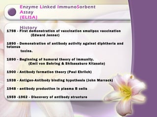
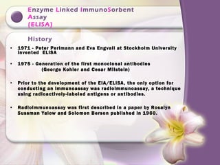
The document provides a history of ELISA (Enzyme-Linked ImmunoSorbent Assay) and describes its components, procedures, types, applications and advantages. It discusses key events in immunology research from the 18th century leading to the development of ELISA in the 1970s. ELISA allows detection and quantification of antigens or antibodies in a simple, sensitive and cost-effective manner using enzyme-labeled antibodies or antigens.