Downloaded 215 times
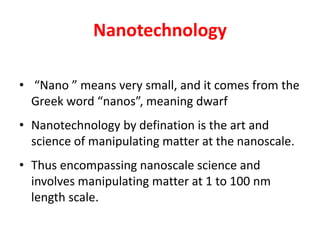
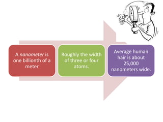
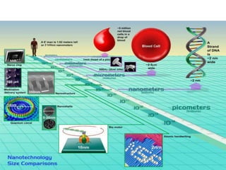
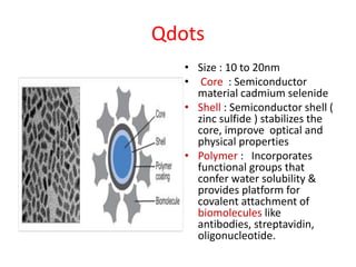
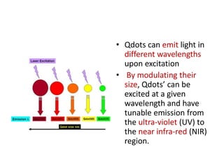
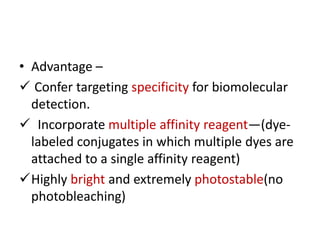
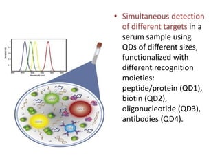


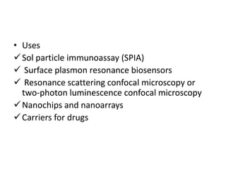

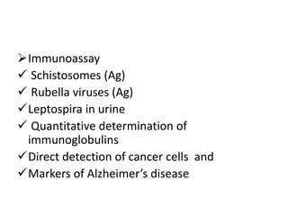
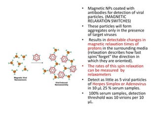
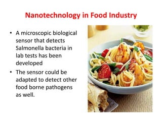
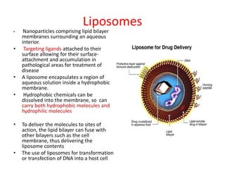
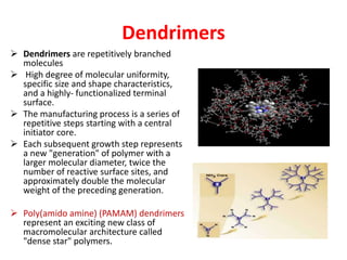
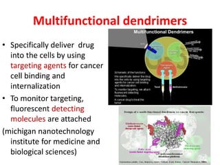


The document discusses various applications of nanotechnology in microbiology. It begins by defining nanotechnology as the manipulation of matter at the nanoscale of 1 to 100 nm. Some key applications discussed include using quantum dots for pathogen detection through fluorescence, using gold and silver nanoparticles in assays like sol particle immunoassays, and using magnetic nanoparticles in detection methods like magnetic relaxation switches that can detect as few as 5 viral particles. The document also discusses nanoparticle-based methods that enable faster, more sensitive detection of pathogens without sample preparation.