Aium image library tim thai chi tiet 2019
•
2 likes•3,370 views

This document contains a collection of fetal echocardiography images from the second and third trimesters. The American Institute of Ultrasound in Medicine thanks GE for providing the ultrasound equipment used to capture some of the images. The images show normal anatomy, views, and measurements needed to assess fetal heart structure and function during routine echocardiography exams. Key structures like the four chambers, valves, great vessels, and ductus venosus are represented.

More Related Content
What's hot
What's hot (19)
Similar to Aium image library tim thai chi tiet 2019
Similar to Aium image library tim thai chi tiet 2019 (20)
More from Võ Tá Sơn
More from Võ Tá Sơn (20)
Recently uploaded
Recently uploaded (20)
Aium image library tim thai chi tiet 2019
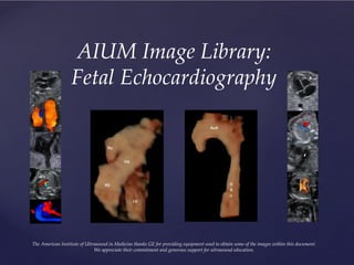
- 1. AIUM Image Library: Fetal Echocardiography The American Institute of Ultrasound in Medicine thanks GE for providing equipment used to obtain some of the images within this document. We appreciate their commitment and generous support for ultrasound education.
- 2. Second Trimester Cardiac Apex Position
- 3. Second Trimester Situs: Breech Presentation
- 4. Second Trimester Situs: Cephalic Presentation AO = aorta LA = left atrium LV = left ventricle MV = mitral valve RA = right atrium RV = right ventricle SP = spine TV = tricuspid valve
- 6. Second Trimester Left Ventricular Outflow Tract (LVOT)
- 7. Second trimester Right Ventricular Outflow Tract (RVOT)
- 8. Second trimester Right Ventricular Outflow Tract RVOT = right ventricular outflow tract
- 9. Second trimester Outflow Tracts: Short Axis View
- 10. Second Trimester Great Vessels: Short Axis View AO = aorta PV = pulmonary vein RV = right ventricle
- 11. 3 Vessel & Trachea View Second Trimester 3 Vessel Views Ductal Arch View AO = aorta PA = pulmonary artery SVC = superior vena cava LPA = left pulmonary artery RPA = right pulmonary artery 3 Vessel View RPA LPA
- 12. Second Trimester 3 Vessel & Trachea (3VT) View PA = pulmonary artery AO = aorta SVC = superior vena cava
- 13. Third Trimester 3 Vessel & Trachea (3VT) View
- 14. Second Trimester Pulmonary Veins (PV)
- 17. Second Trimester Ventricles: Short Axis View LV = left ventricle RV = right ventricle
- 18. Second Trimester Superior & Inferior Vena Cava IVC = inferior vena cava RA = right atrium SVC = superior vena cava
- 19. Second Trimester Color Doppler IVC = inferior vena cava SVC = superior vena cava
- 23. Second Trimester Atrioventricular Valves: Color Doppler MV = mitral valve TV = tricuspid valve
- 25. Second Trimester Semilunar Valves: Color Doppler LVOT = left ventricular outflow tract RVOT = right ventricular outflow tract
- 26. Second Trimester Branching of the Pulmonary Arteries LPA = left pulmonary artery RPA = right pulmonary artery
- 29. 23week gestation Atrioventricular valves: Pulsed Doppler
- 30. Second Trimester Pulmonary Valve: Pulsed Doppler
- 31. Second Trimester Aortic Valve: Pulsed Doppler
- 32. Second Trimester Ductus Venosus: Pulsed Doppler
- 33. Second Trimester Aortic Annulus Measurement in Systole AO = aorta
- 34. Second Trimester Pulmonary Artery Measurement in Systole PA = pulmonary artery
- 35. Second Trimester Atrioventricular Valve Annulus: Measurement in Diastole
- 37. Second Trimester Aortic Arch, Isthmus, Ductus Arteriosus Diameter Measurements
- 38. Second Trimester Main Pulmonary Artery Diameter Measurement MPA = main pulmonary artery
- 40. Second Trimester Ventricular Free Wall Thickness
- 41. Second Trimester Interventricular Septum Thickness IVS = interventricular septum
- 42. Third Trimester Cardiothoracic Ratio Card-circ = cardiac circumference Th-circ = thoracic circumference
- 43. Third Trimester Systolic Dimensions of the Ventricles
- 44. Second Trimester Transverse Dimensions of the Atria
- 45. Third Trimester Foramen Ovale FO = foramen ovale
- 46. Third Trimester Location for Measurement of Branch Pulmonary Arteries