
More Related Content
Similar to The Heart.ppt
Similar to The Heart.ppt (20)
Recently uploaded
Recently uploaded (20)
The Heart.ppt
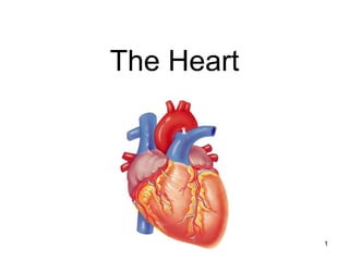
- 1. 1 The Heart
- 2. 2 Overview The right side receives oxygen-poor blood from the body and tissues and then pumps it to the lungs to pick up oxygen and dispel carbon dioxide Its left side receives oxygenated blood returning from the lungs and pumps this blood throughout the body to supply oxygen and nutrients to the body tissues The heart=a muscular double pump with 2 functions
- 3. 3 simplified… Cone shaped muscle Four chambers Two atria, two ventricles Double pump – the ventricles Two circulations Systemic circuit: blood vessels that transport blood to and from all the body tissues Pulmonary circuit: blood vessels that carry blood to and from the lungs
- 4. 4 Heart’s position in thorax
- 5. 5 Heart’s position in thorax In mediastinum – behind sternum and pointing left, lying on the diaphragm It weighs 250-350 gm (about 1 pound) Feel your heart beat at apex (this is of a person lying down)
- 6. 6 CXR (chest x ray) Normal male
- 7. 7 Chest x rays Normal female Lateral (male)
- 8. 8 Coverings of the heart: pericardium Three layered: (1) Fibrous pericardium Serous pericardium of layers (2) & (3) (2) Parietal layer of serous pericardium (3) Visceral layer of serous pericardium = epicardium: on heart and is part of its wall (Between the layers is pericardial cavity)
- 9. 9 How pericardium is formed around heart
- 10. 10 Layers of the heart wall Muscle of the heart with inner and outer membrane coverings Muscle of heart = “myocardium” The layers from out to in: Epicardium = visceral layer of serous pericardium Myocardium = the muscle Endocardium lining the chambers
- 11. 11 Layers of pericardium and heart wall
- 12. 12 Chambers of the heart sides are labeled in reference to the subject facing you Two atria Right atrium Left atrium Two ventricles Right ventricle Left ventricle --------------------------------------------------------------------------------
- 13. 13 Chambers of the heart divided by septae: Two atria-divided by interatrial septum Right atrium Left atrium Two ventricles- divided by interventricular septum Right ventricle Left ventricle
- 14. 14 Valves three tricuspid one bicuspid “Tricuspid” valve RA to RV Pulmonary or pulmonic valve RV to pulmonary trunk (branches R and L) Mitral valve (the bicuspid one) LA to LV Aortic valve LV to aorta (cusp means flap)
- 15. 15 Function of AV valves
- 16. 16 Function of semilunar valves (Aortic and pulmonic valves)
- 17. 17 Pattern of flow (simple to more detailed) Body RA RV Lungs LA LV Boby Body to right heart to lungs to left heart to body Body, then via vena cavas and coronary sinus to RA, to RV, then to lungs via pulmonary arteries, then to LA via pulmonary veins, to LV, then to body via aorta From body via SVC, IVC & coronary sinus to RA; then to RV through tricuspid valve; to lungs through pulmonic valve and via pulmonary arteries; to LA via pulmonary veins; to LV through mitral valve; to body via aortic valve then aorta
- 18. 18 Note positions of valves Valves open and close in response to pressure differences Trabeculae carnae Note papillary muscles, chordae tendinae (heart strings): keep valves from prolapsing (purpose of valve = 1 way flow)
- 19. 19 Relative thickness of muscular walls LV thicker than RV because it forces blood out against more resistance; the systemic circulation is much longer than the pulmonary circulation Atria are thin because ventricular filling is done by gravity, requiring little atrial effort
- 20. 20
- 21. 21
- 22. 22 Heartbeat Systole: contraction Diastole: filling Normal rate: 60-100 Slow: bradycardia Fast: tachycardia ***Note: blood goes to RA, then RV, then lungs, then LA, then LV, then body; but the fact that a given drop of blood passes through the heart chambers sequentially does not mean that the four chambers contract in that order; the 2 atria always contract together, followed by the simultaneous contraction of the 2 ventricles Definition: a single sequence of atrial contraction followed by ventricular contraction
- 23. 23 Heart sounds Called S1 and S2 S1 is the closing of AV (Mitral and Tricuspid) valves at the start of ventricular systole S2 is the closing of the semilunar (Aortic and Pulmonic) valves at the end of ventricular systole Separation easy to hear on inspiration therefore S2 referred to as A2 and P2 Murmurs: the sound of flow Can be normal Can be abnormal
- 24. 24 Places to auscultate Routine places are at right and left sternal border and at apex
- 25. 25 Cardiac muscle (microscopic) Automaticity: inherent rhythmicity of the muscle itself
- 26. 26 “EKG” (or ECG, electrocardiogram) Electrical depolarization is recorded on the body surface by up to 12 leads Pattern analyzed in each lead P wave=atrial depolarization QRS=ventricular depolarization T wave=ventricular repolarization
- 27. 27 Electrical conduction system: (Explanation in next slides) specialized cardiac muscle cells that carry impulses throughout the heart musculature, signaling the chambers to contract in the proper sequence
- 28. 28 Conduction system SA node (sinoatrial) In wall of RA Sets basic rate: 70-80 Is the normal pacemaker Impulse from SA to atria Impulse also to AV node via internodal pathway AV node In interatrial septum
- 29. 29 Conduction continued SA node through AV bundle (bundle of His) Into interventricular septum Divides R and L bundle branches become subendocardial branches (“Purkinje fibers”) Contraction begins at apex
- 30. 30
- 31. 31 12 lead EKG
- 33. 33 Autonomic innervation Sympathetic Increases rate and force of contractions Parasympathetic (branches of Vagus n.) Slows the heart rate http://education.med.nyu.edu/courses/old/physiology/courseware/ekg_pt1/EKGseq.html For a show on depolarization:
- 34. 34
- 35. 35 Embryological development during week 4 (helps to understand heart defects) Day 22, (b) in diagram, heart starts pumping (day 24) (day 28) (day 23)
- 36. 36 Normal and abnormal Congenital (means born with) abnormalities account for nearly half of all deaths from birth defects One of every 150 newborns has some congenital heart defect
- 37. 37 more…
- 38. 38 Use to study