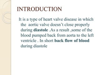
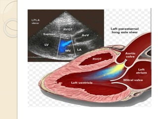
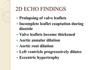
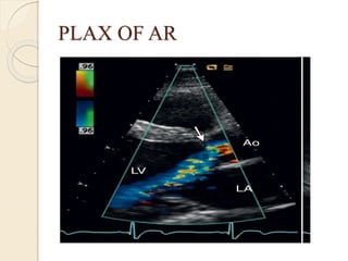
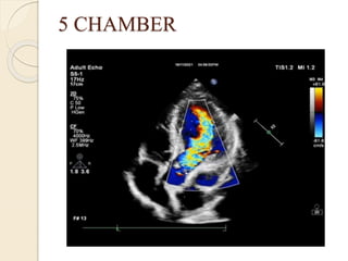
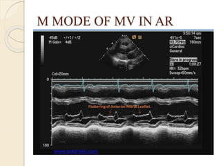
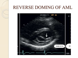
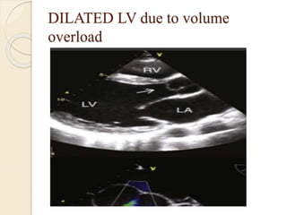
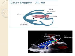
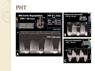
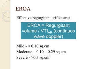
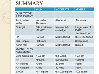
AORTIC
REGURGITATION
TEAM BY
ASHWATHI
BABINA MERLIN
ASWINI
DEPT OF CARDIOLOGY
3RD YEAR
INTRODUCTION
It is a type of heart valve disease in which
the aortic valve doesn’t close properly
during diastole .As a result ,some of the
blood pumped back from aorta to the left
ventricle . In short back flow of blood
during diastole
CAUSES
 Bicuspid aortic valve
Rheumatic
Calcification
Systemic inflammatory disease
Marfan syndrome
Endocarditis
Etc ….
SIGNS & SYMPTOMS
 Heart murmur
 Arrhythmia
 Palpitations
 Angina
 Shortness of breath
 Swollen of ankles and feet
ECHOCARDIOGRAPHIC
EVALUATION
ECHO WINDOWS USED :
• Parasternal long axis view
• Parasternal short axis view (Aortic valve
level)
• Apical five chamber view
• Apical three chamber view
2D ECHO FINDINGS
 Prolapsing of valve leaflets
 Incomplete leaflet coaptation during
diastole
 Valve leaflets become thickened
 Aortic annular dilation
 Aortic root dilation
 Left ventricle progresssively dilates
 Eccentric hypertrophy
PLAX OF AR
5 CHAMBER
INDIRECT SIGNS
 Increased EPSS :
maximal anterior motion of MV
in early diastole and the posterior motion
of IVS is >7mm or >1cm
 High frequency fluterring of AML
 Reversed diastolic curvature of the AML
 Jet lesion on septum or mitral valve
M MODE OF MV IN AR
REVERSE DOMING OF AML
DILATED LV due to volume
overload
LV RESPONSE TO AR
It occurs due to chronic volume overload
from AR
 Left ventricle inner cavity dilation and
becomes spherical
 LV end diastolic dimension 70-75 mm
 LV end systolic dimension 50-55mm
 LV ejection fraction becomes < 50%
EVALUATION OF AORTIC
REGURGITANT SEVERITY
COLOR FLOW IMAGING :
Parasternal views both in long and short
axis view and allow :
 Origin of the regurgitant jet
 Width of the jet
 Cross sectional area of the jet
Color imaging cont …..
 Mild AR
Fills small area of the outflow tract,
central jet fills <25%
 Moderate to Severe AR :
Fills larger percentage of outflow
tract diameter
Eccentric jet traverse the outflow tract
obliquely .
VENA CONTRACTA
Vena contracta is the narrowest diameter
of the flowstream which reflects the
diameter of regurgitant orifice
 Color flow image in PLAX
 Narrowest segment of the jet
 Measure perpendicular to the jet
VC
SEVERITY OF AR (Vena contracta)
SEVERITY VENA
CONTRACTA
MILD AR < 0.3 cm
MODERATE AR 0.3-0.7 cm
SEVERE AR >0.7 cm
CONTINUOUS WAVE DOPPLER
Continuous wave doppler signal of AR is
assessed by placing doppler line in apical
five chamber view across the aortic valve
It is used to compared the density of the
the envelope of the antegrade aortic flow
and the regurgitant jet
Continuous wave doppler cont …
 Mild AR
Velocity of the jet is remains high and
the envelope appears flat slope
 Severe AR
The doppler envelope shows steeper
slope
CW at Aortic Valve in AP5CH
PRESSURE HALF TIME
Doppler velocity- The diastolic deceleration
slope provides a semiquantitative measure
of AR severity
 A Flat slope consist with mild AR
( Pressure Half Time >500ms)
 A steeper slope indicates severe AR
( Pressure Half Time <200ms)
 For moderate AR PHT 500-200ms
PHT
PULSE WAVE DOPPLER
 In severe AR , holodiastolic flow
reversal in abdominal aorta is seen
 In addition to this more proximal
holodiastolic flow reversal is observed in
descending aorta during severe AR
It is also known as aortic flow reversal
AORTIC FLOW REVERSAL
CONTINUITY EQUATION
LVOT
SVLVOT = CSALVOT X VTILVOT
A1 X V1 = A2 X V2
CONTINUITY EQUATION
CONT….
MITRAL VALVE
SVMV = CSA MV X VTIMV
REGURGITANT VOLUME
 It is calculated by using transaortic volume
flow and transluminal volume flow
SV LVOT = CSA LVOT X VTI LVOT
SV MV = CSA MV X VTI LVOT
◦ Mild AR - < 30 ml
◦ Moderate AR - 30 to 44 ml
◦ Moderate to Severe – 45 to 59 ml
◦ Severe - > 60 ml
RV = SVLVOT – SVMV
REGURGITANT FRACTION
It is defined as the percentage of regurgitant
volume
 Mild - < 30%
 Moderate – 30 – 49%
 Severe - > 50%
RF = (Rvolume / SVLVOT) x
100
PISA
Proximal Isovelocity Surface Area :
blood moving back towards
closed aortic valve at the given aliasing
velocity
PISA = Regurgitant flow rate / Valiasing
EROA
Effective regurgitant orifice area
Mild - < 0.10 sq.cm
Moderate – 0.10 – 0.29 sq.cm
Severe - >0.3 sq.cm
EROA = Regurgitant
volume / VTIAR (continuos
wave doppler)
SUMMARY
MILD MODERATE SEVERE
QUALITATIVE
ASSESSMENT
Aortic valve
leaflet
Normal or
Abnormal
Abnormal Abnormal
Color jet Fills small area
of LVOT
Intermediate
central jet
Larger area of
LVOT
,eccenteric jet
LV Normal Mildly dilated Severely dilated
CW Doppler Flat slope --- Steep slope
Aortic root Normal Mildly dilated Dilated
QUANTITATIVE
ASSESSMENT
Vena Contracta < 0.3 cm 0.3-0.7cm >0.7 cm
PHT >500ms 500-200ms <200ms
AR Volume <30ml 30-59ml >60ml
AR Fraction <30% 30-49% >50%
EROA <0.1 sq.cm 0.1-0.29 sq.cm >0.3 sq.cm
THANK YOU FOR THE
ATTENTION ….

AR Aortic Regurgitation.pptx

  • 1.
  • 2.
    INTRODUCTION It is atype of heart valve disease in which the aortic valve doesn’t close properly during diastole .As a result ,some of the blood pumped back from aorta to the left ventricle . In short back flow of blood during diastole
  • 4.
    CAUSES  Bicuspid aorticvalve Rheumatic Calcification Systemic inflammatory disease Marfan syndrome Endocarditis Etc ….
  • 5.
    SIGNS & SYMPTOMS Heart murmur  Arrhythmia  Palpitations  Angina  Shortness of breath  Swollen of ankles and feet
  • 6.
    ECHOCARDIOGRAPHIC EVALUATION ECHO WINDOWS USED: • Parasternal long axis view • Parasternal short axis view (Aortic valve level) • Apical five chamber view • Apical three chamber view
  • 7.
    2D ECHO FINDINGS Prolapsing of valve leaflets  Incomplete leaflet coaptation during diastole  Valve leaflets become thickened  Aortic annular dilation  Aortic root dilation  Left ventricle progresssively dilates  Eccentric hypertrophy
  • 8.
  • 9.
  • 10.
    INDIRECT SIGNS  IncreasedEPSS : maximal anterior motion of MV in early diastole and the posterior motion of IVS is >7mm or >1cm  High frequency fluterring of AML  Reversed diastolic curvature of the AML  Jet lesion on septum or mitral valve
  • 11.
    M MODE OFMV IN AR
  • 12.
  • 13.
    DILATED LV dueto volume overload
  • 14.
    LV RESPONSE TOAR It occurs due to chronic volume overload from AR  Left ventricle inner cavity dilation and becomes spherical  LV end diastolic dimension 70-75 mm  LV end systolic dimension 50-55mm  LV ejection fraction becomes < 50%
  • 15.
    EVALUATION OF AORTIC REGURGITANTSEVERITY COLOR FLOW IMAGING : Parasternal views both in long and short axis view and allow :  Origin of the regurgitant jet  Width of the jet  Cross sectional area of the jet
  • 17.
    Color imaging cont…..  Mild AR Fills small area of the outflow tract, central jet fills <25%  Moderate to Severe AR : Fills larger percentage of outflow tract diameter Eccentric jet traverse the outflow tract obliquely .
  • 18.
    VENA CONTRACTA Vena contractais the narrowest diameter of the flowstream which reflects the diameter of regurgitant orifice  Color flow image in PLAX  Narrowest segment of the jet  Measure perpendicular to the jet
  • 19.
  • 20.
    SEVERITY OF AR(Vena contracta) SEVERITY VENA CONTRACTA MILD AR < 0.3 cm MODERATE AR 0.3-0.7 cm SEVERE AR >0.7 cm
  • 21.
    CONTINUOUS WAVE DOPPLER Continuouswave doppler signal of AR is assessed by placing doppler line in apical five chamber view across the aortic valve It is used to compared the density of the the envelope of the antegrade aortic flow and the regurgitant jet
  • 22.
    Continuous wave dopplercont …  Mild AR Velocity of the jet is remains high and the envelope appears flat slope  Severe AR The doppler envelope shows steeper slope
  • 23.
    CW at AorticValve in AP5CH
  • 24.
    PRESSURE HALF TIME Dopplervelocity- The diastolic deceleration slope provides a semiquantitative measure of AR severity  A Flat slope consist with mild AR ( Pressure Half Time >500ms)  A steeper slope indicates severe AR ( Pressure Half Time <200ms)  For moderate AR PHT 500-200ms
  • 25.
  • 26.
    PULSE WAVE DOPPLER In severe AR , holodiastolic flow reversal in abdominal aorta is seen  In addition to this more proximal holodiastolic flow reversal is observed in descending aorta during severe AR It is also known as aortic flow reversal
  • 27.
  • 28.
    CONTINUITY EQUATION LVOT SVLVOT =CSALVOT X VTILVOT A1 X V1 = A2 X V2
  • 29.
  • 30.
    REGURGITANT VOLUME  Itis calculated by using transaortic volume flow and transluminal volume flow SV LVOT = CSA LVOT X VTI LVOT SV MV = CSA MV X VTI LVOT ◦ Mild AR - < 30 ml ◦ Moderate AR - 30 to 44 ml ◦ Moderate to Severe – 45 to 59 ml ◦ Severe - > 60 ml RV = SVLVOT – SVMV
  • 31.
    REGURGITANT FRACTION It isdefined as the percentage of regurgitant volume  Mild - < 30%  Moderate – 30 – 49%  Severe - > 50% RF = (Rvolume / SVLVOT) x 100
  • 32.
    PISA Proximal Isovelocity SurfaceArea : blood moving back towards closed aortic valve at the given aliasing velocity PISA = Regurgitant flow rate / Valiasing
  • 33.
    EROA Effective regurgitant orificearea Mild - < 0.10 sq.cm Moderate – 0.10 – 0.29 sq.cm Severe - >0.3 sq.cm EROA = Regurgitant volume / VTIAR (continuos wave doppler)
  • 34.
    SUMMARY MILD MODERATE SEVERE QUALITATIVE ASSESSMENT Aorticvalve leaflet Normal or Abnormal Abnormal Abnormal Color jet Fills small area of LVOT Intermediate central jet Larger area of LVOT ,eccenteric jet LV Normal Mildly dilated Severely dilated CW Doppler Flat slope --- Steep slope Aortic root Normal Mildly dilated Dilated QUANTITATIVE ASSESSMENT Vena Contracta < 0.3 cm 0.3-0.7cm >0.7 cm PHT >500ms 500-200ms <200ms AR Volume <30ml 30-59ml >60ml AR Fraction <30% 30-49% >50% EROA <0.1 sq.cm 0.1-0.29 sq.cm >0.3 sq.cm
  • 35.
    THANK YOU FORTHE ATTENTION ….