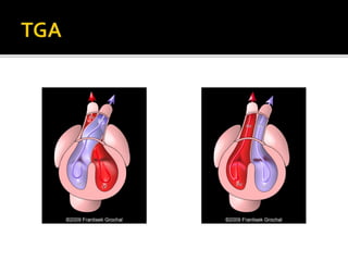
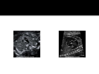
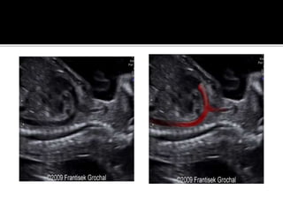
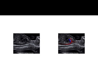
Generally occurs secondary to pulmonary atresia with intact IVS .
Pathophysiology- it develops because of a reduction in the blood flow secondary to inflow impedence from tricuspid atresia or outflow impedence from pulmonary arterial atresia .
Typical findings- a small , hypertrophic RV and a small or absent pulmonary artery
❤️ Chandigarh Call Girls☎️98151-579OO☎️ Call Girl service in Chandigarh ☎️ Ch...
The fetal heart
1.
2. Abnormal heart on screening ultrasound
Hydrops
Polyhydramnios
Fetal arrhythmia
Chromosomal anomalies
Extracardiac anomalies
Family history (CHD, syndromes associated with
CHD)
Maternal disease (diabetes, collagen vascular,
phenylketonuria)
Teratogen exposure
Increased nuchal translucency on first-trimester
screening
Monitoring response to intrauterine therapy
Monitoring fetus at risk for decompensation
(persistent tachyarrhythmia, hydrops)
3. Continuity of aorta (A) with left ventricle (LV);
LA, left atrium; RA, right atrium; RV, right ventricle.
L
A
RA
A
LV
RV
4. A, Normal position of the heart.The heart is predominantly in the left chest, with only the
right atrium in the right chest.There is a normal cardiac axis of
(dashed line) of 40 degrees from the midline (solid line).
5. B, Dextroposition of fetal heart caused by a large, congenital cystic adenomatoid
malformation.Transverse image through the fetal chest shows the heart displaced to the
right, but the apex remaining Leftward.
S
P
R
A
RV
LV
L
A
6. RA
L
V
SP
Four-chamber view of heart. A, Apical four chamber view shows the
interatrial and interventricular septa parallel to the angle of insonation
7. B, Subcostal four-chamber view shows the
interatrial
and interventricular septa perpendicular to the angle
of insonation.
8. C, Apical four-chamber view shows the two
superior pulmonary veins (P)
entering the left atrium (LA);
P
PLA SP
10. Short-axis view of ventricles. Anterior right
ventricle (RV) is normally slightly larger than the
left ventricle
(LV); IVS, interventricular septum.
RV
LV
IVS
11. 9. Short-axis view of great vessels. Aorta
(AO) in center with pulmonary artery (PA) draping anteriorly;
RVOT, right ventricular outflow tract; LA, left atrium; RA, right
atrium; PV, pulmonic valve; FF, foraminal flap; SP, spine.
AO
PARVO
T
RA LA
SP
FF
12. Apical five-chamber view shows continuity
of the aorta (A) with the left ventricle (LV); LA, left
atrium; RA,
right atrium; RV, right ventricle; SP, spine.
A
LV
LA
R
A
RV
13. Three-vessel and trachea view shows the
correct orientation of the main pulmonary artery–ductus arteriosus
confluence (P), the transverse aortic arch (A), and the superior
vena cava (S).This view also shows the two great vessels correctly
positioned on the left side of the trachea (T). SP, Spine.
P
A
S
SP
T
14. Normal aortic arch. Sagittal view shows
the rounded, “candy cane” appearance of aortic arch and
the head
and neck vessels arising from it; LS, left subclavian
artery; LC, left
carotid artery; I, innominate artery; AO, descending aorta.
A
O
LS
LC
I
15. Normal ductal arch. Sagittal view shows
pulmonary artery (PA) draping over the aorta (A) and joining the
ductus arteriosus (D), which then joins the descending aorta (AO);
LA, left atrium.
PA
DA
LA
AO
16. Using M-mode echocardiography
to analyze an arrhythmia: conducted premature
atrial contractions.The cursor is placed simultaneously
through the left ventricle (LV) and right atrium (RA).The
M-mode tracing shows normal atrial beats (A) followed by a
premature atrial contraction (PA).The ventricles show normal
ventricular contraction (V) following each atrial beat and a premature
beat (PV) following the premature atrial contraction. LA,
Left atrium; RV, right ventricle.
LV
R
A
A A A PA A
17. M-mode echocardiography. A, M-mode tracing through the
aortic root shows the aortic valve opening and
closing.The foraminal flap can be seen opening into the left
atrium (LA). RV, Right ventricle; AL, anterior leaflet of aortic
valve; PL,
posterior leaflet of aortic valve
AL
PL
AO
LA
RV
18. M-mode tracing shows opening and closing of the mitral
valve (MV) and tricuspid valve (TV); IVS,
interventricular septum.
TV
IVS
MV
19. Spectral Doppler ultrasound used
to interrogate a normal mitral valve. Spectral Doppler
sample volume is placed distal to the mitral valve in the left ventricle
(LV). A normal mitral valve waveform is appreciated above
the baseline, showing the normal early diastolic (E) and atrial
contraction (A) wave points. LA, Left atrium; RA, right atrium;
RV, right ventricle; SP, spine
RV LV
RA LA
E
A
20. Tricuspid insufficiency. Spectral
Doppler sample volume is placed proximal to the tricuspid valve
in the right atrium (RA).The regurgitant flow (R) can be seen
above the baseline.This implies that the valve has not closed
completely during systole, and therefore blood flow is retrograde
into the right atrium. RV, Right ventricle
R
R
RA
RV
21. Using color Doppler ultrasound to access normal blood
flow. A, Color Doppler ultrasound shows
normal flow through the pulmonary artery (PA).
PA
22. Color Doppler ultrasound to access normal blood flow through the aortic
arch (AA)
and descending aorta (D). Note that flow is continuous through the
descending aorta, but due to angle of 0 degrees in the middle of the
image, an artifact gives the appearance of narrowing, and the color of
flow changes from red to bluE
AA
D
23.
24. Results from an error in the amount of tissue
resorbed or deposited in the interatrial
septum .
5th m/c form of CHD.
m/c CHD in adults .
Incidence -1/1500 live births .
6.7% of CHD in live born infants .
F>M(2times )
25. Association with a variety of cardiac .
Extracardiac and chromosomal abnormalities .
Embryologically interatrial septum starts
developing at 4-6 weeks of gestation.
Septum primum
Ostium primum
Ostium secundum
Septum secundum
Foraminal flap
Foramen ovale
26. 1) OSTIUM SECUNDUM - >80% of all ASDs.
- caused by excessive resorption of the septum
primum ( foraminal flap ) or inadequate growth
of the septum secundum .
2) OSTIUM PRIMUM – 2nd m/c , located low in
the atrial septum near the AV valves .
- may occur in association with AV septal
defect.
3) SINUSVENOSUSASD – rare . A) sinus venosus
ASD OF svc b) sinus venosus ASD of IVC
27. diagnosis is difficult bcz Foramen ovale itself
presents as ASD which is a normal structure .
Foraminal flap is clearly visualised in four
chamber view which has a loose –pocket
configuration appearing either circular or linear
in shape as it opens into the left atrium .
The septum secundum and foramen ovale are
clearly visualised in four chamber view .
The max diameter of foramen ovale differs by
1mm or less from the aortic root diameter at allb
gestational ages .
28. Four-chamber view shows an ostium primum
atrial septal defect (arrow) in a fetus with an atrioventricular
septal defect;
L
A
RA
29. IsolatedVSD –m/c cardiac anomaly , accounts
for 30% of heart defects diagnosed in live
born infants and 9.7% diagnosed in utero .
Types –1)membranous -80%
perimembranous type when involves
adjacent muscular septum.
2)muscularVSD ( inlet ,trabecular and
outlet )
30. Sub costal four chamber view best to evaluate .
1/3rd cases are missed on usg if it is small .
Always evaluate further with color doppler .
VSD on color doppler – typically shows
bidirectional shunting with a systolic rigjht to
left shunt and a late diastolic left to right shunt .
PseudoVSD – in apical 4 chamber view
membranous portion may not be visualised
when angle of insonation is parallel to the
septum .
31. Muscular ventricular septal defect
(VSD). Subcostal four-chamber color Doppler ultrasound view
shows a muscularVSD (arrow) across the interventricular septum
(S); RA, right atrium; RV, right ventricle;
LA
RA
LV
RV
S
32. Spectrum of cardiac abnormalities involving
various degrees of deficiency of the interatrial
and interventricular septa & of the mitral and
tricuspid valves .
Cause – when the endocardial cushoins fail to
fuse properly so previously referred as
endocardial cushion defects or A-V canal defects
.
Almost 2/3rd of the fetuses have additional
cardiac anomalies .
33. About 1/3rd cases associated with left heart
isomerism ( both atria anatomically resemble
left atria ) and of these majority develop
complete heart block .
78% of AVSDs are associated with
chromosomal (trisomy 21 ) or extracardiac
anomalies .
34. COMPLETE
PARTIAL
1) COMPLETE – a single multileaflet valve is
present , variable amount of deficient tissue in
the Atrial and ventricular septa .
- 97% are of complete type .
2) INCOMPLETE – two of the leaflets (bridging
leaflets ) are connected by a narrow bridge of
tissue , resulting in the appearance of 2 valve
orifices . Associated with ostium primum ASD .
35. BALANCEDAVSDs – when theAV junction is
connected to both the ventricles such that blood flow
is evenly distributed .
UNBALANCEDAVSDs – if AV junction connected to
primarily one ventricle as in case of left heart
hypoplastic syndrome .
Sonographically , 4 chamber view is best to diagnose ,
better seen in diastole than systole.
Abnormal valve should be suspected when the normal
offset of theAV valves not seen .
Demonstration of 2 AV valves differentiate between
complete and incomplete forms .
36. Shows an open area of flow across the
atrioventricular septal defects + abnormal AV
valves .
Particulary useful in the detection of valvular
insufficiency .
HOLOSYSTOLICVALVULAR INSUFFICIENCY –
association with fetal hydrops , frequently a left
ventricular to right atrial jet can be identified
across the ostium primum defect before the
onset of HSVI.
37. CARDIAC MALFORMATIONS – septum
secundum ASD , hypoplastic left heart syndrome
,valvular PS , coarction of aorta ,TOF.
Chromosomal anomalies- 25-50% of the
affected fetuses .therefore , karyotyping is
indicated .
EXTRACARDIACANOMALIES – common and
include omphalocele, duodenal
atresia,tracheoesophageal atresia,facial
clefts,cystic hygroma,neural tube defects ,and
MCKDs.
38. Atrioventricular septal defect (AVSD).A, Apical four-chamber
view shows absent atrial septum, resulting
in a single, large atrium (RA-LA).AVSD is seen between the left
ventricle (LV) and right ventricle (RV). A single, multileaflet
atrioventricular
valve is also appreciated.
RA LA
39. Apical four-chamber color Doppler ultrasound
view shows the atrioventricular septal defect
RA LA
LVRV
40. INFERIOR DISPLACEMENT OFTHE
TRICUSPIDVALVE( +TETHERED
ATTACHMENT OFTHE LEAFLETS
+TRICUSPID DYSPLASIA +RIGHT
VENTRICULAR DYSPLASIA ) .
7% of cardiac anomalies , may be associated
with structural cardiac defects as pulmonary
atresia or stenosis, arrhythmias and
chromosomal anomalies .
41. Readily detected in utero.
Sonographic diagnosis rests on recognition of
apical displacement of the tricuspid valve into
the right ventricle- the most reliable sign ,an
enlaged RA containing a portion of the atrialised
RV ,and a reduction in the size of functional RV
.d/d- tricuspid valvular dysplasia.
It may cause substantial cardiac dysfunction in
utero frequently with hydrops ,cardiomegaly
and tachyarrhytmias .
42. Examine with spectral and color doppler USG
for demostrating tricuspid valve regugitation
.
Arrhythmias –SVT-common
43. LV
RV
LA
RA
Gray-scale image shows tricuspid valve (arrows) displaced
inferiorly, resulting in an “atrialized” right ventricle (RV) and
enlarged right atrium (RA); LV, left ventricle; LA, left atrium
44. Generally occurs secondary to pulmonary
atresia with intact IVS .
Pathophysiology- it develops because of a
reduction in the blood flow secondary to
inflow impedence from tricuspid atresia or
outflow impedence from pulmonary arterial
atresia .
Typical findings- a small , hypertrophic RV
and a small or absent pulmonary artery .
45.
46. Hypoplastic right ventricle. Apical
four-chamber view shows small, right ventricular chamber
(RV);
RA, right atrium; LV, left ventricle; LA, left atrium; SP, spine.
LV
L
A
RV
R
A
SP
47. 8% of all CHDs .
Small left ventricle due to decresed blood
flow in and out of left ventricle .
Primary abnormalities –aortic stenosis,aortic
atresias ,and mitral valve atresias .80% cases
associated with COARCTATION OF AORTA .
Color doppler shows absence of flow through
the mitral and aortic valves .
48. Hypoplastic left heart syndrome.
The left atrium (LA) and left ventricle (LV) are small; RA, right
atrium; RV, right ventricle; SP, spine.
RV
LV
SP
RA
LA
49. Two atria empty into a single ventricle via 2
A-V valves or a common AV valve .
2% of CHDs.
Occurs due to failure of the interventricular
septum to develop .
50. Consists of 1)VSD –PERIMEMBRANOUS
TYPE 2 ) OVERRIDING AORTA 3)
HYPERTROPHY OF RV (rarely seen inutero )
4) STENOSIS OFTHE RV OUTFLOWTRACT.
5-10% OF CHDs.
diagnosis has been made before 15 weeks
gestation usingTVS .
51. Tetralogy of Fallot.The aorta (AO) overrides
both the right ventricle (RV) and the left ventricle (LV). A
ventricular septal defect (arrows) is also appreciated. IVS,
Interventricular
septum.
RV
LV
IVS
AO
52. 1.3% of fetal cardiac anomalies .
Characterised by a single large vessel arising
from the base of the heart .almost all cases have
VSD.The truncal valve may have 2-6 cusps and
generally overrides the ventricular septum .
4 types by collett and edwards –
1) type 1 – has a pulmonary artery that bifurcates
into rt and lt branches after it arises from the
ascending portion of truncal vessel .
53. 2) type 2 – has rt and lt pul arteries arising
separately from the posterior truncus .
3) type 3- pul arteries that arise from the sides of
the proximal truncus .
4) type 4- has systemic collateral vessels from
the descending aorta as the source of flow .
the single large truncal artery with overriding of
the ventricular septum and an associatedVSD
identified on 4 chamber and outflow tract views
.
54. Truncus arteriosus.The single truncal
artery (TA) overrides both the right ventricle (RV) and left ventricle
(LV). A ventricular septal defect (arrow) is present. No
pulmonary artery was seen, helping to differentiate from
tetralogy
of Fallot. SP, Spine.
T
A
RV
LV
55.
56. <1% of all CHDs .
Occurs when more than 50% of both the
aorta and the pul artery arise from the RV .
Types -1) aorta post and to the right of the pul
artery
2) aorta and pul artery parallel with aorta to
the right ( taussig –bing type )
3) aorta and pul artery parallel with the aorta
anterior and to the left .
58. TYPES- 1) COMPLETE OR DEXTROTRANSPOSITION
(D-TGA) -80%
-AV concordance ( atria and ventricles are correctly
paired ) withVA discordance .
-further classified into 2 types depending on the
absence (70%) or presence ofVSD .+ pulmoary
stenosis .
2) CONGENITALLYCORRECTED OR
LEVOTRANSPOSITION (L-TGA) -20% .
IN both the types ,VENTRICULOARTERIAL (V-A)
DISCORDANCE is present ( aorta arises from RV and
pulmonary artery from LV)
59. Aorta origin – RV
-Receives – systemic blood
-Output to systemic circulation .
pulmonary artery –
origin –LV
receives – pulmonary venous blood
returns it to lungs .
Generally , the aortic root lies anterior and
slightly to the right of pul outflow tract .
60. The great vessels exit the heart in parallel
rather than crossing in normal fashion .
Optimally seen in a long axis or short axis
view of the great vessels .
In 3 vessel view only one great vessel (aorta )
is visualised .
61. A-V DISCORDANCEWITHV-A DISCORDANCE .
Aorta – origin – left sided morphologic RV and is
anterior and to the left of the pulmonary artery
Pulmonary artery – origin – right sided
morphologic left ventricle .
VSD AND PULMONIC STENOSIS –seen in 50%
of cases .
Malformation and inferior displacement of the
morphologic tricuspid valve may be present .
Pathophysiologically the blood flow is normal .
62. Complete transposition of great
arteries.The aorta (A) is anterior to the pulmonary artery (P).
This abnormal arrangement results in both vessels running parallel
to each other in this short-axis view.
A
P
63. Congenitally corrected transposition
of great arteries. An apical four-chamber view shows
the morphologic right ventricle (RV) and morphologic left ventricle
(LV) located on the incorrect sides of the heart.This is
evidenced by identifying the atrioventricular valve leaflet insertion
(arrow) on the left side of the heart in a more apical location than
the right-sided atrioventricular valve leaflet insertion.
RV LV
LA
RA
71. 1) PULVEINS ->VERTICALVEIN -> INNOMINATEV-> SVC
2) PULVEINS -> CORONARY SINUS -> RA
3) PULVEINS -> RA
4) PULVEINS -> PORTALVEIN -> IVCVIA DUCTUS
VENOSUS .
Embryologically thought to result from failure of
obliteration of the normal connections between the
primitive pulmonary vein and the splanchic
,umbilical,vitelline and cardinal veins .
TAPVR associated with AVSDs ,polysplenia,and asplenia
syndromes
72. Antenatal sonographic diagnosis is difficult bcz
the anomalous veins are extremely small and
variable in their course .
Often the first sign –mild right ventricular and
pulmonary arterial prominence
Color doppler –helpful in detecting and
following the anomalous connection
Spectral doppler for flow across the valves .a
ratio of right flow /left flow >2 times is highly
suspicious forTAPVR .
73. TAPVR- suspected when no pulmonary veins are seen
entering the left atrium .
A small LA bcz of decreased blood return + lack of
normal incorporation of the common pulmonary vein
into the left atrium –also suggestive ofTAPVR.
Approx 1/3rd pts have varied + cardiac anomalies .
Right atrium isomerism is common.
Extracardiac anomalies – gut malrotation ,and midline
liver and stomach .
PAPVR difficult to diagnose –can only be diagnosed
when pul veins are seen entering in both the atria.
74. Normal pulmonary venous anatomy. A, Four-chamber color
Doppler ultrasound view shows two superior
pulmonary veins (P) entering the left atrium (LA).
RV
LV
RA
LA
P
P
75. Subcostal four-chamber view using pulsed Doppler ultrasound
shows normal waveform
and direction of pulmonary venous flow into the left atrium
RA
LA
RV
LV
S D
76. Total anomalous pulmonary venous
return. Apical four-chamber view shows anomalous insertion of
all four pulmonary veins (P) into the right atrium (RA); RV, right
ventricle; LA, left atrium; LV, left ventricle.
LV
LA RA
RV
P
77. Narrowing of the aortic lumen usually occuring
between the insertion of the ductus arteriosus
and the left subclavian artery .
Severity ranges from a slight narrowing at the
distal end of the arch to severe hypoplasia of the
entire arch
6% prenatal incidence
90% cases associated with other cardiac
anomalies including abnormal aortic valve (
bicuspid or stenotic) ,VSD, DORV,AVSD,
Chromosomal abnormalities in 5%.
78. 3 embryologic theories to explain it –
1) a primary developmental defect with
failure of connection of the 4th and 6th aortic
arches with the deescending aorta
2) aberrant ductal tissue at the level of aortic
arch .
3)decreased blood flow through the aortic
isthmus .
Sonographic diagnosis is difficult
79. Ventricular size discrepancy with a prominent RV and relatively
small LV with a right to left ventricle diameter ratio > 2SD above
the norm suggests it .
A discrepancy in pulmonary artery to ascending aorta diameter
that falls >2SD above the normal ratio of 1.18 to0.06 suggestive of
coarctation .
Role of color doppler – detect the area of narrowing
Spectral doppler can identify increased flow distal to the narrowed
segment .
Many donot manifest before birth ,presents after birth due to
closure of ductus arteriosus .wait until 6-12 weeks after birth for
clinical and echocardiographic signs after closure of ductus
arteriosus .
If suspected follow upto 1 year of age .
80. Coarctation of aorta. Spectral Doppler
tracing shows increased velocity through the aortic arch.
81. Rare malformation in which cardia is located
outside the thoracic cavity .
Results from failure of fusion of lateral body fold
in the thoracic region .
4 types-
1.thoracic (60%)-the heart is displaced from the
thoracic cavity through the sternal defect .
2,abdominal (30%)-the heart is displaced into the
abdominal cavity through the diaphragmatic
defect .
82. 3)thoracoabdominal (7%)-the heart is
displaced from the chest through a defect in
the lower sternum with an associated
diaphragmatic or ventral abdominal wall
defect ( PENTOLOGY OF CANTRELL )
4) cervical (3%)- the heart is displaced into
the neck area .
84. MOST cases of ectopia cordis are isolated .
-Sonographic diagnosis can be made as early
as 10 weeks of gestation .
- carries a poor prognosis , most infants die
within few days after birth.
85. Thoracic ectopia cordis.The heart is
located outside the thorax; LA, left atrium; LV, left
ventricle;
RA, right ventricle; RV, right ventricle.
LV