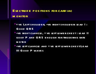
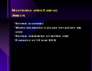
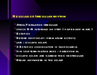
Cardiac monitoring and Rhythm recognition Rhythm in lead II ( most prominent P wave with sufficient amplitude to trigger the rate meter.
Electrode positions for cardiac monitor +ve Left shoulder, -ve right shoulder: lead I : Good QRS -ve right clavicle, +ve left lower chest : lead II good P and QRS enough for triggering rate meter -ve left clavicle and +ve left lower chest:Lead III Good P waves
Monitoring after Cardiac Arrests Rhythm monitores Watch for arching if jell and not jelpads are used Rhythm interpreted by rhythm strip Diagnosis by 12 lead ECG
How to read a rhythm strip ? Ventricular rate Rhythm QRS regular or irregular Is Atrial activity present Relationship Atrium vs Ventricle QRS width normal or prolonged
Ventricular rate < 60/min  Bradychardia > 100/min Tachycardia 1second = 5 large squares Number of large squares between beats . Divide into 300
Regular or Irregular rhythm Atrial Fibrillation irregular check R-R intervals on strip ( paper and mark ) Ectopics Before next beat : premature ectopic late : escape beats 3 Ectopics consecutive is tachycardia If in self terminating runs : paroxysmal escape beats are narrow then ventricular Broad anywhere in the heart
Is Atrial Activity present lead V1 will record the best recording of Atrial activity Slow rate : vagal stimulus or Adenosine Atrial flutter rate 300/min Irregularity of Atrial Fibrillation Shape of the P wave P upright II and AVF : sinoatrial node P wave inverted in II and AVF then retrograde conduction
Relationship Atrial and Ventricular rhythm 1st degree block : PR > 0.21 seconds 2nd Degree Block AV Wenckebach ( Mobits I ): progressive prolonging PR  until P dropped Mobitz II : blocked P wave without PR prolonging (*permanent pacing) Fixed 2:1 unlickely to progress  3rd Degree : Atrioventricular dissociation : Atrial Fibrillation
Conduction Ventricles RBBB: Broad QRS; R wave in V1 and secondary ST/T changes LBBB: Broad QRS; R in I and absence septal q inV6 ( R wave ) Hemiblock : LAHB:Absence of other causes of left axis deviation with LAD  LPHB: RAD
Causes Left Axis Deviation inferior MI Wolff-Parkinson-White syndrome Ostium primum ASD Hyperkalaemia
Causes Right Axis Deviation Right ventricular Hypertrophy Pulmonary Embolism Cor Pulmonale W-P-W syndrome Ostium secundum Anterolateral MI
Rhythms Associated with cardiac arrest  VF Asystole EMD / PEA Cardiac output loss severe bradycardias ventricular tachycardia
Ventricular Fibrillation ventricular myocardium depolarises at random. All coordination lost Confused polymorphic ventricular tachycardia ( torsades de pointes ) AF in the presence of AV nodal bypass
Polymorphic Ventricular Tachcardia tachycardia rate varies from beat to beat complexes have pointed appearances arise from background Long QT syndrome or abnormal T repolarisation Treatment : B blockers, magnesium and pacing  can initiate VF
Atrial Fibrillation with pre-excitation  rapid, broad achycardias variable QRS complexes W-P-W syndrome : QRS show delayed start More organised and lacks chaotic activity of variable amplitude
Ventricular Tachycardia broad complex tachycardia with rate 120 - 270/min QRS configuration constant
Asystole if suspected the gain controll should be turned up check not fine VF
Bradycardias 3rd Degree: Complete heart block : P =90/min or AF ;  QRS=20 - 50/min
Agonal rhythm slow, wide irregular ventricle complexes of varying morphology becomes broader before all electrical activity is lost

Rhythms

  • 1.
    Cardiac monitoring andRhythm recognition Rhythm in lead II ( most prominent P wave with sufficient amplitude to trigger the rate meter.
  • 2.
    Electrode positions forcardiac monitor +ve Left shoulder, -ve right shoulder: lead I : Good QRS -ve right clavicle, +ve left lower chest : lead II good P and QRS enough for triggering rate meter -ve left clavicle and +ve left lower chest:Lead III Good P waves
  • 3.
    Monitoring after CardiacArrests Rhythm monitores Watch for arching if jell and not jelpads are used Rhythm interpreted by rhythm strip Diagnosis by 12 lead ECG
  • 4.
    How to reada rhythm strip ? Ventricular rate Rhythm QRS regular or irregular Is Atrial activity present Relationship Atrium vs Ventricle QRS width normal or prolonged
  • 5.
    Ventricular rate <60/min Bradychardia > 100/min Tachycardia 1second = 5 large squares Number of large squares between beats . Divide into 300
  • 6.
    Regular or Irregularrhythm Atrial Fibrillation irregular check R-R intervals on strip ( paper and mark ) Ectopics Before next beat : premature ectopic late : escape beats 3 Ectopics consecutive is tachycardia If in self terminating runs : paroxysmal escape beats are narrow then ventricular Broad anywhere in the heart
  • 7.
    Is Atrial Activitypresent lead V1 will record the best recording of Atrial activity Slow rate : vagal stimulus or Adenosine Atrial flutter rate 300/min Irregularity of Atrial Fibrillation Shape of the P wave P upright II and AVF : sinoatrial node P wave inverted in II and AVF then retrograde conduction
  • 8.
    Relationship Atrial andVentricular rhythm 1st degree block : PR > 0.21 seconds 2nd Degree Block AV Wenckebach ( Mobits I ): progressive prolonging PR until P dropped Mobitz II : blocked P wave without PR prolonging (*permanent pacing) Fixed 2:1 unlickely to progress 3rd Degree : Atrioventricular dissociation : Atrial Fibrillation
  • 9.
    Conduction Ventricles RBBB:Broad QRS; R wave in V1 and secondary ST/T changes LBBB: Broad QRS; R in I and absence septal q inV6 ( R wave ) Hemiblock : LAHB:Absence of other causes of left axis deviation with LAD LPHB: RAD
  • 10.
    Causes Left AxisDeviation inferior MI Wolff-Parkinson-White syndrome Ostium primum ASD Hyperkalaemia
  • 11.
    Causes Right AxisDeviation Right ventricular Hypertrophy Pulmonary Embolism Cor Pulmonale W-P-W syndrome Ostium secundum Anterolateral MI
  • 12.
    Rhythms Associated withcardiac arrest VF Asystole EMD / PEA Cardiac output loss severe bradycardias ventricular tachycardia
  • 13.
    Ventricular Fibrillation ventricularmyocardium depolarises at random. All coordination lost Confused polymorphic ventricular tachycardia ( torsades de pointes ) AF in the presence of AV nodal bypass
  • 14.
    Polymorphic Ventricular Tachcardiatachycardia rate varies from beat to beat complexes have pointed appearances arise from background Long QT syndrome or abnormal T repolarisation Treatment : B blockers, magnesium and pacing can initiate VF
  • 15.
    Atrial Fibrillation withpre-excitation rapid, broad achycardias variable QRS complexes W-P-W syndrome : QRS show delayed start More organised and lacks chaotic activity of variable amplitude
  • 16.
    Ventricular Tachycardia broadcomplex tachycardia with rate 120 - 270/min QRS configuration constant
  • 17.
    Asystole if suspectedthe gain controll should be turned up check not fine VF
  • 18.
    Bradycardias 3rd Degree:Complete heart block : P =90/min or AF ; QRS=20 - 50/min
  • 19.
    Agonal rhythm slow,wide irregular ventricle complexes of varying morphology becomes broader before all electrical activity is lost