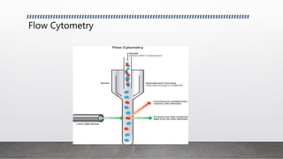
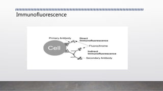
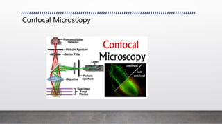
Advanced Techniques in
Animal Cell Culture
Kishan Sharma
M.Sc. 2nd Year
Biotechnology
Manipal University Jaipur
Introduction
• Cell culture refers to the process by which cells are grown in a controlled
artificial environment.
• Cells can be maintained in vitro outside of their original body by this process
which is quite simple compared to organ and tissue culture.
• In a animal cell culture technique, cells are removed from an animal, and grown
subsequently in a favourable environment.
Introduction
• For animal cell culture the cells are taken from the organ of an experimental
animal.
• The cells may be removed directly or by mechanical or enzymatic action.
• The cells can also be obtained by previously made cell line or cell strain.
• Examples of cells used to culture are lymphocytes, cells from cardiac and skeletal
tissues, cells from liver, breast, skin, and kidney and different types of tumor cells.
There are various techniques for animal cell culture
• Western Blotting
• Flow Cytometry
• Transfection
• Immunofluorescence
• Confocal Microscopy
Western Blotting
• Western blotting, also known as immunoblotting or protein blotting, is a core
technique in cell and molecular biology. In most basic terms, it is used to detect
the presence of a specific protein in a complex mixture extracted from cells or
tissue.
• Electrophoretic transfer of proteins from sodium dodecyl sulfate-polyacrylamide
gels to nitrocellulose membrane.
Flow Cytometry
• Flow cytometry is a scientific method used to measure and characterize cells in a
fluid as it passes through one or multiple lasers.
• In flow cytometry, cells are fluorescently labelled using antibodies conjugated to
fluorochromes, which emit light of different wavelengths.
• Common characteristics measured in a flow cytometry experiment are cell size,
relative granularity and relative fluorescence.
Flow Cytometry
Transfection
• Transfection is the process of introducing naked or purified nucleic acids into
cells.
• Transfection of animal cells typically involves opening transient pores in the cell
membrane to allow the uptake of material.
• Transfection can be carried out using calcium phosphate by electroporation or
by cell squeezing.
• introduction of genetic material, DNA or RNA, from a virus or bacteriophage into
cells, resulting in an infection.
Transfection
Immunofluorescence
• Immunofluorescence (IF) is a common laboratory technique that is used in evaluation
of cells in suspension, cultured cells and tissue for the detection of specific proteins.
• In Immunofluorescence techniques, antibodies are chemically conjugated to
fluorescent dyes such as fluorescein isothiocyanate (FITC) or tetramethyl rhodamine
isothiocyanate (TRITC).
• These labelled antibodies bind (directly or indirectly) to the antigen of interest which
allows for antigen detection through fluorescence techniques.
Immunofluorescence
Confocal Microscopy
• Confocal microscopy, most frequently confocal laser scanning microscopy (CLSM),
is an optical imaging technique for increasing optical resolution and contrast of a
micrograph by means of adding a spatial pinhole placed at the confocal plane of
the lens to eliminate out-of-focus light.
Confocal Microscopy
THANKYOU

Advanced techniques in animal cell culture

  • 1.
    Advanced Techniques in AnimalCell Culture Kishan Sharma M.Sc. 2nd Year Biotechnology Manipal University Jaipur
  • 2.
    Introduction • Cell culturerefers to the process by which cells are grown in a controlled artificial environment. • Cells can be maintained in vitro outside of their original body by this process which is quite simple compared to organ and tissue culture. • In a animal cell culture technique, cells are removed from an animal, and grown subsequently in a favourable environment.
  • 3.
    Introduction • For animalcell culture the cells are taken from the organ of an experimental animal. • The cells may be removed directly or by mechanical or enzymatic action. • The cells can also be obtained by previously made cell line or cell strain. • Examples of cells used to culture are lymphocytes, cells from cardiac and skeletal tissues, cells from liver, breast, skin, and kidney and different types of tumor cells.
  • 4.
    There are varioustechniques for animal cell culture • Western Blotting • Flow Cytometry • Transfection • Immunofluorescence • Confocal Microscopy
  • 5.
    Western Blotting • Westernblotting, also known as immunoblotting or protein blotting, is a core technique in cell and molecular biology. In most basic terms, it is used to detect the presence of a specific protein in a complex mixture extracted from cells or tissue. • Electrophoretic transfer of proteins from sodium dodecyl sulfate-polyacrylamide gels to nitrocellulose membrane.
  • 6.
    Flow Cytometry • Flowcytometry is a scientific method used to measure and characterize cells in a fluid as it passes through one or multiple lasers. • In flow cytometry, cells are fluorescently labelled using antibodies conjugated to fluorochromes, which emit light of different wavelengths. • Common characteristics measured in a flow cytometry experiment are cell size, relative granularity and relative fluorescence.
  • 7.
  • 8.
    Transfection • Transfection isthe process of introducing naked or purified nucleic acids into cells. • Transfection of animal cells typically involves opening transient pores in the cell membrane to allow the uptake of material. • Transfection can be carried out using calcium phosphate by electroporation or by cell squeezing. • introduction of genetic material, DNA or RNA, from a virus or bacteriophage into cells, resulting in an infection.
  • 9.
  • 10.
    Immunofluorescence • Immunofluorescence (IF)is a common laboratory technique that is used in evaluation of cells in suspension, cultured cells and tissue for the detection of specific proteins. • In Immunofluorescence techniques, antibodies are chemically conjugated to fluorescent dyes such as fluorescein isothiocyanate (FITC) or tetramethyl rhodamine isothiocyanate (TRITC). • These labelled antibodies bind (directly or indirectly) to the antigen of interest which allows for antigen detection through fluorescence techniques.
  • 11.
  • 12.
    Confocal Microscopy • Confocalmicroscopy, most frequently confocal laser scanning microscopy (CLSM), is an optical imaging technique for increasing optical resolution and contrast of a micrograph by means of adding a spatial pinhole placed at the confocal plane of the lens to eliminate out-of-focus light.
  • 13.
  • 14.