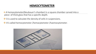
SUBMITTED BY-
AARTI PAL
M. PHARM (PHARMACOLOGY)
SUBMITTED TO-
MRS.PRABHJEET KAUR BAMRAH
(ASSISTANT PROFESSOR )
INSTITUTE OF PHARMACEUTICAL SCIENCES,KUK
A PRESENTATION ON CELL COUNTING, CRYOPRESERVATION,
CHARACTERIZATION OF CELLS AND THEIR APPLICATION
CONTENTS:
1. CELL COUNTING TECHNIQUE
a) Haemocytometer
b) Coulter counter method
2. CRYOPRESERVATION
3. CHARACTERIZATION OF CELLS
4. APPLICATION OF CELL CULTURE
5. REFERENCE
 A hemocytometer(Neubauer’s chamber) is a square chamber carved into a
piece of thick glass that has a specific depth.
 it is used to calculate the density of cells in suspensions.
 It’s called hemocytometer /hemacytometer /haemocytometer.
HEMOCYTOMETER
Hemo
Cyto
Meter
Blood
Cell
Measuring
Measuring blood cells
Invented by:
Louis-Charles Malassez
Hemocytometer:
Haemocytometers
 RBC Pipette
 WBC Pipette
 Neubauer chamber
 Cover slip
Neubauer chamber
PROCEDURE:
 Ensure cleanliness of all the instruments.
 Take diluting fluid (Turk’s for WBC and Hayem’s fluid for RBC).
 Finger prick
 Fill the pipette till 0.5 mark with 2nd drop of blood.
 Wipe the tip and fill the diluting fluid till marking 101 for RBC and
11 for WBC holding pipette horizontally.
 Roll the pipette horizontally between the palm to ensure mixing
of blood.
 Place coverslip and charge the Neubauer chamber after
discarding first few drops from pipette by touching edge of cover
slip.
 after 1 minute focus, under low power- high power and count the
cells.
Counting of viable cells and non-viable cells using Haemocytometer:
Cell Dye(Tryptan)
Non viable cells Viable cells
Intact membrane
permeability
Increased membrane
permeability
Stained blue Exclude permeation of dye,hence not stained
Loaded onto the Haemocytometer Viable cells counted
Counting of anchorage dependent cells using haemocytometer:
Anchorage dependent cells
Citric acid (0.1mol/L)
+
Crystal violet (0.1%)
Cells in the sample firmly attached to the substratum
Cell lysis
Release of nuclei
Stained purple
Cells counted using Haemocytometer
Counting Rules:
COULTER COUNTER METHOD
A popular instrument for measuring the volume of
particles is the Coulter counter.
Instrument measures particle volume which can be
expressed as dx: the diameter of a sphere that has the
same volume as the particle.
Principle:
This instrument operates on the principal that when a
particle suspended in a conducting liquid passes
through a small orifice on either side of which are
electrodes ; a change in electric resistance occurs.
.
 As the particle travels through the orifice, it displaces its own volume of electrolyte and
this result in an increased resistance between the two electrodes.
 The change in resistance which is related to the particle volume causes a voltage pulse
that is amplified and fed to a pulse height analyzer calibrated in term of particle size.
 The instrument record electronically all those particles producing pulses that are within
two threshold value of the Analyzer .
 By systematically varying the threshold settings and counting the number of particles in a
constant sample size , it is possible to obtain a particle -size distribution.
 The instrument is capable of counting particles at the rate of approximately 4000 per
second , and So both Gross Count and particle size distribution are obtained in a relatively
Working:
 In practice ,a known volume of a dilute suspension is pumped through the
orifice.
 Provided the suspension is sufficiently dilute the particle passed through
essentially one at a time.
 A constant voltage is applied across the electrode to produce a current.
CRYOPRESERVATION:
Cryo is Greek word. (krayos - frost)
It literally means preservation in "frozen state."
The principle to bring plant cells or tissue to a zero metabolism and non - dividing
state by reducing the temperature in the presence of cryoprotectant.
It can be done :
Over solid carbon dioxide (at -79 degree)
Low temperature deep freezer (at -80 degree)
In vapor phase nitrogen (at -150 degree)
In liquid nitrogen (at -196 degree)
◦
1. Cryopreservation - generally involves storage in liquid nitrogen.
2. Cold storage - it involves storage in low and non freezing
temperature.
3. Low pressure - it involves partially reducing the atmospheric
pressure of surrounding.
4. Low oxygen storage - it involves reducing the oxygen level but
maintaining the pressure.
Various methods of storage:
Methods of Cryopreservation:
Application
Freezing of cell cultures.
Maintenance of disease free stock
Seed Bank
 Gene Bank
Storage of rare germplasm.
Characterization of cell lines:
1. Monitoring of cell lines for genetic stability: on repeated subculturing there is a
tendency that cell lines may undergo genetic changes moreover the cell may get
contaminated or cross contaminated with some other cells. for example :HeLa cells grow
faster than most of the cells so if cell culture get contaminated by HeLa cells, heLa will
outnumber the originally intended cells. Hence, characterization of cell lines is necessary
to ensure the growth of normal and desired cell lines. The method used for
characterization of cell lines are:
A. Karyotyping: Using this method a change in the chromosomes or any damage inflicted to the
chromosomes can be analyzed . Karyotype refer to the distribution of chromosomes ,which
is a characteristics feature of every species.
The step involved in karyotyping include:
(a) The Growth of cells is optimized
(b) The cells are blocked in the mitosis stage by addition of Colchicine which act as a mitotic
inhibitor these halt the cell division and cells are arrested in metaphase .
(c) cells are smeared on the slide and allowed to dry.
(d) Suitable dye such as Giemsa or acetic orcein is used to stain the chromosomes
(e) The chromosome are then counted under the microscope. A photomicrograph may
also be taken.
(f) Fixative agent, methanol: acetic acid (3:1 v/v) is added .
(g) The separation between chromosomes is maximized by putting hypotonic solution,
which causes swelling of cells.
B. Obtaining pattern of Isozymes(Zymography):
The pattern analysis of isozymes for certain enzymes can be used to establish the identity of
cell lines. all isozymes of a given enzyme are separated using gel electrophoresis or
Zymogram (banding pattern) is obtained .Zymogram is a characteristic feature of every cell
line. Generally, glucose 6 phosphate dehydrogenase , lactate dehydrogenase , and nucleoside
phosphorylase are employed to obtain Zymogram.
C. Fluorescent labeled antibodies:
Fluorescent labeled antibody, which is specific to the antigens present on the cell
membrane can be used to identify a cell line. for example ; fluorescein isothiocyanate is a
fluorescent dye, which is attached to antibody. under microscope, the cells with
fluorochrome on their surface shows fluorescence and can thus be identified.
2. Monitoring cell lines for cell contamination : cell culture are at high risk of getting
contaminated with both chemical substances as well as microorganism .
(a) chemical contamination : it is comparatively more difficult to detect due to
invisibility of the chemical in the cultures the most common sources of chemical
contamination include – endotoxins, plasticizers, metals ion , chemical disinfectant ,etc.
(b) biological contamination : the animal culture media are highly susceptible to
biological contaminant microorganisms. such as: yeast, Bacteria ,and fungi however
these contamination are easy to identify because of visible effect produced by these
microorganism which include changes in turbidity and PH of the culture.
Applications of Animal cell culture:
Reference:
D A Gilbert et.al "Application of DNA fingerprints for cell-line individualization"Am J Hum Genet. 1990
September; 47(3): 499-514
Freshney R.lan, "Culture of Animal Cells: A Manual of Basic Technique"(2010):239-260
J.G.Dauwerse, J.Wiegant, A.K.Raap, M.H.Breuning and G.J.B.van Ommen "Multiple colors by fluorescence in
situ hybridization using ratio-labelled DNA probes create a molecular karyotype" Human Molecular Genetics,
Vol. 1, No.8: 593-598
Thomas Ried, Evelin Schlock, Yi Ning and Johannes Wienberg "Chromosome painting: a useful art" Human
Molecular Genetics, 1998, Vol. 7, No. 10 Review 1619-1626.
https://www.slideshare.net/Vijayanarasimha1/cell-lines

haemocytometer (AARTI PAL).pptx M Pharmacy

  • 1.
    SUBMITTED BY- AARTI PAL M.PHARM (PHARMACOLOGY) SUBMITTED TO- MRS.PRABHJEET KAUR BAMRAH (ASSISTANT PROFESSOR ) INSTITUTE OF PHARMACEUTICAL SCIENCES,KUK A PRESENTATION ON CELL COUNTING, CRYOPRESERVATION, CHARACTERIZATION OF CELLS AND THEIR APPLICATION
  • 2.
    CONTENTS: 1. CELL COUNTINGTECHNIQUE a) Haemocytometer b) Coulter counter method 2. CRYOPRESERVATION 3. CHARACTERIZATION OF CELLS 4. APPLICATION OF CELL CULTURE 5. REFERENCE
  • 3.
     A hemocytometer(Neubauer’schamber) is a square chamber carved into a piece of thick glass that has a specific depth.  it is used to calculate the density of cells in suspensions.  It’s called hemocytometer /hemacytometer /haemocytometer. HEMOCYTOMETER
  • 4.
  • 5.
    Haemocytometers  RBC Pipette WBC Pipette  Neubauer chamber  Cover slip
  • 6.
  • 7.
    PROCEDURE:  Ensure cleanlinessof all the instruments.  Take diluting fluid (Turk’s for WBC and Hayem’s fluid for RBC).  Finger prick  Fill the pipette till 0.5 mark with 2nd drop of blood.  Wipe the tip and fill the diluting fluid till marking 101 for RBC and 11 for WBC holding pipette horizontally.  Roll the pipette horizontally between the palm to ensure mixing of blood.  Place coverslip and charge the Neubauer chamber after discarding first few drops from pipette by touching edge of cover slip.  after 1 minute focus, under low power- high power and count the cells.
  • 8.
    Counting of viablecells and non-viable cells using Haemocytometer: Cell Dye(Tryptan) Non viable cells Viable cells Intact membrane permeability Increased membrane permeability Stained blue Exclude permeation of dye,hence not stained Loaded onto the Haemocytometer Viable cells counted
  • 9.
    Counting of anchoragedependent cells using haemocytometer: Anchorage dependent cells Citric acid (0.1mol/L) + Crystal violet (0.1%) Cells in the sample firmly attached to the substratum Cell lysis Release of nuclei Stained purple Cells counted using Haemocytometer
  • 10.
  • 11.
    COULTER COUNTER METHOD Apopular instrument for measuring the volume of particles is the Coulter counter. Instrument measures particle volume which can be expressed as dx: the diameter of a sphere that has the same volume as the particle. Principle: This instrument operates on the principal that when a particle suspended in a conducting liquid passes through a small orifice on either side of which are electrodes ; a change in electric resistance occurs. .
  • 12.
     As theparticle travels through the orifice, it displaces its own volume of electrolyte and this result in an increased resistance between the two electrodes.  The change in resistance which is related to the particle volume causes a voltage pulse that is amplified and fed to a pulse height analyzer calibrated in term of particle size.  The instrument record electronically all those particles producing pulses that are within two threshold value of the Analyzer .  By systematically varying the threshold settings and counting the number of particles in a constant sample size , it is possible to obtain a particle -size distribution.  The instrument is capable of counting particles at the rate of approximately 4000 per second , and So both Gross Count and particle size distribution are obtained in a relatively Working:  In practice ,a known volume of a dilute suspension is pumped through the orifice.  Provided the suspension is sufficiently dilute the particle passed through essentially one at a time.  A constant voltage is applied across the electrode to produce a current.
  • 13.
    CRYOPRESERVATION: Cryo is Greekword. (krayos - frost) It literally means preservation in "frozen state." The principle to bring plant cells or tissue to a zero metabolism and non - dividing state by reducing the temperature in the presence of cryoprotectant. It can be done : Over solid carbon dioxide (at -79 degree) Low temperature deep freezer (at -80 degree) In vapor phase nitrogen (at -150 degree) In liquid nitrogen (at -196 degree)
  • 14.
    ◦ 1. Cryopreservation -generally involves storage in liquid nitrogen. 2. Cold storage - it involves storage in low and non freezing temperature. 3. Low pressure - it involves partially reducing the atmospheric pressure of surrounding. 4. Low oxygen storage - it involves reducing the oxygen level but maintaining the pressure. Various methods of storage:
  • 15.
  • 21.
    Application Freezing of cellcultures. Maintenance of disease free stock Seed Bank  Gene Bank Storage of rare germplasm.
  • 22.
    Characterization of celllines: 1. Monitoring of cell lines for genetic stability: on repeated subculturing there is a tendency that cell lines may undergo genetic changes moreover the cell may get contaminated or cross contaminated with some other cells. for example :HeLa cells grow faster than most of the cells so if cell culture get contaminated by HeLa cells, heLa will outnumber the originally intended cells. Hence, characterization of cell lines is necessary to ensure the growth of normal and desired cell lines. The method used for characterization of cell lines are: A. Karyotyping: Using this method a change in the chromosomes or any damage inflicted to the chromosomes can be analyzed . Karyotype refer to the distribution of chromosomes ,which is a characteristics feature of every species. The step involved in karyotyping include: (a) The Growth of cells is optimized (b) The cells are blocked in the mitosis stage by addition of Colchicine which act as a mitotic inhibitor these halt the cell division and cells are arrested in metaphase . (c) cells are smeared on the slide and allowed to dry.
  • 23.
    (d) Suitable dyesuch as Giemsa or acetic orcein is used to stain the chromosomes (e) The chromosome are then counted under the microscope. A photomicrograph may also be taken. (f) Fixative agent, methanol: acetic acid (3:1 v/v) is added . (g) The separation between chromosomes is maximized by putting hypotonic solution, which causes swelling of cells. B. Obtaining pattern of Isozymes(Zymography): The pattern analysis of isozymes for certain enzymes can be used to establish the identity of cell lines. all isozymes of a given enzyme are separated using gel electrophoresis or Zymogram (banding pattern) is obtained .Zymogram is a characteristic feature of every cell line. Generally, glucose 6 phosphate dehydrogenase , lactate dehydrogenase , and nucleoside phosphorylase are employed to obtain Zymogram.
  • 24.
    C. Fluorescent labeledantibodies: Fluorescent labeled antibody, which is specific to the antigens present on the cell membrane can be used to identify a cell line. for example ; fluorescein isothiocyanate is a fluorescent dye, which is attached to antibody. under microscope, the cells with fluorochrome on their surface shows fluorescence and can thus be identified. 2. Monitoring cell lines for cell contamination : cell culture are at high risk of getting contaminated with both chemical substances as well as microorganism . (a) chemical contamination : it is comparatively more difficult to detect due to invisibility of the chemical in the cultures the most common sources of chemical contamination include – endotoxins, plasticizers, metals ion , chemical disinfectant ,etc. (b) biological contamination : the animal culture media are highly susceptible to biological contaminant microorganisms. such as: yeast, Bacteria ,and fungi however these contamination are easy to identify because of visible effect produced by these microorganism which include changes in turbidity and PH of the culture.
  • 25.
  • 26.
    Reference: D A Gilbertet.al "Application of DNA fingerprints for cell-line individualization"Am J Hum Genet. 1990 September; 47(3): 499-514 Freshney R.lan, "Culture of Animal Cells: A Manual of Basic Technique"(2010):239-260 J.G.Dauwerse, J.Wiegant, A.K.Raap, M.H.Breuning and G.J.B.van Ommen "Multiple colors by fluorescence in situ hybridization using ratio-labelled DNA probes create a molecular karyotype" Human Molecular Genetics, Vol. 1, No.8: 593-598 Thomas Ried, Evelin Schlock, Yi Ning and Johannes Wienberg "Chromosome painting: a useful art" Human Molecular Genetics, 1998, Vol. 7, No. 10 Review 1619-1626. https://www.slideshare.net/Vijayanarasimha1/cell-lines