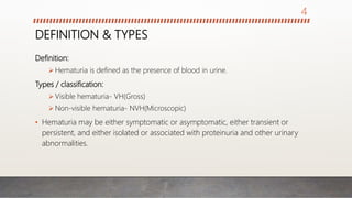
4. DEFINITION & TYPES
Definition:
Hematuria is defined as the presence of blood in urine.
Types / classification:
Visible hematuria- VH(Gross)
Non-visible hematuria- NVH(Microscopic)
• Hematuria may be either symptomatic or asymptomatic, either transient or
persistent, and either isolated or associated with proteinuria and other urinary
abnormalities.
4
5. DEFINITION & TYPES
CLASSIFICATION BASED ON TIMING
Initial hematuria: presence of visible blood in the initial portion of urinary stream
during micturition. Usually of a urethral pathology.
Terminal hematuria: presence of blood in the terminal portion of urinary stream
during micturition. Usually of a bladder neck pathology.
Total hematuria: presence of blood in the entire portion of urinary stream during
micturition. Usually of kidney, ureter, bladder and prostate pathology.
5
7. OTHER CAUSES OF RED URINE
Several medications and food can cause red/orange discolouration of the urine;
DRUGS;
Rifampicin,
Isoniazid,
Phenazopyridine,
Chlorpromazine,
Thioridazine,
Laxatives containing a phenolphthalein component.
FOODS;
Beetroot, Fava beans, Carotene-containing food (e.g. carrots, winter squash)
7
8. HISTORY/SYMPTOMS
Duration:
• When did the bleeding start?
• Continuous or intermittent?
Colour:
• Bright red /dark red?
• Are there clots in it?
• Is it a small amount or profuse?
Blood thinning agents:
• Anticoagulants like heparin & warfarin?
• Antiplatelet agents like aspirin and
clopidogrel?
Timing:
• Initial →Urethral pathology
• Total → bladder or upper tract
• Terminal →prostate or bladder neck
Pain:
• Painful →stones and obstruction
• Painless→ Renal and bladder tumors
Drugs & food:
• Drugs→ Rifampicin, Pyridium
• Food → Beetroot, Rhubarb
Associated symptoms:
• Frequency/nocturia/dysuria
8
9. PHYSICAL EXAM/SIGNS
Abdominal examination:
• Kidney mass→Bimanually palpable
• Bladder mass→ usually not palpable,
but the bladder itself may be palpable
secondary to urinary retention.
• Renal angle tenderness→ may be
positive in kidney pathology
Digital Rectal Examination:
• For prostate pathologies
• An exquisitely tender prostate is a
feature of prostatitis
• A smooth enlarged prostate is benign
prostatic hyperplasia
• A hard craggy prostate is probably
malignant→ Ca Prostate.
Vaginal examination in females:
• Reveal posterior bladder wall tumor
9
10. INVESTIGATIONS
• Decision making in investigating hematuria is usually guided by the history and
findings on physical examination.
• However there are 4 traditional tests in investigating hematuria.
Renal ultrasound
Urine cytology
Intravenous urography
Cystoscopy
• What informs these 4 traditional tests ?
20-25% of patients with macroscopic hematuria has urological cancer.
10
11. INVESTIGATIONS
5-10% of patients with microscopic haematuria has urological cancer.
• Renal ultrasound and intravenous urography are good in detecting upper tract
malignancy, while urine cytology and cystoscopy are for the lower tract
malignant lesions.
• Others tests:
CT Scan
Ureteroscopy: if no cause of hematuria is found after the 4T Tests
Retrograde pyelography: as above
Renal Angiography: for suspected case of vascular malformation
11
12. OTHER ANCILLARY INVESTIGATIONS
Urine examination:
• Dipstick; shows minute traces of blood in the
urine, presence of protein and nitrites from
bacteria breakdown of urea, suggesting
infection
• Microscopy & culture; Microscopy confirms
presence of RBCs, WBCs and organisms in
infection. Culture confirms the organism and its
antibiotic sensitivity
Urine Cytology:
• Urothelial tumors shed cells into the urine that
can be seen on staining and microscopy. Early
morning urine specimen.
Full blood count:
• Anemia in severe hematuria
• Polycythemia in Renal cell carcinoma because of
release of erythropoietin
Urea & Electrolytes:
• Renal failure will not occur until 60% of kidney function
is lost, so normal urea & electrolyte does not rule out
kidney damage.
PSA: Prostsate specific antigen
• Up to 4ngm/ml→ Normal
• 5 to 10ngm/ml → BPH & 25% chance of Ca prostate
• >10ngm/ml → BPH & 50% chance of Ca prostate
• >30ngm/ml → 80% chance of Ca prostate.
12
13. TREATMENT
GOALS:
To stabilize the patient.
Prevent and or treat infection.
Adequate monitoring of the patient.
Establish the cause of the bleeding.
Stop the bleeding.
Treat the cause.
13
14. TREATMENT
• Stabilize the patient.
If hematuria is significant, secure intravenous access, collect blood samples for
ancillary tests and ensure hemodynamic stability.
Catheterize the patient for better monitoring with a wide bore 3-way catheter. This
also prevents clot retention with saline irrigation.
• Treat and prevent infection with antibiotics.
• Monitor vital signs regularly, packed cell volume and transfuse if necessary.
• Find the cause of the bleeding by investigations and treat accordingly.
14
15. CONCLUSION & REFERENCES
• Hematuria is a urological emergency
that needs to be evaluated to know
the underlying cause.
• Malignancy should be ruled out in
the case of gross hematuria.
REFERNCES
• Bailey and love’s short practice of surgery
27th edition
• Slideshare.net/urological emergencies
15
18. INTRODUCTION
• A urologic emergency refers to any urologic condition that requires urgent
medical attention from a urologist or an emergency room and immediate
treatment.
• Compared to other surgical fields, there are relatively few emergencies in
urology.
• It includes different conditions ranging from kidney injury to testicular torsion.
18
20. URETERIC COLIC
• One of the commonest urological emergencies
• Most often due to passage of stone formed in the kidney, down through the
ureter.
• It is a common cause of acute abdomen
• The chief cause of pain in the urinary tract is distention from increased
intraluminal pressure.
20
21. URETERIC COLIC
INTRALUMINAL OBSTRUCTION
Impacted stone
Blood clot
Mucosal edema
INTRAMURAL OBSTRUCTION
Stricture
malignancy
EXTRAMURAL OBSTRUCTION
Ureteral ligation
Local extension of Ca prostate or Ca
cervix causing pressure on one or
both ureteric orifices
Compression from an abscess or
inflammatory mass
Pelvic hematoma following trauma
Retroperitoneal fibrosis
Pregnancy
ETIOLOGY
21
22. URETERIC COLIC
PRESENTATION
• The classic presentation for a patient with ureteric stone is the sudden onset of
severe colicky pain, originating in the flank and radiating inferiorly and anteriorly.
• The patient cannot get comfortable, and may roll around in agony.
• Associated with nausea or vomiting, usually afebrile
22
24. URETERIC COLIC
INVESTIGATION
• FBC
• Urinalysis
• Urine M/C/S
• Pregnancy test
• Urea & electrolyte
• KUB (Kidneys, ureters, bladder scan)
• IVU
• CT
TREATMENT
• Pain relief
NSAIDS
Narcotic analgesics
• IV access & adequate hydration
• Anti emetics if there is severe vomiting
• Antibiotics if there is fever or you suspect
an infection
24
25. URETERIC COLIC
• Watchful waiting with analgesics &
hydration supplements
95% of stones measuring 5mm or less pass
spontaneously
• Indications for intervention to relieve
obstruction:
Pain unrelieved by analgesics
Signs and symptoms of sepsis
Persistent nausea & vomiting
Bilateral ureteral obstruction
Solitary kidney
• Temporary relief of the obstruction:
Insertion of a JJ stent or percutaneous
nephrostomy tube
• Definitive treatment of a ureteric stone
ESWL (Extracorporeal shock
wave lithotripsy)
Ureteroscope
PCNL (Percutaneous nephrolithotomy)
Open surgery
25
26. ACUTE SCROTUM
• It is an emergency situation reguiring
prompt evaluation, differential
diagnosis, and sometimes immediate
surgical exploration.
Differential diagnosis of acute scrotum:
Testicular torsion
Epididymo-orchitis
Epididymitis
Orchitis
Torsion of testicular appendages
Strangulated hernia
26
27. TESTICULAR TORSION
• A testicular torsion is a twist of the spermatic cord resulting in strangulation of
the blood supply to the testis and epididymis.
• If not treated emergently (within 4 to 6 hours after onset of pain), complete
infarction of the testis results, followed by atrophy of the testis.
• Testicular salvage decreases as duration of torsion increases
• Testicular torsion occurs most frequently between the ages of 10-30 (peak
incidence 13-15 years of age), but any age group may be affected.
27
28. TESTICULAR TORSION
• 50% of cases of torsion occur during sleep.
Testicular torsion is broadly classified into two
types.
Extra-vaginal.
• This form is seen in neonates.
• The entire testis and tunica twist in a vertical axis
on the spermatic cord as a consequence of
incomplete fixation of the gubernaculum to the
scrotal wall, which allows free rotation within the
scrotum.
Intra-vaginal (Bell-clapper)
• This form is more commonly found in
adolescents and adults.
• Occurs when the testicle rotates on the
spermatic cord within the tunica vaginalis
• Because this anomaly is bilateral, there is a
significant risk for a contralateral metachronous
torsion.
• Spasm of the cremaster muscle causes the
right testis to rotate clockwise and the left
counterclockwise as observed from the foot of
the bed.
28
30. TESTICULAR TORSION
PRESENTATION
• Sudden onset of severe scrotal pain
• Pain is referred to ipsilateral lower quadrant of abdomen
• Nausea & vomiting
• Mild fever
• There may be dysuria but other urinary symptoms are usually absent
• There is quite often a history of previous, brief episodes of similar pain, with
spontaneous resolution.
30
31. TESTICULAR TORSION
EXAMINATION
• The testis is usually slightly swollen and
very tender to touch.
• It may be high-riding and may be in a
horizontal position due to twisting of the
cord.
• Prehn's sign, although not always
reliable.
• It is necessary to consider an acutely
painful swollen testis in an adolescent
as torsion until it is proven otherwise at
surgery.
• Scrotal exploration takes precedence
over all forms of investigations.
Investigation
• Doppler ultrasound
• Radionuclide scanning
31
32. TESTICULAR TORSION
TREATMENT
• Scrotal exploration should be undertaken as a matter of urgency.
• Option depends on findings at exploration.
• Bilateral orchidopexy if the affected testis is still viable.
• Orchidectomy of the nonviable testis and orchidopexy of the contralateral testis
if the affected testis is gangrenous.
32
33. ACUTE URINARY RETENTION
• Painful inability to void, with relief of pain following drainage of the bladder by
catheterization.
PATHOPHYSIOLOGY:
Low bladder pressure (i.e. impaired bladder contractility)
Increased urethral resistance (i.e. bladder outlet obstruction (BOO)
Interruption of sensory or motor innervation of bladder
Central failure of coordination of bladder contraction with external sphincter
relaxation
33
34. ACUTE URINARY RETENTION
IN MEN IN WOMEN BOTH
• BOO
• Urethral stricture
• Acute urethritis
• Acute prostatitis
• Phimosis
• Malignant enlargement of
prostate.
• Prostatic abscess
• Pelvic prolapse (cystocele,
rectocele, uterine).
• Urethral stricture.
• Urethral diverticulum.
• Post-surgery for stress
incontinence.
• Pelvic masses (e.g. ovarian
masses).
• Blood clot
• Urethral calculus
• Rupture of urethra
• Neurogenic
• Fecal impaction
• Spinal anesthesia
ETIOLOGY
34
35. ACUTE URINARY RETENTION
MANAGEMENT
• Central to the diagnosis is the
presence of a large volume of urine,
which when drained by
catheterization, leads to resolution of
the pain.
• Volumes of 500-800ml are typical.
Volumes <500ml should lead one to
question the diagnosis. Volumes
>800ml may be defined as acute-on-
chronic retention.
Initial management
Urethral catheterization to relieve pain
Suprapubic catheterization if urethral
route not possible.
Definitive management
Treatment of the primary cause after
appropriate investigations.
35
36. PRIAPISM
• Priapism is a painful persistent prolonged erection not related to sexual
stimulation, lasting >4-6hours.
• Incidence has two peaks, at ages 5-10years and 20-50 years.
• The two broad categories of priapism are:
Low flow (most common)
High flow
36
37. PRIAPISM
• Low-flow (ischemic):
Due to veno-occlusion
Manifests as a painful, rigid erection, with
absent or low cavernosal blood flow.
Ischaemic priapism beyond 4hrs requires
emergency intervention.
Blood gas analysis shows hypoxia and
acidosis.
Etiology: hematological diseases, malignant
infiltration of the corpora cavernosa (e.g.
advanced bladder cancer), or drugs.
• High-flow (non ischemic)
Due to unregulated arterial blood flow
Manifests as painless, semi-rigid erection.
Blood gas analysis shows similar results to
arterial blood.
Etiology: perineal trauma which creates an
arteriovenous fistula.
37
38. PRIAPISM
MANAGEMENT
• The diagnosis of priapsim is usually obvious from the history and examination of
the erect, tender penis (in low-flow priapism)
• Blood testing:
Full blood count
Genotype to exclude sickle cell
Cavernous blood samples to determine type of priapism.
• Color Doppler ultrasound scan of cavernosal artery and corpora cavernosa.
Reduced blood flow in ischaemic priapism; ruptured artery with pooling of
blood around injured area in non-ischaemic priapism.
38
39. PRIAPISM
TREATMENT
LOW FLOW PRIAPISM
MEDICAL:
Aspiration of blood from corpora, and
intracavernosal injection of alpha-
adrenergic agonist (phenylephrine 10mg
in 19ml saline, injected in 0.5-1ml aliquots
every 5 min until detumescence occurs).
Monitor BP and pulse during drug.
Use of antiandrogen: indicated only in
patients with recurrent or stuttering
priapism with no sexual concerns.
Oral Terbutaline: used in the early
phase of priapism caused by
intracavernosal injection. Efficacy not
certain.
39
40. PRIAPISM
SURGICAL
Distal cavernosal glandular shunt
I. Winter’s procedure
II. Ebbehoj procedure
III. Al-Ghorab procedure.
Proximal shunt procedure.
HIGH FLOW PRIAPISM (Not an
emergency)
Early stages may respond to a cool
bath or icepack (causing vasospasm
and arterial thrombosis).
Delayed presentations require
arteriography and embolization of the
internal pudendal artery.
40
41. PHIMOSIS
• Occurs only in uncircumcised male patients.
• It is defined as the failure of retraction of the foreskin.
• The patient usually complains of erythema, itching, or pain on intercourse. Most
commonly, there is a mild associated infection (balanoposthitis).
• Treatment
1. Treat infection with antibiotics.
2. Dorsal slit
3. Circumcision
41
42. PARAPHIMOSIS
• A condition in which the foreskin becomes trapped in a retracted position
behind the glans.
• Most commonly, this occurs in a patient with preexisting phimosis.
• With time, the entrapped foreskin becomes edematous, and the glans itself
becomes engorged with severe pain and tenderness.
42
43. PARAPHIMOSIS
TREATMENT
• Ice bags for firm compression of the glans, gentle manual reduction and
injection of a solution of hyaluronidase in normal saline may help reduce the
swelling
• If manipulation fails, incision of the constricting ring under local anesthesia
should be performed.
• Once the inflammation and edema have subsided (3 to 4 days), elective
circumcision is indicated.
43
44. FOURNIER’S GANGRENE
• Necrotizing fascitis of external genitalia, perineal or perianal region
• Polymicrobial from GIT or GU
• Affects all ages and both genders
• Life threatening with a mortality rate of 13-22%
• 50-60% of infections stem from lower GIT or GU source
• Risk factors: HIV, DM, alcohol, perineal trauma etc
• Organisms include E.coli, bacteroides and staphylococci
• Most likely culprit for an infection of colorectal origin is clostridium.
44
45. FOURNIER’S GANGRENE
PATHOPHYSIOLOGY
It begins locally with skin infection and spreads
down the fascial plane
Its results into inflammation and ischaemia then
necrosis later
Low oxygen content and necrosis potentiate the
effects of the anaerobic bacteria and cause
rapid dissemination of the infection.
DIAGNOSIS
Patients present with genital induration,
pain, erythema and crepitus
Diagnosis is straightforward when the
lesions are found
Plain radiographs or CT may demonstrate
air in the perineal tissues
Retrograde urethrogram reveals
suspected periurethral infection
Perirectal infection source suspected,
proctoscopy may be revealing
45
46. FOURNIER’S GANGRENE
MANAGEMENT
• It relies on aggressive medical and surgical approach
• Rapid fluid resuscitation and broad-spectrum antibiotics
• Surgical debridement, aggressive wound care and redebridement
• Suprapubic catheter and faecal diversion may be needed
• Genital skin is highly elastic and grafts are not required unless over 60% of the
skin is removed.
46
47. RENAL ARTERIAL EMBOLI
• The main renal arteries are most frequently involved by systemic emboli from
the left atrium in association with atrial fibrillation, artificial heart valves, the
vegetations of endocarditis, or a mural thrombus from a myocardial infarct.
• Iatrogenic emboli are being increasingly seen because of the widespread use of
invasive vascular procedures.
• The intrarenal arteries are end arteries, so their occlusion leads to a wedge-
shaped infarction of the renal parenchyma.
47
48. RENAL ARTERIAL EMBOLI
• SYMPTOMS
Acute flank pain; radiates to the groin
Nausea and vomiting
Fever
Hematuria ( gross or microscopic )
• It could be asymptomatic.
48
49. RENAL ARTERIAL EMBOLI
INVESTIGATIONS
Contrast enhanced CT scan; If it fails to
visualize all or part of the kidney in a
patient with a vascular or cardiac problem
raises the suspicion.
Selective renal angiography; confirms the
diagnosis.
Glutamic oxaloacetic transaminase level,
followed by a prolonged elevation of
lactate dehydrogenase suggests renal
infarction.
TREATMENT
The treatment of choice is systemic
anticoagulant (heparin).
Intra-arterial fibrinolytic agents
(streptokinase), if instituted promptly
within 4 to 6 hours, can lead to a
significant recovery of renal function.
49
50. RENAL VEIN THROMBOSIS
• Renal vein thrombosis is rare and primarily observed in children with severe
dehydration or in adults with nephrotic syndrome, renal tumours, or
hypercoagulable states and after surgery or trauma to the renal vessels.
• Thrombosis of the longer left renal vein may also involve ureteric, gonadal,
adrenal and phrenic branches that drain into the left renal vein.
50
51. RENAL VEIN THROMBOSIS
DIAGNOSIS
Urinalysis: Gross or microscopic
hematuria caused by focal renal
infarction is invariably found.
Full blood count: Thrombocytopenia
is also a consistent finding in the
acute setting, and its absence
should make one suspect the renal
vein thrombosis is resolving.
CT scan shows a large kidney with
delayed or absent enhancement of
the parenchyma.
USS usually shows an enlarged
hypoechoic kidney with a renal vein
or vena cava thrombus.
Selective renal venography remains
the definitive test.
51
52. RENAL VEIN THROMBOSIS
TREATMENT
• In adults, early heparinization and selective intravenous fibrinolysis
(streptokinase or urokinase) have yielded promising results.
• Surgical thrombectomy is reserved for cava thrombosis.
• In a small subset of patients, nonfunction, renal hypertension, or chronic renal
infection may necessitate delayed nephrectomy.
52
53. MALIGNANT URETERIC OBSTRUCTION
• Locally advanced prostate cancer, bladder or ureteric cancer may cause
unilateral or bilateral ureteric obstruction. Locally advanced non-urological
malignancies can also obstruct the ureters (e.g. cervical cancer, rectal cancer,
lymphoma).
• The patient presents either with symptoms and signs of renal failure, or anuria
without a palpable bladder.
53
54. MALIGNANT URETERIC OBSTRUCTION
Investigation
Renal ultrasound will demonstrate
bilateral hydronephrosis and an empty
bladder.
Treatment
Short term treatment
• Correct life-threatening hyperkalaemia
• Bilateral percutaneous nephrostomy or
• Bilateral ureteric stenting.
Long term treatment
• Urinary diversion by formation of ileal
conduit.
• Ureteric re-implantation.
• Insertion of short permanent metallic
ureteric stents.
54
55. CONCLUSION & REFERENCES
• Early intervention is necessary to
prevent further progession and
reduce the rate of mortalities
REFERENCES
• Bailey and love’s short practice of surgery
27th edition
• Slideshare.net/urological emergencies
55
Editor's Notes
Although the maneuver is not always reliable, elevation of the testicle increases pain in torsion and decreases pain in epididymorchitis
Stuttering (intermittent) priapism is a recurrent form of ischemic priapism in which unwanted painful erections occur repeatedly with intervening periods of detumescence. This historical term identifies a patient whose pattern of recurrent ischemic priapism encourages the clinician to seek options for prevention of future episodes.
WINTER’S PROCEDURE; The procedure creates a shunt between the engorged corpora cavernosa and the corpus spongiosum of the glans penis. The blood is now able to drain away more freely resulting in detumescence.