Downloaded 136 times


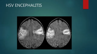


This document discusses acute encephalitis in India. It defines acute encephalitis and acute encephalitis syndrome. Japanese encephalitis virus is a major cause of AES in India, transmitted via Culex mosquitoes between pigs, birds and humans. The document outlines the epidemiology, clinical features, diagnosis and management of AES. It emphasizes the importance of vaccination and vector control in prevention and control of AES in India.