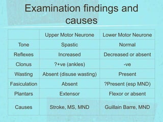
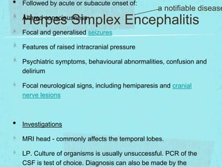
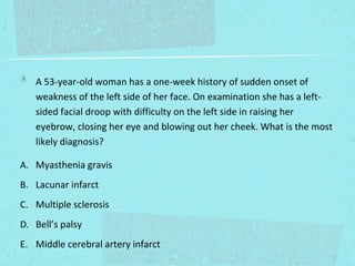
Trickier Neurology
Clinical Syndromes for the AMK
By Olivia Jagger
Academic Education F2
Examination findings and
causes
Upper Motor Neurone

Lower Motor Neurone

Tone

Spastic

Normal

Reflexes

Increased

Decreased or absent

Clonus

?+ve (ankles)

-ve

Wasting

Absent (disuse wasting)

Present

Fasiculation

Absent

?Present (esp MND)

Plantars

Extensor

Flexor or absent

Causes

Stroke, MS, MND

Guillain Barre, MND
A 34-year old woman presents to A+E with a two day history of
increasing headache and confusion. She has a temperature of 38.2
degrees. Whilst in the emergency department she has a tonic-clonic
seizure. MRI shows increased signal in the temporal lobes on T2
imaging. What is the most likely cause?
A. Alcohol intoxication
B. Severe pneumonia
C. Viral encephalitis
D. Subarachnoid haemorrhage
E. Hepatic encephalopathy
•

Followed by acute or subacute onset of:

a notifiable disease

Herpes Simplex Encephalitis

Altered consciousness

Focal and generalised seizures
Features of raised intracranial pressure

Psychiatric symptoms, behavioural abnormalities, confusion and
delirium
Focal neurological signs, including hemiparesis and cranial
nerve lesions

•

Investigations
MRI head - commonly affects the temporal lobes.

LP. Culture of organisms is usually unsuccessful. PCR of the
CSF is test of choice. Diagnosis can also be made by the
A 53-year-old woman has a one-week history of sudden onset of
weakness of the left side of her face. On examination she has a leftsided facial droop with difficulty on the left side in raising her
eyebrow, closing her eye and blowing out her cheek. What is the most
likely diagnosis?
A. Myasthenia gravis
B. Lacunar infarct
C. Multiple sclerosis
D. Bell’s palsy
E. Middle cerebral artery infarct
A 51 year old gentleman is referred by his GP for worsening back
pain. He works as an electrician and is concerned that he is unable to
work due to the pain. On questioning, he has had 'niggling pain' in his
lower back for a few months but this morning, 4 hours prior to
presentation he notes that all of a sudden the pain became far worse,
very sharp in nature with no radiation. He notes that he wants to pass
urine but feels unable to and is complaining of saddle anaesthesia.

•

Which is the most appropriate diagnosis?

sciatica
mechanical back pain
cauda equina syndrome
syringomyelia
prolapsed disc
A 51 year old gentleman is referred by his GP for worsening back
pain. He works as an electrician and is concerned that he is unable to
work due to the pain. On questioning, he has had 'niggling pain' in his
lower back for a few months but this morning, 4 hours prior to
presentation he notes that all of a sudden the pain became far worse,
very sharp in nature with no radiation. He notes that he wants to pass
urine but feels unable to and is complaining of saddle anaesthesia.

•

Which is the most appropriate diagnosis?
Thoraco-lumbar spine Xrays
MRI spine
Bloods including erythrocyte sedimentation rate and C-reactive
protein
CT head
Nerve conduction studies
Cauda Equina - neurological
emergency!
Commonest cause is central disc prolapse

Compression of central sacral or lumbar roots
Sciatica

Loss of bladder / bowel control / sexual dysfunction
Saddle sensory loss

Perianal sensory loss and sphincter disturbance
Lower limb weakness (decreased/lost reflexes,
paraesthesia)
Urgent lumbar MRI and surgical referral for decompression
•

•

A 25-year-old man develops sudden weakness of the legs followed a
few days later by weakness in the arms associated with ‘pins and
needles’. He is normally well apart from 3 days of diarrhoea about 2
weeks before. On examination he has weakness in all four limbs and
his reflexes are absent.

What is the most appropriate diagnosis?

Peripheral neuropathy
Brainstem lesion

Spinal Cord Compression
Guillain-Barre syndrome
Brown-Sequard syndrome
•

•

A 25-year-old man develops sudden weakness of the legs followed a
few days later by weakness in the arms associated with ‘pins and
needles’. He is normally well apart from 3 days of diarrhoea about 2
weeks before. On examination he has weakness in all four limbs and
his reflexes are absent.

What is the most appropriate investigation?

Vital Capacity
CSF

Viral blood screen
Nerve conduction tests
Chest Xray
Guillain-Barre Syndrome Presentation
Autoimmue attack v myelin antigens
60% have antecedent infection (EBV, CMV, campylobacter)

Syndrome about 2 weeks later
Sensory symptoms in toes (paraesthesia), followed by
weakness in legs (areflexia and later wasting)
Weakness ascends over 10-14 days (can involve face and
respiratory muscles)
can be associated with back pain
GBS - Investigations and
Management
CSF: raised protein, absence of WBCs
Nerve conduction studies normal in early stages
? recent infection (viral screen)
Monitor Vital Capacity (+ / - ventilation)
Best nursed on HDU / ITU
ECG (occ heart block)
IVIg if evolving significant weakness
Analgesia
5% mortality; 10% permanent disability
A 67-year-old woman had a 3 month history of weakness of his left
leg and a 1 month history of weakness in both arms. He felt things
were getting progressively worse. On examination he had wasting
and fasciculations in all four arms. He was weak in all four limbs with
brisk reflexes throughout with upgoing plantars. He had a normal
sensory examination. What is the most likely diagnosis?

•

What is the most appropriate diagnosis?
Parkinson’s disease
Multiple sclerosis
Motor neurone disease
Myasthenia gravis
Huntington’s Chorea
Motor Neurone Disease - Signs
Combination of UMN and LMN signs (selective of MOTOR
neurones)
Wasting / weakness small muscles hands (LMN)
Fasiculations upper limbs / shoulders (LMN)

leg weakness / brisk reflexes / extensor plantars (spastic) (UMN)
Bulbar / pseudobulbar palsy - affects speech / swallow
(LMN/UMN)
Motor Neurone Disease Diagnosis and Management
Diagnosis mainly clinical (Absence of sensory symptoms / signs,
CK moderately increased, NCS show denervation)
Aetiology unknown, 10% dominantly inherited

Life expectancy on average 4 years
Management - supportive (SALT, OT, PEG, +/- Riluzole)
A 25-year-old woman develops a painful visual disturbance in her left
eye. She had previously had an episode of ataxia a year earlier lasting
a few months.

What is the most appropriate diagnosis?
Parkinson’s disease
Multiple sclerosis
Motor neurone disease
Myasthenia gravis
Huntington’s Chorea
Multiple Sclerosis

Inflammatory demyelinating CNS disorder - Disease of the white matter

relapsing/remitting, secondary progressive, primary progressive

Acute attack:

-

Optic neuritis (optic nerve)

-

Transverse myelitis (spinal cord)

-

Diplopia (brainstem)
Late disease: spastic paraplegia, incontinance, dementia, cerebella
ataxia, bilateral intranuclear opthalmoplegia
Multiple Sclerosis

Investigations: Brain MRI / LP (oligoclonal banding)

Acute Management: steroids
Disease modifying agents: Betainterferons, Natalizumab

Other treatments: PT, OT, antispasmotic agents (baclofen),
??Vit D
A 50-year-old woman presents with difficulty keeping her head
up. She has proximal weakness in the arms which is fatigable.
She comments that he speech becomes very slurred towards the
end of the day and she is finding it difficult to swallow.

•

What is the most appropriate diagnosis?
Stroke
Parkinson’s disease

Multiple Sclerosis
Myasthenia Gravis

Huntington’s disease
Myasthenia Gravis - presentation

• Aetiology
✴Autoimmune condition. Antibodies attack the postsynaptic

acetylcholine receptors at the neuromuscular junction, leading to
decreased conduction at NMJ and muscle fatigue/ weakness.

✴Disease has a fluctuating pattern of ‘crises’ and remittances.
✴Very strong association with disorders of the THYMUS. In 75% of
patients with Myasthenia Gravis there is hyperplasia of the thymus,
and in 10% this becomes a thymoma.

• Symptoms
✴Weakness in muscles, particularly the ocular, bulbar, proximal
skeletal and axial. (Respiratory muscles can be involved).

✴Fatigability is a key symptom!
day.

Symptoms get worse at the end of the
•Investigations
MG -tests for serum acetylcholine receptor antibodies
Investigation / Treatment
✴Blood
✴Nerve conduction studies
✴CT/ MRI scans - to image the thymus
✴Vital Capacity (Spirometry / peak flow) may need respiratory
support

✴Ice cube test - place over ptosis for 2mins, eye opens. Though
that low temperatures improve neuromuscular transmission.

• Treatment
✴Acetylcholinesterase inhibitors (neostigime) increase the amount
of Acetylcholinesterase available to functioning receptors.
A 66-year-old man is finding that he has more difficulty getting up
and moving about for the past year. He is annoyed by a tremor in
his hands, but the tremor goes away when he performs routine
tasks using his hands. His friends remark that he seems more
sullen and doesn't smile at them, but only stares with a fixed
expression on his face. He has not suffered any loss of mental
ability.

•

Which of the following conditions is he most likely to have?
Parkinson’s disease
Recurrent TIA’s
Huntington’s disease
Myasthenia Gravis
Motor Neurone Disease
Parkinsons - clinical features

Pathology: loss of dopaminergic neurons from the substania
nigra. Surviving neurons often contain Lewy bodies (collections
of proteins - α-synuclein).

Aetiology: most commonly idiopathic (can be drug induced and
several genetic ‘parkinsons plus’ syndromes)
Clinical features: TRIAD 1) resting tremor 2) rigidity 3)
bradykinesia
Other typical signs/symptoms: Festinating gait - stooped,
shuffling, narrow based, slow to start then accelerates, with
swinging arms, Hypomimia (and reduced blinking rate - glabellar
tap), Hypographia, Depression, Hallucinations, Lewy body
dementia, Anosmia.
Power and reflexes are normal (extra-pyramidal)
Parkinsons - treatment

Dopamine cannot cross the BBB, but its precursor L-DOPA can,
and once it has, it is converted to dopamine by DOPA
decarboxylase. Acts by increasing levels of free dopamine in the
brain.

Short half-life. Therefore, L-Dopa is given with a decarboxylase
inhibitor (carbidopa, entacapone or benserazide) to prevent
peripheral breakdown and prolong the half-life of L-dopa,
reducing the dose needed by about 90%!

•
•
•
•
•

Side Effects
Efficacy decreases over time
‘On-off effect’ at the end of the dose
Nausea / GI Upset
Dyskinesias
Other neuro conditions you
should be familiar with ...
✴ Unilateral
✴ Visual disturbance – zigzag lines
Migraine - Classical Features
✴ Changes in sensation of face
✴ Vomiting/nausea/abdominal pain
✴ Aura/halo
✴ Daytime onset
✴ Relieved by sleep
✴ Have to lie in a quiet, dark room (photophobia and noise adversion)
✴ FH migraine
✴ Are they on COCP? (CI in migraines)
✴ Can have focal signs (i.e. arm/leg weakness)
•

Meningitis

a notifiable disease!

Signs and Symptoms
Photophobia
Stiff neck
Rash (non-blanching)

Treatment

•Start antibiotics immediately if suspecte
•At GP/in ambulance - IM Benzylpenicilli
•At Hospital - IV Ceftriaxone

Systemically unwell
Contact with meningitis

•

Investigations
CT head prior to LP to ensure no raised intracranial pressure
LP - increased protein (inflammation) and WCC (infection) and
low glucose (gobbled up by bacteria). May culture an organism
(commonly Neisseria meningitidis)
✴ Occipital
✴ SEVERE - peak intensity within seconds
Subarachnoid Haematoma
✴ Associated vomiting
✴ Occasionally seizures
✴ FH of berry aneurysms (related to Polycystic Kidneys)
• Investigation
✴ CT angiogram
✴ LP (if normal CT) looking for xanthochromia broken down RBC in
CSF

•

Treatment:
✴Worse in the mornings
Space-occupying lesion
✴Night time wakening
✴Worse when lying flat / coughing / bending down
✴Associated with vomiting
✴Visual changes
✴Facial nerve palsy
✴Seizures
✴Papilloedema
• Tumours
✴Abnormal movements – abnormal gait/loss of coordination
•

Bells Palsy
Signs and Symptoms
Rapid onset unilateral facial nerve weakness.
Patient unable to show teeth, crew up eye and raise eyebrows on affected
side
Severe cases may also present with notable loss of taste sensation
(anterior 2/3 of tongue), intolerance of high-pitched noises, mild dysarthria.
Lower Motor Neuron Signs (LMN)

Can be distinguished from an UMN lesion (e.g. a stroke) by testing if the
forehead is affected

◦ Forehead normal – UMN lesion – due to bilateral cortical representation
and innervation of the forehead

◦ Forehead affected – LMN lesion
Good Luck!
Remember, some of the possible answers you will
know and be able to exclude / rule in
Think what signs the question scenario is describing
UMN or LMN
Some extra reading, look up Stroke and Epilepsy.
Any questions to olivia.jagger@nhs.net

Neurology mc qs

  • 1.
    Trickier Neurology Clinical Syndromesfor the AMK By Olivia Jagger Academic Education F2
  • 2.
    Examination findings and causes UpperMotor Neurone Lower Motor Neurone Tone Spastic Normal Reflexes Increased Decreased or absent Clonus ?+ve (ankles) -ve Wasting Absent (disuse wasting) Present Fasiculation Absent ?Present (esp MND) Plantars Extensor Flexor or absent Causes Stroke, MS, MND Guillain Barre, MND
  • 3.
    A 34-year oldwoman presents to A+E with a two day history of increasing headache and confusion. She has a temperature of 38.2 degrees. Whilst in the emergency department she has a tonic-clonic seizure. MRI shows increased signal in the temporal lobes on T2 imaging. What is the most likely cause? A. Alcohol intoxication B. Severe pneumonia C. Viral encephalitis D. Subarachnoid haemorrhage E. Hepatic encephalopathy
  • 4.
    • Followed by acuteor subacute onset of: a notifiable disease Herpes Simplex Encephalitis Altered consciousness Focal and generalised seizures Features of raised intracranial pressure Psychiatric symptoms, behavioural abnormalities, confusion and delirium Focal neurological signs, including hemiparesis and cranial nerve lesions • Investigations MRI head - commonly affects the temporal lobes. LP. Culture of organisms is usually unsuccessful. PCR of the CSF is test of choice. Diagnosis can also be made by the
  • 5.
    A 53-year-old womanhas a one-week history of sudden onset of weakness of the left side of her face. On examination she has a leftsided facial droop with difficulty on the left side in raising her eyebrow, closing her eye and blowing out her cheek. What is the most likely diagnosis? A. Myasthenia gravis B. Lacunar infarct C. Multiple sclerosis D. Bell’s palsy E. Middle cerebral artery infarct
  • 6.
    A 51 yearold gentleman is referred by his GP for worsening back pain. He works as an electrician and is concerned that he is unable to work due to the pain. On questioning, he has had 'niggling pain' in his lower back for a few months but this morning, 4 hours prior to presentation he notes that all of a sudden the pain became far worse, very sharp in nature with no radiation. He notes that he wants to pass urine but feels unable to and is complaining of saddle anaesthesia. • Which is the most appropriate diagnosis? sciatica mechanical back pain cauda equina syndrome syringomyelia prolapsed disc
  • 7.
    A 51 yearold gentleman is referred by his GP for worsening back pain. He works as an electrician and is concerned that he is unable to work due to the pain. On questioning, he has had 'niggling pain' in his lower back for a few months but this morning, 4 hours prior to presentation he notes that all of a sudden the pain became far worse, very sharp in nature with no radiation. He notes that he wants to pass urine but feels unable to and is complaining of saddle anaesthesia. • Which is the most appropriate diagnosis? Thoraco-lumbar spine Xrays MRI spine Bloods including erythrocyte sedimentation rate and C-reactive protein CT head Nerve conduction studies
  • 8.
    Cauda Equina -neurological emergency! Commonest cause is central disc prolapse Compression of central sacral or lumbar roots Sciatica Loss of bladder / bowel control / sexual dysfunction Saddle sensory loss Perianal sensory loss and sphincter disturbance Lower limb weakness (decreased/lost reflexes, paraesthesia) Urgent lumbar MRI and surgical referral for decompression
  • 9.
    • • A 25-year-old mandevelops sudden weakness of the legs followed a few days later by weakness in the arms associated with ‘pins and needles’. He is normally well apart from 3 days of diarrhoea about 2 weeks before. On examination he has weakness in all four limbs and his reflexes are absent. What is the most appropriate diagnosis? Peripheral neuropathy Brainstem lesion Spinal Cord Compression Guillain-Barre syndrome Brown-Sequard syndrome
  • 10.
    • • A 25-year-old mandevelops sudden weakness of the legs followed a few days later by weakness in the arms associated with ‘pins and needles’. He is normally well apart from 3 days of diarrhoea about 2 weeks before. On examination he has weakness in all four limbs and his reflexes are absent. What is the most appropriate investigation? Vital Capacity CSF Viral blood screen Nerve conduction tests Chest Xray
  • 11.
    Guillain-Barre Syndrome Presentation Autoimmueattack v myelin antigens 60% have antecedent infection (EBV, CMV, campylobacter) Syndrome about 2 weeks later Sensory symptoms in toes (paraesthesia), followed by weakness in legs (areflexia and later wasting) Weakness ascends over 10-14 days (can involve face and respiratory muscles) can be associated with back pain
  • 12.
    GBS - Investigationsand Management CSF: raised protein, absence of WBCs Nerve conduction studies normal in early stages ? recent infection (viral screen) Monitor Vital Capacity (+ / - ventilation) Best nursed on HDU / ITU ECG (occ heart block) IVIg if evolving significant weakness Analgesia 5% mortality; 10% permanent disability
  • 13.
    A 67-year-old womanhad a 3 month history of weakness of his left leg and a 1 month history of weakness in both arms. He felt things were getting progressively worse. On examination he had wasting and fasciculations in all four arms. He was weak in all four limbs with brisk reflexes throughout with upgoing plantars. He had a normal sensory examination. What is the most likely diagnosis? • What is the most appropriate diagnosis? Parkinson’s disease Multiple sclerosis Motor neurone disease Myasthenia gravis Huntington’s Chorea
  • 14.
    Motor Neurone Disease- Signs Combination of UMN and LMN signs (selective of MOTOR neurones) Wasting / weakness small muscles hands (LMN) Fasiculations upper limbs / shoulders (LMN) leg weakness / brisk reflexes / extensor plantars (spastic) (UMN) Bulbar / pseudobulbar palsy - affects speech / swallow (LMN/UMN)
  • 15.
    Motor Neurone DiseaseDiagnosis and Management Diagnosis mainly clinical (Absence of sensory symptoms / signs, CK moderately increased, NCS show denervation) Aetiology unknown, 10% dominantly inherited Life expectancy on average 4 years Management - supportive (SALT, OT, PEG, +/- Riluzole)
  • 16.
    A 25-year-old womandevelops a painful visual disturbance in her left eye. She had previously had an episode of ataxia a year earlier lasting a few months. What is the most appropriate diagnosis? Parkinson’s disease Multiple sclerosis Motor neurone disease Myasthenia gravis Huntington’s Chorea
  • 17.
    Multiple Sclerosis Inflammatory demyelinatingCNS disorder - Disease of the white matter relapsing/remitting, secondary progressive, primary progressive Acute attack: - Optic neuritis (optic nerve) - Transverse myelitis (spinal cord) - Diplopia (brainstem) Late disease: spastic paraplegia, incontinance, dementia, cerebella ataxia, bilateral intranuclear opthalmoplegia
  • 18.
    Multiple Sclerosis Investigations: BrainMRI / LP (oligoclonal banding) Acute Management: steroids Disease modifying agents: Betainterferons, Natalizumab Other treatments: PT, OT, antispasmotic agents (baclofen), ??Vit D
  • 19.
    A 50-year-old womanpresents with difficulty keeping her head up. She has proximal weakness in the arms which is fatigable. She comments that he speech becomes very slurred towards the end of the day and she is finding it difficult to swallow. • What is the most appropriate diagnosis? Stroke Parkinson’s disease Multiple Sclerosis Myasthenia Gravis Huntington’s disease
  • 20.
    Myasthenia Gravis -presentation • Aetiology ✴Autoimmune condition. Antibodies attack the postsynaptic acetylcholine receptors at the neuromuscular junction, leading to decreased conduction at NMJ and muscle fatigue/ weakness. ✴Disease has a fluctuating pattern of ‘crises’ and remittances. ✴Very strong association with disorders of the THYMUS. In 75% of patients with Myasthenia Gravis there is hyperplasia of the thymus, and in 10% this becomes a thymoma. • Symptoms ✴Weakness in muscles, particularly the ocular, bulbar, proximal skeletal and axial. (Respiratory muscles can be involved). ✴Fatigability is a key symptom! day. Symptoms get worse at the end of the
  • 21.
    •Investigations MG -tests forserum acetylcholine receptor antibodies Investigation / Treatment ✴Blood ✴Nerve conduction studies ✴CT/ MRI scans - to image the thymus ✴Vital Capacity (Spirometry / peak flow) may need respiratory support ✴Ice cube test - place over ptosis for 2mins, eye opens. Though that low temperatures improve neuromuscular transmission. • Treatment ✴Acetylcholinesterase inhibitors (neostigime) increase the amount of Acetylcholinesterase available to functioning receptors.
  • 22.
    A 66-year-old manis finding that he has more difficulty getting up and moving about for the past year. He is annoyed by a tremor in his hands, but the tremor goes away when he performs routine tasks using his hands. His friends remark that he seems more sullen and doesn't smile at them, but only stares with a fixed expression on his face. He has not suffered any loss of mental ability. • Which of the following conditions is he most likely to have? Parkinson’s disease Recurrent TIA’s Huntington’s disease Myasthenia Gravis Motor Neurone Disease
  • 23.
    Parkinsons - clinicalfeatures Pathology: loss of dopaminergic neurons from the substania nigra. Surviving neurons often contain Lewy bodies (collections of proteins - α-synuclein). Aetiology: most commonly idiopathic (can be drug induced and several genetic ‘parkinsons plus’ syndromes) Clinical features: TRIAD 1) resting tremor 2) rigidity 3) bradykinesia Other typical signs/symptoms: Festinating gait - stooped, shuffling, narrow based, slow to start then accelerates, with swinging arms, Hypomimia (and reduced blinking rate - glabellar tap), Hypographia, Depression, Hallucinations, Lewy body dementia, Anosmia. Power and reflexes are normal (extra-pyramidal)
  • 24.
    Parkinsons - treatment Dopaminecannot cross the BBB, but its precursor L-DOPA can, and once it has, it is converted to dopamine by DOPA decarboxylase. Acts by increasing levels of free dopamine in the brain. Short half-life. Therefore, L-Dopa is given with a decarboxylase inhibitor (carbidopa, entacapone or benserazide) to prevent peripheral breakdown and prolong the half-life of L-dopa, reducing the dose needed by about 90%! • • • • • Side Effects Efficacy decreases over time ‘On-off effect’ at the end of the dose Nausea / GI Upset Dyskinesias
  • 25.
    Other neuro conditionsyou should be familiar with ...
  • 26.
    ✴ Unilateral ✴ Visualdisturbance – zigzag lines Migraine - Classical Features ✴ Changes in sensation of face ✴ Vomiting/nausea/abdominal pain ✴ Aura/halo ✴ Daytime onset ✴ Relieved by sleep ✴ Have to lie in a quiet, dark room (photophobia and noise adversion) ✴ FH migraine ✴ Are they on COCP? (CI in migraines) ✴ Can have focal signs (i.e. arm/leg weakness)
  • 27.
    • Meningitis a notifiable disease! Signsand Symptoms Photophobia Stiff neck Rash (non-blanching) Treatment •Start antibiotics immediately if suspecte •At GP/in ambulance - IM Benzylpenicilli •At Hospital - IV Ceftriaxone Systemically unwell Contact with meningitis • Investigations CT head prior to LP to ensure no raised intracranial pressure LP - increased protein (inflammation) and WCC (infection) and low glucose (gobbled up by bacteria). May culture an organism (commonly Neisseria meningitidis)
  • 28.
    ✴ Occipital ✴ SEVERE- peak intensity within seconds Subarachnoid Haematoma ✴ Associated vomiting ✴ Occasionally seizures ✴ FH of berry aneurysms (related to Polycystic Kidneys) • Investigation ✴ CT angiogram ✴ LP (if normal CT) looking for xanthochromia broken down RBC in CSF • Treatment:
  • 29.
    ✴Worse in themornings Space-occupying lesion ✴Night time wakening ✴Worse when lying flat / coughing / bending down ✴Associated with vomiting ✴Visual changes ✴Facial nerve palsy ✴Seizures ✴Papilloedema • Tumours ✴Abnormal movements – abnormal gait/loss of coordination
  • 30.
    • Bells Palsy Signs andSymptoms Rapid onset unilateral facial nerve weakness. Patient unable to show teeth, crew up eye and raise eyebrows on affected side Severe cases may also present with notable loss of taste sensation (anterior 2/3 of tongue), intolerance of high-pitched noises, mild dysarthria. Lower Motor Neuron Signs (LMN) Can be distinguished from an UMN lesion (e.g. a stroke) by testing if the forehead is affected ◦ Forehead normal – UMN lesion – due to bilateral cortical representation and innervation of the forehead ◦ Forehead affected – LMN lesion
  • 31.
    Good Luck! Remember, someof the possible answers you will know and be able to exclude / rule in Think what signs the question scenario is describing UMN or LMN Some extra reading, look up Stroke and Epilepsy. Any questions to olivia.jagger@nhs.net