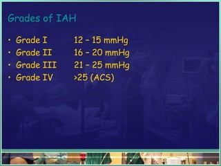
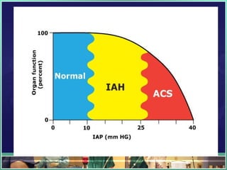
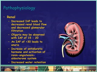
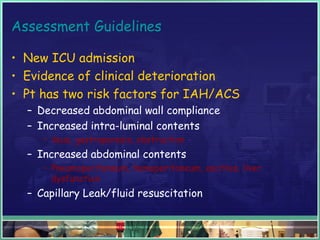
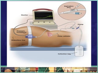
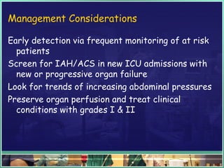
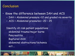
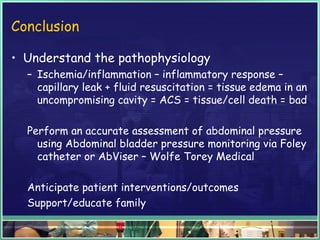
Abdominal Compartment Syndrome
By
Maged Abulmagd,MD,EDIC
? What it is ?
A disease process that dramatically increases
organ failure and death for medical and surgical
ICU patients
What is a normal intra-abdominal
pressure
or
IAP
This is the pressure within the abdominal cavity
5 – 7 mmHg is normal in a critically ill adult
Intra – abdominal Hypertension
IAH
• Defined as sustained or repeatedly elevated
abdominal pressure
• >12 and is graded
Grades of IAH
• Grade I 12 – 15 mmHg
• Grade II 16 – 20 mmHg
• Grade III 21 – 25 mmHg
• Grade IV >25 (ACS)
IAH
• Sustained pressure, >12 that has significant
effects on abdominal organs and cardiac output
with subsequent dysfunction of both abdominal
and extra-abdominal organs
Understanding Abdominal Compartment Syndrome
• APP – Abdominal perfusion pressure
• MAP – Mean arterial pressure
• IAP – Intra-abdominal pressure
• APP = MAP – IAP
• A critical IAP that leads to organ failure is
variable by patient & a single threshold cannot
be applied globally to all patients
• APP is superior to IAP, arterial pH, base deficit
& lactate in predicting organ failure & patient
outcomes
Definition of ACS
• A sustained IAP > 20 mmHg (with or without an
APP of <60 mmHg) that is associated with new
organ dysfunction/failure
• Adverse physiological effects caused by massive
interstitial and retroperitoneal swelling which
leads to organ or multi-organ failure
• Historically IAPs as high as 40 mmHg had been
acceptable; therefore, most clinicians are
concerned when IAP reaches 20 – 25 mmHg
Abdominal Compartment Syndrome
• Primary ACS – associated with injury or disease
in abdomen/pelvis requiring early surgical or
interventional radiological screening
• Secondary ACS is from conditions not
originating in the abdomen/pelvis
• Recurrent ACS is the redevelopment of ACS
following previous surgical or medical treatment
of primary or secondary ACS
Common Causes of ACS
• Primary causes
– Abdominal trauma with
bleeding
– Pancreatitis
– Ruptured abdominal aortic
aneurysm
– Retroperitoneal hematoma
– Obstructions/ileus
– Pneumoperitoneum
– Abcesses
– Visceral edema
Common Causes
• Secondary Causes
– Acute respiratory distress
syndrome
– Major trauma or burns
– Massive fluid resuscitation
– Hypothermia <33 degrees
Celsius
– Acidosis with pH < 7.2
– Hypotension
– Massive blood transfusion > 10
units
– Coagulopathy
– Sepsis
Common Causes
• Chronic Causes
– Obesity
– Liver failure with ascities
– Malignancies
Ischemia Inflammatory
response
Capillary leak
Tissue Edema
(Including bowel wall and mesentery)
Intra-abdominal hypertension
Fluid
resuscitation
Physiologic Insult/Critical Illness
Pathophysiological Consequences
of ACS
• Cardiovascular
– Reduced Cardiac Output
• Compression of the
inferior vena cava and
portal vein
• Reduced blood return to
the heart
• Afterload increased from
mechanical compression of
vascular beds and
vasoconstriction
Pathophysiology
• Cardiovascular
– Reduced Stroke volume
– Tachycardia
– Increased pressure on
great vessels making
hemodynamic monitoring
challenging with falsely
elevated and misguiding
pressures
– Increased risk for
thromboembolic events
secondary to venous stasis
Pathophysiology
Pulmonary
– Reduced lung compliance
secondary to diaphragmatic
elevation leads to
– Hypoventilation and
ventilation-perfusion
mismatch
– Increased work of
breathing
– Hypoxia and hypercarbia
– Respiratory failure
Pathophysiology
• Respiratory
– Increased peak airway
secondary to decreased
lung compliance
– Increased risk of
barotrauma
Pathophysiology
• Renal
– Increased IAP leads to
decreased renal blood flow
and decreased glomerular
filtration
– Oliguria may be observed
with IAP of 15 - 20
– An IAP of >30 leads to
anuria
– Increase of antidiuretic
hormone and activation of
renin-angiotensin-
aldosterone system
– Increased water retention
Pathophysiology
• Abdominal Visceral
– Reduced blood flow which
leads to intestinal ischemia
– Decreased blood flow to all
abdominal organs
Pathophysiology
• Central Nervous System
– Increased thoracic and
central venous pressure
leads to
– Decreased cerebral outflow
of blood
– Increased intracranial
pressure which leads to
decreased cerebral
perfusion pressure
Measuring Intra-Abdominal Pressure
Importance of accurate measurement
• Physical examination yields low levels of
detection of IAH/ACS
• Early detection and intervention reduces
morbidity and mortality.
• Diagnosis is dependent on frequent and accurate
measurement of IAP (watching trends)
• Cost effective, safe and accurate
Assessment Guidelines
• New ICU admission
• Evidence of clinical deterioration
• Pt has two risk factors for IAH/ACS
– Decreased abdominal wall compliance
– Increased intra-luminal contents
• ileus, gastroparesis, obstruction
– Increased abdominal contents
• Pneumoperitoneum, hemoperitoneum, ascities, liver
dysfunction
– Capillary Leak/fluid resuscitation
IAH/ACS Assessment algorithm from
World Society of Abdominal Compartment
Syndrome (WSACS)
www.wsacs.org
Excellent references
Types of Measurements
• Direct Pressure via intraperitoneal catheters
• Indirect Pressure
– Gastric Measure
– IVC
– Rectal
– Urinary bladder pressure – Gold Standard
Urinary Bladder Pressure
Most technically reliable
Correlate closely with pressures measured directly
in the abdominal cavity
Reliably reproducible
Transduced through a Foley catheter
Intermittent Monitoring
• Open Systems
• Closed Systems
Equipment needed for open measurement
• Disposable transducer
• 12” pressure monitoring
tubing
• 4-way stopcock
• Red dead-ender
• 60 cc, lure-lock syringe,
sterile
• Sterile normal saline
• Clamp, non-sterile
• Level
Procedure for open, intermittent monitoring
• Collect and gather all
supplies
• Attach stopcock to end of
sterile transducer
• Important to maintain
sterile technique to avoid
contamination and
potential infectious
process
Procedure for open, intermittent monitoring
• Attach pressure tubing to
the remaining end of the
transducer
Procedure for open, intermittent monitoring
• Fill 60 cc syringe with 40
cc of sterile normal saline
• Attach syringe to side
port of the stopcock
• Flush stopcock, pressure
tubing and transducer
with the normal saline
ensuring all air is removed
Procedure for open, intermittent monitoring
• Clamp the urinary drain tubing distal to the sampling port
• Cleanse the sampling port with alcohol
• Using sterile technique attach the pressure tubing to
the LuerLok connecting sampling port of the urinary
catheter
Procedure for open, intermittent monitoring
• Instill 25 cc of sterile normal saline into urinary
catheter via the sampling port (Larger vol. of NS can result in
falsely elevated IAP measurements)
• Briefly release the clamp to allow fluid from the bladder
to fill tubing and reclaim
• Read the IAP as a mean pressure at end expiration 30 –
60 seconds after instillation.
• Perform with patient supine
• Notify MD for sustained IAP greater than 12 mmHg
unless otherwise ordered.
Disadvantages with open, intermittent monitoring
• Collecting a number of items
• Correct assembly
• Risk of infection every time system is accessed
Closed Monitoring
• AbViser, Wolfe Tory Medical, SLC, UT
• Pre-assembled kit
• Adapts to Foley catheter and any transducer
• Reduces risk of infection
• Readily available, easily assessable data
Measuring Bladder Pressure
• Position patient flat & supine
• Read Mean pressure
• End Expiration
Management Considerations
Early detection via frequent monitoring of at risk
patients
Screen for IAH/ACS in new ICU admissions with
new or progressive organ failure
Look for trends of increasing abdominal pressures
Preserve organ perfusion and treat clinical
conditions with grades I & II
Management Considerations
Early surgical consultations for at risk patients
Early intervention for ACS or Grade III
Anticipate emergent surgical interventions to
prevent tissue damage/death
Management Considerations
• Anticipate patient to return with an alternative
surgical closure or “open” abdomen.
• The abdominal contents will not be sutured into
the abdominal cavity
• Alternative closures vary from surgeon to
surgeon
Examples:
The “Bogata Bag” – A 3 L IV bag, open and
sterilized and applied to the abdominal opening
Management Considerations
• KCI Vac Pac
• Sponge overlies abd.
Dressing/contents
• Attached to
continuous suction
canister
• Covered over with
occlusive dressing
Management Considerations
• Ioban Dressing
• An occlusive dressing
with iodine
impregnation
• Surgical towels will
overlie abdominal
contents with JP
drains – Ioban
overlies abdomen
Another Excellent Reference,
IAH/ACS Management Algorithm from
WSACS
www.wsacs.org
Conclusion
• Know the difference between IAH and ACS
– IAH = Abdominal pressure >12 and graded via severity
– ACS = Abdominal pressures > 20 – 25
Identify At risk patient populations
abdominal trauma/major burns
Pancreatitis
Ruptured AAA
abdominal obstructions/ischemia
ect….
Conclusion
• Understand the pathophysiology
– Ischemia/inflammation – inflammatory response –
capillary leak + fluid resuscitation = tissue edema in an
uncompromising cavity = ACS = tissue/cell death = bad
Perform an accurate assessment of abdominal pressure
using Abdominal bladder pressure monitoring via Foley
catheter or AbViser – Wolfe Torey Medical
Anticipate patient interventions/outcomes
Support/educate family
Case Study - 63 Y.O. male pt with pancreatitis is admitted
to the ICU. Pt has history of gallbladder disease, COPD
and ETOH abuse. He has been without ETOH reportedly
for approximately 24 hrs. VS upon admission are T 38.0,
HR 130, BP 90/62, MAP 61, RR 30 – 34 & O2 sat of 91%
on 100% NRB, wt approximately 125 kg. His breathing is
labored and he has c/o SOB. He is also mildly agitated &
resistive to O2 therapy with Bi-Pap. His lung sounds are
diminished bilaterally. Denies recent increase in cough.
His abdomen is firm and distended. States unknown last
BM but + for N/V.
• He has a Foley catheter in place with
approximately 100 cc of dark, amber urine in the
collection chamber. Lab values show H&H of
10.2/31.0, wbc 20, K 5.0, Na 142, Foley was
placed approximately 4 hours ago in the ED. His
peripheral arterial pulses are weak and thready
and his BLE show signs of PVD. He is currently
receiving bolus # 3 of NS.
Does this patient need IAP monitoring?
Is he at risk?
What could you use as a reference if you were
unsure?
After consulting with your attending MD, it is
decided that a baseline ABP reading would be
appropriate for this patient. Your initial ABP is
15mmHg.
Does this value represent intra-abdominal
hypertension or abdominal compartment
syndrome?
What is his APP based on his MAP and IAP?
What grade would you give this value?
Why is this patient at risk?
How would you proceed?
After reporting the findings to the resident,
serial ABP readings are ordered Q6 HR. His SBP
continues to remain low with a map consistently <
65 & his respiratory status continues to
deteriorate. The resident also orders another
fluid bolus.
• With what you have learned about IAH /ACS
management, what clinical suggestions could you
collaborate on to advocate for your patient?
•  
After collaboration with the medical team the decision is
made to intubate as his O2 sats continue to drop and RR
rate cont. to increase. After intubation and appropriate
sedation, the patient continues to have an increasingly
firm abdomen, increased HR and decreased SBP and map
<60 despite added norepinephrine. He is also now vented
with a respiratory rate of 24 – 30 and has become
increasingly agitated. His urine output for the last 2
hours is 30 ml. You repeat the ABP prior to the 4 hr
interval and you notice that his ABP value has risen to 20
after two separate measurements. What could you
expect at this point?
• www.abdominalcompartmentsyndrome.org
• The World Society of Abdominal Compartment
Syndrome, www.wsacs.org 

Abdominal compartment syndrome

  • 1.
  • 2.
    ? What itis ? A disease process that dramatically increases organ failure and death for medical and surgical ICU patients
  • 3.
    What is anormal intra-abdominal pressure or IAP This is the pressure within the abdominal cavity 5 – 7 mmHg is normal in a critically ill adult
  • 4.
    Intra – abdominalHypertension IAH • Defined as sustained or repeatedly elevated abdominal pressure • >12 and is graded
  • 5.
    Grades of IAH •Grade I 12 – 15 mmHg • Grade II 16 – 20 mmHg • Grade III 21 – 25 mmHg • Grade IV >25 (ACS)
  • 6.
    IAH • Sustained pressure,>12 that has significant effects on abdominal organs and cardiac output with subsequent dysfunction of both abdominal and extra-abdominal organs
  • 7.
    Understanding Abdominal CompartmentSyndrome • APP – Abdominal perfusion pressure • MAP – Mean arterial pressure • IAP – Intra-abdominal pressure • APP = MAP – IAP • A critical IAP that leads to organ failure is variable by patient & a single threshold cannot be applied globally to all patients • APP is superior to IAP, arterial pH, base deficit & lactate in predicting organ failure & patient outcomes
  • 8.
    Definition of ACS •A sustained IAP > 20 mmHg (with or without an APP of <60 mmHg) that is associated with new organ dysfunction/failure • Adverse physiological effects caused by massive interstitial and retroperitoneal swelling which leads to organ or multi-organ failure • Historically IAPs as high as 40 mmHg had been acceptable; therefore, most clinicians are concerned when IAP reaches 20 – 25 mmHg
  • 10.
    Abdominal Compartment Syndrome •Primary ACS – associated with injury or disease in abdomen/pelvis requiring early surgical or interventional radiological screening • Secondary ACS is from conditions not originating in the abdomen/pelvis • Recurrent ACS is the redevelopment of ACS following previous surgical or medical treatment of primary or secondary ACS
  • 11.
    Common Causes ofACS • Primary causes – Abdominal trauma with bleeding – Pancreatitis – Ruptured abdominal aortic aneurysm – Retroperitoneal hematoma – Obstructions/ileus – Pneumoperitoneum – Abcesses – Visceral edema
  • 12.
    Common Causes • SecondaryCauses – Acute respiratory distress syndrome – Major trauma or burns – Massive fluid resuscitation – Hypothermia <33 degrees Celsius – Acidosis with pH < 7.2 – Hypotension – Massive blood transfusion > 10 units – Coagulopathy – Sepsis
  • 13.
    Common Causes • ChronicCauses – Obesity – Liver failure with ascities – Malignancies
  • 14.
    Ischemia Inflammatory response Capillary leak TissueEdema (Including bowel wall and mesentery) Intra-abdominal hypertension Fluid resuscitation Physiologic Insult/Critical Illness
  • 16.
    Pathophysiological Consequences of ACS •Cardiovascular – Reduced Cardiac Output • Compression of the inferior vena cava and portal vein • Reduced blood return to the heart • Afterload increased from mechanical compression of vascular beds and vasoconstriction
  • 17.
    Pathophysiology • Cardiovascular – ReducedStroke volume – Tachycardia – Increased pressure on great vessels making hemodynamic monitoring challenging with falsely elevated and misguiding pressures – Increased risk for thromboembolic events secondary to venous stasis
  • 18.
    Pathophysiology Pulmonary – Reduced lungcompliance secondary to diaphragmatic elevation leads to – Hypoventilation and ventilation-perfusion mismatch – Increased work of breathing – Hypoxia and hypercarbia – Respiratory failure
  • 19.
    Pathophysiology • Respiratory – Increasedpeak airway secondary to decreased lung compliance – Increased risk of barotrauma
  • 20.
    Pathophysiology • Renal – IncreasedIAP leads to decreased renal blood flow and decreased glomerular filtration – Oliguria may be observed with IAP of 15 - 20 – An IAP of >30 leads to anuria – Increase of antidiuretic hormone and activation of renin-angiotensin- aldosterone system – Increased water retention
  • 21.
    Pathophysiology • Abdominal Visceral –Reduced blood flow which leads to intestinal ischemia – Decreased blood flow to all abdominal organs
  • 22.
    Pathophysiology • Central NervousSystem – Increased thoracic and central venous pressure leads to – Decreased cerebral outflow of blood – Increased intracranial pressure which leads to decreased cerebral perfusion pressure
  • 23.
  • 24.
    Importance of accuratemeasurement • Physical examination yields low levels of detection of IAH/ACS • Early detection and intervention reduces morbidity and mortality. • Diagnosis is dependent on frequent and accurate measurement of IAP (watching trends) • Cost effective, safe and accurate
  • 25.
    Assessment Guidelines • NewICU admission • Evidence of clinical deterioration • Pt has two risk factors for IAH/ACS – Decreased abdominal wall compliance – Increased intra-luminal contents • ileus, gastroparesis, obstruction – Increased abdominal contents • Pneumoperitoneum, hemoperitoneum, ascities, liver dysfunction – Capillary Leak/fluid resuscitation
  • 26.
    IAH/ACS Assessment algorithmfrom World Society of Abdominal Compartment Syndrome (WSACS) www.wsacs.org Excellent references
  • 27.
    Types of Measurements •Direct Pressure via intraperitoneal catheters • Indirect Pressure – Gastric Measure – IVC – Rectal – Urinary bladder pressure – Gold Standard
  • 28.
    Urinary Bladder Pressure Mosttechnically reliable Correlate closely with pressures measured directly in the abdominal cavity Reliably reproducible Transduced through a Foley catheter
  • 30.
    Intermittent Monitoring • OpenSystems • Closed Systems
  • 31.
    Equipment needed foropen measurement • Disposable transducer • 12” pressure monitoring tubing • 4-way stopcock • Red dead-ender • 60 cc, lure-lock syringe, sterile • Sterile normal saline • Clamp, non-sterile • Level
  • 32.
    Procedure for open,intermittent monitoring • Collect and gather all supplies • Attach stopcock to end of sterile transducer • Important to maintain sterile technique to avoid contamination and potential infectious process
  • 33.
    Procedure for open,intermittent monitoring • Attach pressure tubing to the remaining end of the transducer
  • 34.
    Procedure for open,intermittent monitoring • Fill 60 cc syringe with 40 cc of sterile normal saline • Attach syringe to side port of the stopcock • Flush stopcock, pressure tubing and transducer with the normal saline ensuring all air is removed
  • 35.
    Procedure for open,intermittent monitoring • Clamp the urinary drain tubing distal to the sampling port • Cleanse the sampling port with alcohol • Using sterile technique attach the pressure tubing to the LuerLok connecting sampling port of the urinary catheter
  • 36.
    Procedure for open,intermittent monitoring • Instill 25 cc of sterile normal saline into urinary catheter via the sampling port (Larger vol. of NS can result in falsely elevated IAP measurements) • Briefly release the clamp to allow fluid from the bladder to fill tubing and reclaim • Read the IAP as a mean pressure at end expiration 30 – 60 seconds after instillation. • Perform with patient supine • Notify MD for sustained IAP greater than 12 mmHg unless otherwise ordered.
  • 37.
    Disadvantages with open,intermittent monitoring • Collecting a number of items • Correct assembly • Risk of infection every time system is accessed
  • 38.
    Closed Monitoring • AbViser,Wolfe Tory Medical, SLC, UT • Pre-assembled kit • Adapts to Foley catheter and any transducer • Reduces risk of infection • Readily available, easily assessable data
  • 40.
    Measuring Bladder Pressure •Position patient flat & supine • Read Mean pressure • End Expiration
  • 41.
    Management Considerations Early detectionvia frequent monitoring of at risk patients Screen for IAH/ACS in new ICU admissions with new or progressive organ failure Look for trends of increasing abdominal pressures Preserve organ perfusion and treat clinical conditions with grades I & II
  • 42.
    Management Considerations Early surgicalconsultations for at risk patients Early intervention for ACS or Grade III Anticipate emergent surgical interventions to prevent tissue damage/death
  • 43.
    Management Considerations • Anticipatepatient to return with an alternative surgical closure or “open” abdomen. • The abdominal contents will not be sutured into the abdominal cavity • Alternative closures vary from surgeon to surgeon Examples: The “Bogata Bag” – A 3 L IV bag, open and sterilized and applied to the abdominal opening
  • 45.
    Management Considerations • KCIVac Pac • Sponge overlies abd. Dressing/contents • Attached to continuous suction canister • Covered over with occlusive dressing
  • 46.
    Management Considerations • IobanDressing • An occlusive dressing with iodine impregnation • Surgical towels will overlie abdominal contents with JP drains – Ioban overlies abdomen
  • 47.
    Another Excellent Reference, IAH/ACSManagement Algorithm from WSACS www.wsacs.org
  • 48.
    Conclusion • Know thedifference between IAH and ACS – IAH = Abdominal pressure >12 and graded via severity – ACS = Abdominal pressures > 20 – 25 Identify At risk patient populations abdominal trauma/major burns Pancreatitis Ruptured AAA abdominal obstructions/ischemia ect….
  • 49.
    Conclusion • Understand thepathophysiology – Ischemia/inflammation – inflammatory response – capillary leak + fluid resuscitation = tissue edema in an uncompromising cavity = ACS = tissue/cell death = bad Perform an accurate assessment of abdominal pressure using Abdominal bladder pressure monitoring via Foley catheter or AbViser – Wolfe Torey Medical Anticipate patient interventions/outcomes Support/educate family
  • 50.
    Case Study -63 Y.O. male pt with pancreatitis is admitted to the ICU. Pt has history of gallbladder disease, COPD and ETOH abuse. He has been without ETOH reportedly for approximately 24 hrs. VS upon admission are T 38.0, HR 130, BP 90/62, MAP 61, RR 30 – 34 & O2 sat of 91% on 100% NRB, wt approximately 125 kg. His breathing is labored and he has c/o SOB. He is also mildly agitated & resistive to O2 therapy with Bi-Pap. His lung sounds are diminished bilaterally. Denies recent increase in cough. His abdomen is firm and distended. States unknown last BM but + for N/V.
  • 51.
    • He hasa Foley catheter in place with approximately 100 cc of dark, amber urine in the collection chamber. Lab values show H&H of 10.2/31.0, wbc 20, K 5.0, Na 142, Foley was placed approximately 4 hours ago in the ED. His peripheral arterial pulses are weak and thready and his BLE show signs of PVD. He is currently receiving bolus # 3 of NS.
  • 52.
    Does this patientneed IAP monitoring? Is he at risk? What could you use as a reference if you were unsure?
  • 53.
    After consulting withyour attending MD, it is decided that a baseline ABP reading would be appropriate for this patient. Your initial ABP is 15mmHg.
  • 54.
    Does this valuerepresent intra-abdominal hypertension or abdominal compartment syndrome? What is his APP based on his MAP and IAP?
  • 55.
    What grade wouldyou give this value? Why is this patient at risk? How would you proceed?
  • 56.
    After reporting thefindings to the resident, serial ABP readings are ordered Q6 HR. His SBP continues to remain low with a map consistently < 65 & his respiratory status continues to deteriorate. The resident also orders another fluid bolus. • With what you have learned about IAH /ACS management, what clinical suggestions could you collaborate on to advocate for your patient? •  
  • 57.
    After collaboration withthe medical team the decision is made to intubate as his O2 sats continue to drop and RR rate cont. to increase. After intubation and appropriate sedation, the patient continues to have an increasingly firm abdomen, increased HR and decreased SBP and map <60 despite added norepinephrine. He is also now vented with a respiratory rate of 24 – 30 and has become increasingly agitated. His urine output for the last 2 hours is 30 ml. You repeat the ABP prior to the 4 hr interval and you notice that his ABP value has risen to 20 after two separate measurements. What could you expect at this point?
  • 58.
    • www.abdominalcompartmentsyndrome.org • TheWorld Society of Abdominal Compartment Syndrome, www.wsacs.org