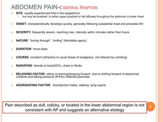
ACUTE PANCREATITIS
Abbas Qaddomi ( intern)
1
INTRODUCTION
 Acute pancreatitis(AP) is an inflammatory condition
of the pancreas characterized clinically by
abdominal pain and elevated levels of pancreatic
enzymes in the blood.
 AP was the most common gastroenterology
discharge diagnosis with a cost of 2.6 billion dollars.
2
 Worldwide the incidence of acute pancreatitis
ranges from 5 to 80/100,000 population
3
ETIOLOGY
 Gallstones : reflux of bile into the pancreatic duct
due to transient obstruction of the ampulla during
passage of gallstones
 gallstone obstructing the pancreatic duct, leading to
ductal hypertension.
4
 Alcohol : amount for many years
 Generation of toxic metabolites such as
acetaldehyde and fatty acid ethyl esters
 Sensitization of acinar cells to cholecystokinin
(CCK)-induced premature activation of zymogens
 Ethanol also increases the protein content of
pancreatic juice, decreases bicarbonate levels, and
trypsin inhibitor concentration
ETIOLOGY
5
 Hereditary Pancreatitis

 Tumors
 Hyperlipidemia
 Iatrogenic
ETIOLOGY
6
7
PATHOPHYSIOLOGY
 Acinar cell injury: auto-digestion of pancreatic
substance by inappropriately activated enzymes (
trypsinogen )
 Intrapancreatic Events : Leukocyte
chemoattraction, release of cytokines, and
oxidative stress
 Systemic Events systemic inflammatory response
syndrome (SIRS) Organ failure
8
ABDOMEN PAIN-CARDINAL SYMPTOM
 SITE: usually experienced first in the epigastrium
 but may be localized to either upper quadrant or felt diffusely throughout the abdomen or lower chest
 ONSET: characteristically develops quickly, generally following substantial meal and precedes NV
 SEVERITY: frequently severe, reaching max. intensity within minutes rather than hours
 NATURE: “boring through”, “knifing” (illimitable agony)
 DURATION: hours-days
 COURSE: constant (refractory to usual doses of analgesics, not relieved by vomiting)
 RADIATION: directly to back(50%), chest or flanks
 RELEIVING FACTOR: sitting or leaning/stooping forward due to shifting forward of abdominal
contents and taking pressure off from inflamed pancreas
 AGGRAVATING FACTOR: food/alcohol intake, walking, lying supine
Pain described as dull, colicky, or located in the lower abdominal region is not
consistent with AP and suggests an alternative etiology
9
OTHER MANIFESTATIONS
 Nausea, frequent and effortless vomiting, anorexia,diarrhea
 Due to reflex pylorospasm
 More intense in necrotizing than in edematous pancreatitis
 Persistent retching
 despite empty stomach
 Hiccups
 Due to gastric distension/diaphragmatic irritation
 Fever
 Low grade, seen in infective pancreatitis
 Weakness, Anxiety, Sweating
 Indicates severe attack
10
ABDOMEN EXAMINATION
 Physical findings vary depending upon the severity of acute
pancreatitis
 Tenderness + Rebound tenderness:
 epigastrium/upper abdomen
 Distension:
 Ileus(BS decreased or absent)
 ascites with shifting dullness
 Guarding -upper abdomen
 tensing of the abdominal wall muscles to guard inflamed organs
within the abdomen from the pain of pressure upon them
 Rigidity(involuntary stiffness)-unusual
 Tensing of the abdominal wall muscles to guard inflamed organs
even if patient not touched
11
DIAGNOSTIC CRITERIA
 Most often established by the presence of 2 of the 3 following
criteria
(1) abdominal pain consistent with the disease,
(2) serum amylase and/or lipase greater than three times the upper limit
of normal, and/or
(3) characteristic findings from abdominal imaging.
_____________________________________________________
____
Transabdominal ultrasound should be performed in all patients with
acute pancreatitis
CT and/or MRI of the pancreas should be reserved for patients
 in whom the diagnosis is unclear(typical pain with normal enzymes)
 who fail to improve clinically within the first 48–72 h after hospital
admission (e.g., persistent pain, fever, nausea, unable to begin oral
feeding)
 to evaluate complications
 Genetic testing may be considered in young patients ( < 30 years
old) if no cause is evident and a family history of pancreatic
disease is present
12
CLASSIFICATION OF ACUTE PANCREATITIS
13
ASSESSMENT OF DISEASE SEVERITY
 systemic inflammatory response syndrome (SIRS)
 modified Marshall scoring system
 SOFA
 APACHE II
 Ranson's criteria
 BISAP 14
15
16
17
INDICATIONS FOR ICU ADMISSION
 Patients with severe acute pancreatitis
 Patients with acute pancreatitis and one or more of the following
parameters:
 •Pulse <40 or >150 beats/minute
 •Systolic arterial pressure <80 mmHg or mean arterial pressure
<60 mmHg or diastolic arterial pressure >120 mmHg
 •Respiratory rate >35 breaths/minute
 •Serum sodium <110 mmol/L or >170 mmol/L
 •Serum potassium <2.0 mmol/L or >7.0 mmol/L
 •PaO2 <50 mmHg
 •pH <7.1 or >7.7
 •Serum glucose >800 mg/dL
 •Serum calcium >15 mg/dL
 •Anuria
 •Coma 18
INITIAL MANAGEMENT
 Aggressive hydration during 12-24
 Decrease morbidity and mortality
 Hematocrit and BUN has been widely recommended as
surrogate markers for successful hydration
 No absolute numbers recommended
 Goal to decrease Hct and BUN and maintain normal cr
 In elderly and cardiac/renal comorbidities hydration is
monitored by
 Central venous pressure via CV line or
 Intrathoracic blood volume index
 Better/more accurate correlate with cardiac index than CVP
19
PAIN CONTROL
 should be treated with analgesics. uncontrolled pain
can contribute to the hemodynamic instability
 Attention to adequate fluid resuscitation should be
the first priority in addressing abdominal pain, as
hypovolemia from vascular leak and
hemoconcentration can cause ischemic pain and
resultant lactic acidosis.
20
 should be administered intravenously
 mild pain , NSAID (metamizole 2g/8h IV)
 severe pain opioid analgesia
 Morphine is to be avoided, due to its potential to
cause sphincter of Oddi spasm
21
PAIN CONTROL
ROLE OF ERCP
 most gallstones that cause AP readily pass to the
duodenum and are lost in the stool
 should be performed early in the course (within 24
hours of admission) for patients with:
 gallstone pancreatitis
 Cholangitis
 common bile duct obstruction (visible stone on
imaging)
 dilated common bile duct
 increasing liver tests without cholangitis
22
PREVENTING POST-ERCP
PANCREATITIS
 Over the past 15 years, the risk of post-ERCP
pancreatitis has decreased to 2 – 4 % and the risk
of severe AP to < 1 / 500
 guidewire cannulation
 pancreatic duct stents
 rectal NSAIDs
23
THE ROLE OF ANTIBIOTICS IN AP
 Prophylactic antibiotics are not recommended in
patients with acute pancreatitis, regardless of the
type or disease severity
 Infected necrosis should be considered in patients
with pancreatic or extrapancreatic necrosis who
deteriorate or fail to improve after 7 – 10 days of
hospitalization
24
 As SIRS may be indistinguishable from sepsis
syndrome, so if infection is suspected, antibiotics
should be given while source of infection is being
investigated
 Once blood and other cultures are found negative,
antibiotics should be discontinued
 The antibiotics shown to penetrate and used in
clinical trials include carbapenems, quinolones,
metronidazole, and high-dose cephalosporins
25
 Routine administration of antifungal agents along
with prophylactic or therapeutic antibiotics is not
recommended
26
NUTRITION
 Clinical and experimental studies showed that
bowel rest is associated with intestinal mucosal
atrophy and increased infectious complications
because of bacterial translocation from the gut
27
 In mild AP oral feedings can be started immediately if
there is no nausea/vomiting, and the abdominal
pain/tenderness/Ileus has resolved(amylase return to
normal, patient feel hunger
 low-fat soft diet has been shown to be safe compared
with clear liquids,
28
NUTRITION
NUTRITION
IN SEVERE AP
 In severe AP, enteral nutrition is recommended to
prevent infectious complications.
 Parenteral nutrition should be avoided, unless the
enteral route is not available, not tolerated, or not
meeting caloric requirements
 Nasogastric delivery and nasojejunal delivery of
enteral feeding appear comparable in efficacy and
safety
29
 high protein, low fat, semi-elemental feeding
formulas
 Signs that the formula is not tolerated include
increased abdominal pain, vomiting (with
nasogastric feeding), bloating, or diarrhea
 Parenteral
30
NUTRITION
IN SEVERE AP
ROLE OF SURGERY IN AP
 Cholecystectomy should be performed after recovery in
all patients with gallstone pancreatitis including those
who have undergone an endoscopic sphincterotomy
 In case of mild gallstone AP, cholecystectomy should be
performed before discharge to prevent a recurrence of
AP
 clinical suspicion of CBD stones is high ------- ERCP
 clinical suspicion of CBD stones is moderate -------MRCP or
EUS
 clinical suspicion of CBD stones is low ---------- intraoperative
cholangiogram 31
 In case of necrotizing biliary AP, in order to prevent
infection, cholecystectomy is to be deferred until active
inflammation subsides and fluid collections resolve or
stabilize
 Failure to perform a cholecystectomy is associated with
a 25 to 30 percent risk of recurrent acute pancreatitis,
cholecystitis, or cholangitis within 6 to 18 weeks
 population-based study found that cholecystectomy
performed for recurrent attacks of AP with no stones /
sludge on ultrasound and no significant elevation of liver
tests during the attack of AP was associated with a > 50
% recurrence of AP
32
 If patient unfit for surgery(comorbid/elderly), biliary
sphincherotomy alone may be effective to reduce
further attacks of AP
33
Sterile necrosis infected necrosis
Asymptomatic Does not mandate
intervention regardless of
size, location and
extension
surgical, radiologic, and/or
endoscopic drainage should
be delayed preferably for
more than 4 weeks
• to allow liquefaction of
the contents and the
development of a
fibrous wall around the
necrosis
• Initially treated with
antibiotics
Stable
Symptomatic
(associated
with GOO or
bile
obstruction)
minimally invasive
methods of
necrosectomy are
preferred to open
necrosectomy
Urgent debridement unstable
34
35
 Thank you
36

Acute pancreatitis

  • 1.
  • 2.
    INTRODUCTION  Acute pancreatitis(AP)is an inflammatory condition of the pancreas characterized clinically by abdominal pain and elevated levels of pancreatic enzymes in the blood.  AP was the most common gastroenterology discharge diagnosis with a cost of 2.6 billion dollars. 2
  • 3.
     Worldwide theincidence of acute pancreatitis ranges from 5 to 80/100,000 population 3
  • 4.
    ETIOLOGY  Gallstones :reflux of bile into the pancreatic duct due to transient obstruction of the ampulla during passage of gallstones  gallstone obstructing the pancreatic duct, leading to ductal hypertension. 4
  • 5.
     Alcohol :amount for many years  Generation of toxic metabolites such as acetaldehyde and fatty acid ethyl esters  Sensitization of acinar cells to cholecystokinin (CCK)-induced premature activation of zymogens  Ethanol also increases the protein content of pancreatic juice, decreases bicarbonate levels, and trypsin inhibitor concentration ETIOLOGY 5
  • 6.
     Hereditary Pancreatitis  Tumors  Hyperlipidemia  Iatrogenic ETIOLOGY 6
  • 7.
  • 8.
    PATHOPHYSIOLOGY  Acinar cellinjury: auto-digestion of pancreatic substance by inappropriately activated enzymes ( trypsinogen )  Intrapancreatic Events : Leukocyte chemoattraction, release of cytokines, and oxidative stress  Systemic Events systemic inflammatory response syndrome (SIRS) Organ failure 8
  • 9.
    ABDOMEN PAIN-CARDINAL SYMPTOM SITE: usually experienced first in the epigastrium  but may be localized to either upper quadrant or felt diffusely throughout the abdomen or lower chest  ONSET: characteristically develops quickly, generally following substantial meal and precedes NV  SEVERITY: frequently severe, reaching max. intensity within minutes rather than hours  NATURE: “boring through”, “knifing” (illimitable agony)  DURATION: hours-days  COURSE: constant (refractory to usual doses of analgesics, not relieved by vomiting)  RADIATION: directly to back(50%), chest or flanks  RELEIVING FACTOR: sitting or leaning/stooping forward due to shifting forward of abdominal contents and taking pressure off from inflamed pancreas  AGGRAVATING FACTOR: food/alcohol intake, walking, lying supine Pain described as dull, colicky, or located in the lower abdominal region is not consistent with AP and suggests an alternative etiology 9
  • 10.
    OTHER MANIFESTATIONS  Nausea,frequent and effortless vomiting, anorexia,diarrhea  Due to reflex pylorospasm  More intense in necrotizing than in edematous pancreatitis  Persistent retching  despite empty stomach  Hiccups  Due to gastric distension/diaphragmatic irritation  Fever  Low grade, seen in infective pancreatitis  Weakness, Anxiety, Sweating  Indicates severe attack 10
  • 11.
    ABDOMEN EXAMINATION  Physicalfindings vary depending upon the severity of acute pancreatitis  Tenderness + Rebound tenderness:  epigastrium/upper abdomen  Distension:  Ileus(BS decreased or absent)  ascites with shifting dullness  Guarding -upper abdomen  tensing of the abdominal wall muscles to guard inflamed organs within the abdomen from the pain of pressure upon them  Rigidity(involuntary stiffness)-unusual  Tensing of the abdominal wall muscles to guard inflamed organs even if patient not touched 11
  • 12.
    DIAGNOSTIC CRITERIA  Mostoften established by the presence of 2 of the 3 following criteria (1) abdominal pain consistent with the disease, (2) serum amylase and/or lipase greater than three times the upper limit of normal, and/or (3) characteristic findings from abdominal imaging. _____________________________________________________ ____ Transabdominal ultrasound should be performed in all patients with acute pancreatitis CT and/or MRI of the pancreas should be reserved for patients  in whom the diagnosis is unclear(typical pain with normal enzymes)  who fail to improve clinically within the first 48–72 h after hospital admission (e.g., persistent pain, fever, nausea, unable to begin oral feeding)  to evaluate complications  Genetic testing may be considered in young patients ( < 30 years old) if no cause is evident and a family history of pancreatic disease is present 12
  • 13.
    CLASSIFICATION OF ACUTEPANCREATITIS 13
  • 14.
    ASSESSMENT OF DISEASESEVERITY  systemic inflammatory response syndrome (SIRS)  modified Marshall scoring system  SOFA  APACHE II  Ranson's criteria  BISAP 14
  • 15.
  • 16.
  • 17.
  • 18.
    INDICATIONS FOR ICUADMISSION  Patients with severe acute pancreatitis  Patients with acute pancreatitis and one or more of the following parameters:  •Pulse <40 or >150 beats/minute  •Systolic arterial pressure <80 mmHg or mean arterial pressure <60 mmHg or diastolic arterial pressure >120 mmHg  •Respiratory rate >35 breaths/minute  •Serum sodium <110 mmol/L or >170 mmol/L  •Serum potassium <2.0 mmol/L or >7.0 mmol/L  •PaO2 <50 mmHg  •pH <7.1 or >7.7  •Serum glucose >800 mg/dL  •Serum calcium >15 mg/dL  •Anuria  •Coma 18
  • 19.
    INITIAL MANAGEMENT  Aggressivehydration during 12-24  Decrease morbidity and mortality  Hematocrit and BUN has been widely recommended as surrogate markers for successful hydration  No absolute numbers recommended  Goal to decrease Hct and BUN and maintain normal cr  In elderly and cardiac/renal comorbidities hydration is monitored by  Central venous pressure via CV line or  Intrathoracic blood volume index  Better/more accurate correlate with cardiac index than CVP 19
  • 20.
    PAIN CONTROL  shouldbe treated with analgesics. uncontrolled pain can contribute to the hemodynamic instability  Attention to adequate fluid resuscitation should be the first priority in addressing abdominal pain, as hypovolemia from vascular leak and hemoconcentration can cause ischemic pain and resultant lactic acidosis. 20
  • 21.
     should beadministered intravenously  mild pain , NSAID (metamizole 2g/8h IV)  severe pain opioid analgesia  Morphine is to be avoided, due to its potential to cause sphincter of Oddi spasm 21 PAIN CONTROL
  • 22.
    ROLE OF ERCP most gallstones that cause AP readily pass to the duodenum and are lost in the stool  should be performed early in the course (within 24 hours of admission) for patients with:  gallstone pancreatitis  Cholangitis  common bile duct obstruction (visible stone on imaging)  dilated common bile duct  increasing liver tests without cholangitis 22
  • 23.
    PREVENTING POST-ERCP PANCREATITIS  Overthe past 15 years, the risk of post-ERCP pancreatitis has decreased to 2 – 4 % and the risk of severe AP to < 1 / 500  guidewire cannulation  pancreatic duct stents  rectal NSAIDs 23
  • 24.
    THE ROLE OFANTIBIOTICS IN AP  Prophylactic antibiotics are not recommended in patients with acute pancreatitis, regardless of the type or disease severity  Infected necrosis should be considered in patients with pancreatic or extrapancreatic necrosis who deteriorate or fail to improve after 7 – 10 days of hospitalization 24
  • 25.
     As SIRSmay be indistinguishable from sepsis syndrome, so if infection is suspected, antibiotics should be given while source of infection is being investigated  Once blood and other cultures are found negative, antibiotics should be discontinued  The antibiotics shown to penetrate and used in clinical trials include carbapenems, quinolones, metronidazole, and high-dose cephalosporins 25
  • 26.
     Routine administrationof antifungal agents along with prophylactic or therapeutic antibiotics is not recommended 26
  • 27.
    NUTRITION  Clinical andexperimental studies showed that bowel rest is associated with intestinal mucosal atrophy and increased infectious complications because of bacterial translocation from the gut 27
  • 28.
     In mildAP oral feedings can be started immediately if there is no nausea/vomiting, and the abdominal pain/tenderness/Ileus has resolved(amylase return to normal, patient feel hunger  low-fat soft diet has been shown to be safe compared with clear liquids, 28 NUTRITION
  • 29.
    NUTRITION IN SEVERE AP In severe AP, enteral nutrition is recommended to prevent infectious complications.  Parenteral nutrition should be avoided, unless the enteral route is not available, not tolerated, or not meeting caloric requirements  Nasogastric delivery and nasojejunal delivery of enteral feeding appear comparable in efficacy and safety 29
  • 30.
     high protein,low fat, semi-elemental feeding formulas  Signs that the formula is not tolerated include increased abdominal pain, vomiting (with nasogastric feeding), bloating, or diarrhea  Parenteral 30 NUTRITION IN SEVERE AP
  • 31.
    ROLE OF SURGERYIN AP  Cholecystectomy should be performed after recovery in all patients with gallstone pancreatitis including those who have undergone an endoscopic sphincterotomy  In case of mild gallstone AP, cholecystectomy should be performed before discharge to prevent a recurrence of AP  clinical suspicion of CBD stones is high ------- ERCP  clinical suspicion of CBD stones is moderate -------MRCP or EUS  clinical suspicion of CBD stones is low ---------- intraoperative cholangiogram 31
  • 32.
     In caseof necrotizing biliary AP, in order to prevent infection, cholecystectomy is to be deferred until active inflammation subsides and fluid collections resolve or stabilize  Failure to perform a cholecystectomy is associated with a 25 to 30 percent risk of recurrent acute pancreatitis, cholecystitis, or cholangitis within 6 to 18 weeks  population-based study found that cholecystectomy performed for recurrent attacks of AP with no stones / sludge on ultrasound and no significant elevation of liver tests during the attack of AP was associated with a > 50 % recurrence of AP 32
  • 33.
     If patientunfit for surgery(comorbid/elderly), biliary sphincherotomy alone may be effective to reduce further attacks of AP 33
  • 34.
    Sterile necrosis infectednecrosis Asymptomatic Does not mandate intervention regardless of size, location and extension surgical, radiologic, and/or endoscopic drainage should be delayed preferably for more than 4 weeks • to allow liquefaction of the contents and the development of a fibrous wall around the necrosis • Initially treated with antibiotics Stable Symptomatic (associated with GOO or bile obstruction) minimally invasive methods of necrosectomy are preferred to open necrosectomy Urgent debridement unstable 34
  • 35.
  • 36.

Editor's Notes

  • #3 severe acute upper abdominal pain but requires biochemical or radiologic evidence to establish the diagnosis.
  • #4 بيختلف الانسدنس من بين الاعراق The median age at onset depends on the etiology. [12] The following are median ages of onset for various etiologies:Alcohol-related - 39 years Biliary tract–related - 69 years Trauma-related - 66 years Drug-induced etiology - 42 years ERCP-related - 58 years AIDS-related - 31 years Vasculitis-related - 36 years
  • #6 Sensitization of the pancreas to the toxic effects of coxsackie virus B3 Th e diagnosis should not be entertained unless a person has a history of over 5 years of heavy alcohol consumption “ Heavy” alcohol consumption is generally considered to be > 50 g per day, but is often much higher
  • #7 AD mutations of the cationic trypsinogen gene cause premature activation of trypsinogen to trypsin and cause abnormalities of ductal secretion, periampullary tumor episode of acute pancreatitis can be the first clinical manifestation of the tumor. free fatty acids are released from serum triglycerides in toxic concentrations by the action of pancreatic lipase within pancreatic capillaries Serum triglyceride concentrations above 1000 mg/dL
  • #9 trypsin inhibitor (PSTI or SPINK1), which can bind and inactivate about 20 percent of the trypsin activity autolysis of prematurely activated trypsin mesotrypsin and enzyme Y, which lyses and inactivates trypsin
  • #13 Amylase within few hours last 3-5 days May remain normal in 1/5 pt May be normal in alcohol and hyperlibedemia ap False increase macroamlysemia, inflammation, renal failure Lipase may false increase appendicitis, cholecystitis, renal failure In dm need increase 3-5 times
  • #14 Local complications are defined as peripancreatic fluid collections pancreatic and peripancreatic necrosis (sterile or infected) pseudocysts, and walled-off necrosis (sterile or infected).
  • #20 250 – 500 ml per hour of isotonic crystalloid solution should be provided to all patients, unless cardiovascular, renal, or other related comorbid aim to decrease BUN provides micro- and macrocirculatory support to prevent serious complications such as pancreatic necrosis LR --- more lyte balance and بزيد البي اتش بقلل تفعيل التربسينوجين pancreatitis due to hypercalcemia, lactated Ringer's is contraindicated
  • #22 Fentanyl is being increasingly used due to its better safety profile, especially in renal impairment 20-50 microg buprenorphine, pentazocine, procaine hydrochloride, and meperidine
  • #23 In the absence of common bile duct obstruction, ERCP is not indicated for (mild or severe) gallstone pancreatitis without cholangitis. When in doubt about bile duct obstruction in the absence of cholangitis, liver tests can be rechecked in 24 to 48 hours to determine if they improve or a magnetic resonance cholangiopancreatography (MRCP) or endoscopic ultrasound (EUS) could be performed to determine if there are stones in the common early ERCP within 24 hof admission decreases morbidity and mortality in patients with AP complicated by biliary sepsis early ERCP reduced pancreatitis-related complications but not mortality in patients predicted to have severe pancreatitis
  • #24 guidewire inserted through a catheter) decreases the risk of pancreatitis ( 100 ) by avoiding hydrostatic injury to the pancreas diclofenac or indomethacin at a dose of 100mg
  • #25 (i) initial CT-guided fi ne-needle aspiration (FNA) for Gram stain and culture to guide use of appropriate antibiotics or (ii) empiric use of antibiotics after obtaining necessary cultures for infectious agents,
  • #28 Multiple studies have shown that patients provided oral feeding early in the course of AP have a shorter hospital stay, decreased infectious complications, decreased morbidity, and decreased mortality
  • #29 More recent data suggest that early refeeding when patients are subjectively hungry, regardless of resolution of abdominal pain and normalization of pancreatic enzymes 24 to 48 hours after the onset of pancreatitis
  • #30 no significant differences in APACHE II scores, CRP levels, pain, or analgesic requirements Ng risk of aspiration
  • #31 start at 25 cc per hour and advance as tolerated to at least 30 even in the presence of ileus
  • #32 require complex decision making between the surgeon and gastroenterologist. In these patients, cholecystectomy is typically delayed until (i) a later time in the typically prolonged hospitalization, (ii) as part of the management of the pancreatic necrosis if present, or (iii) aft er discharge
  • #35 Minimally invasive approach: laparoscopic surgery(ant or retroperitoneal approach), percutaneous radiologic catheter drainage or debridement, video-assisted or small incision-based left retroperitoneal debridement, and endoscopy