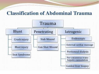
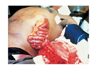
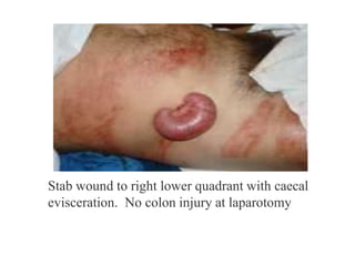
ABDOMINAL TRAUMA : AN OVERVIEW
SURGICAL DEPARTMENT
Dr. Ndung’u (General Surgeon)
Dr. Anthony (Medical Officer)
CASE STUDY
Introduction
• Abdominal trauma is regularly
encountered in the emergency/casualty
department
• One of the leading cause of death and
disability
• Identification of serious intra-abdominal
injuries is often challenging
• Many injuries may not manifest during
the initial assessment and
treatment period
Epidemiology
• Peak incidence of abdominal
Trauma 15 - 30yr
• Prevalence: 13% of all injuries
Blunt Abdominal Trauma
◦ Greater mortality than PAT (more difficult to diagnose, commonly associated
with trauma to multiple organs/systems)
◦ Most commonly injured organs?
- spleen > liver, intestine is the most likely hollow viscus.
◦ Most common causes?
- MVA (50 - 75% of cases) > blows to abdomen (15%) > falls (6 - 9%)
Penetrating Abdominal Trauma
◦ Stabbing 3x more common than firearm wounds
◦ Gun shot wound cause 90% of the deaths
◦ Most commonly injured organs?
- small intestine > colon > liver
Pathophysiology of injury
Penetrating Abdominal Trauma
Stab Wounds
◦ Knives, ice picks, pens, coat hangers,
broken bottles
◦ Liver, small bowel, spleen
Gunshot wounds
◦ small bowel, colon and liver
◦ Often multiple organ injuries,
bowel perforations
Pathophysiology of injury
• Iatrogenic injury
-Endoscopic /Laparoscopic surgical
procedures
-Bag-mask ventilations
-Inadvertent esophageal intubation
-External cardiac compressions
-Heimlich manoeuvre
Pathophysiology of injury
Penetrating Trauma
Penetrating abdominal trauma has a slightly higher mortality rate
Second most common cause of abdominal injury
Gunshot Injury
Prehospital Care
• The goal of prehospital is to deliver the pt
to hospital for definitive care as rapidly
as possible. „Scoop and Run‟
• Maintain airway & start I V line
• Care of spinal cord
• Communicate to medical unit
• Rapid transport of patient to
emergency/trauma centre
Initial Assessment and Resuscitation
Primary Survey –ATLS approach
ABCDE pattern: Airway, Breathing, Circulation, Disability (neurologic
status), and Exposure.
A -intubation may be required if patient is shocked, hypotensive or unconscious or
in need for ventilation. *with cervical precaution.
B -watch for hemothorax in both blunt and penetrating thoracoabdominal injuries.
C -start with 2 L crystalloid (If active bleeding you must find source and stop the
bleeding)
D –May seen associated with thorocolumbar #
E -Watch for other injury
Emergency Care
• I V fluids
• Control external bleeding
• Dressing of wounds
• Protect eviscerated organs with a sterile
dressing
• Stabilize an impaled object in place
• Give high flow oxygen
• Immobilize the patient with a fractured pelvis
• Keep the patient warm
• Analgesics
Secondary Survey
History for all trauma patients:
-Not necessary making an accurate diagnosis
S.A.M.P.L.E
S: Symptoms:
pain,vomiting,hematuria,hematochezia,dyspnea,respiratory
distress…
A: Allergies
M : Medications
L : Last meals
E : Events (mechanism of injury)
Secondary Survey
• General & Systemic Examination - to
identify all occult injuries .
• Special attention to Back, Axilla , Perineum
• PR - sphincter tone ,bleeding ,perforation
, high riding prostate
• Foley‟s catheter- monitor urine output
• Nasogastric tube
Inspection : abrasions, contusion, lacerations,
deformity, entrance and exit wounds to
determine path of injury…
(grey Turner, Kehr, Balance, Cullen, seat belt sign)
Palpation: elicits superficial , deep , or rebound
tenderness; involuntary muscle guarding
Percussion : subtle signs of peritonitis; tympany
in gastric dilatation or free air; dullness with
hemoperitoneum.
Auscultation : bowel sounds may be decrease
( late finding).
Physical examination
Grey-Turner sign : bluid discoloration of
lower flanks, lower back; associated with
retroperitoneal bleeding of
pancrease,kidney or pelvic fracture.
Cullen sign : bluish discoloration around
umbilicus, indicates peritoneal bleeding,
often pancreatic hemorrhage.
Kehr sign: shoulder pain while supine
;caused by diaphragmatic irritation(splenic
injury, free air, intra-abdominal bleeding)
Balance sign : dull percussion in LUQ.Sign
of splenic injury; blood accumulation in
subcapsular or extracapsular spleen
In the trauma patient, a ‘normal’ physical exam of the
abdomen doesn’t equate to much. You NEED to do further
testing.
-Hematocrit – below 30% increases the likelihood of intra-
abdominal injury.
-Leukocyte count – In BAT,the white blood cell (WBC) count is
nonspecific and of little value. Catecholamine release due to
trauma can cause demargination and may elevate the WBC to
12,000 to 20,000/mm3 with a moderate left shift. Solid or hollow
viscus injury can cause comparable elevations
-Pancreatic enzymes – Normal serum amylase and lipase
concentrations cannot exclude significant pancreatic injury . And
while elevated concentrations raise the possibility of pancreatic
injury,
-Liver function tests – Hepatic injury is associated with
elevations in liver transaminase concentrations
-Urinalysis – Gross hematuria suggests serious renal /bladder
injury and mandates further investigation
-Base deficit and lactate - Base deficit less than -6 was
associated with intra-abdominal hemorrhage and the need for
laparotomy and blood transfusion
Investigations
• FAST
• X-Ray Chest & Abdomen
• USG
• CT Scan
• Paracentasis
• Diagnostic Peritoneal Lavage
• Diagnostic Laparoscopy
Focused Assessment with Sonography in
Trauma (FAST)
• First used in 1996
• Rapid ,Accurate
• Sensitivity 86- 99%
• Can detect 100 mL of blood
• Cost effective
• Four different views- Pericardiac
Perihepatic
Perisplenic
Peripelvic space
• Eliminates unnecessary CT scans
• Helps in management plan
Plain X-Ray Chest & Abdomen
• Pneumotharax, Haemothorax
• Free air under diaphragm
• Nasogastric tube, bowel loops in the chest
• Elevation of the both /Single diaphragm
• Lower Ribs # -Liver /Spleen Injury
• Ground GlassAppearance –
Massive Hemoperitoneum
• Obliteration of Psoas Shadow –Retroperitoneal
Bleeding
• #vertebra
USG
Advantage
• Easy & Early to Diagnose
• Noninvasive
• No Radiation Exposure
Resuscitation/Emergency
room
Used in initial Evaluation
Low cost
Disadvantage
. Examiner Dependent
• Obesity
• Gas interposition
• Low Sensitivity for free fluid
less 500 mL
• False –Negative
retroperitoneal & Hallow
viscus injury
Paracentasis
• Four quadrant aspiration of abdomen
• APositive tap – blood , air , bile
stained fluid
• Negative tap doesn‟t rule out injury.
• False negatives are as high as 22-60%
Diagnostic Peritoneal Lavage
• First described in 1965
• Rapid &Accurate test used to identify
intra-abdominal injuries
• Predictive value of greater than 90%
• The RBC count for lavage fluid is >
1,00,000/cu m.m.
• A WBC count > 500/cu m.m.
• Test is highly sensitive to presence of
intraperitoneal blood
• However specificity is low
Diagnostic Peritoneal Lavage
Indications
• Unexplained Shock
• Altered sensorium (Head
injury , Drug)
• General anesthesia for extra-
abdominal procedures
Contraindications
• Clear indication for
Exploratory Laparotomy
• Relative
-Previous Expl. Laparotomy
-Pregnancy
-Obesity
CT Scan
•Gold Standard
•Haemodynamically Stable
• Provides excellent imaging of
pancreas, duodenum and Genitourinary system
•Standard for detection of solid organs injury.
• Determines the source and amount of bleeding
•Can reveal other associated injuries e.g.
Vertebral & Pelvic # & injury in the thoracic
cavity .
•High Specificity-95%
CT Scan
Contraindication:
• Clear indication for Laparotomy
• Haemodynamically Unstable
• Allergy to contrast media
DIAGNOSTIC LAPAROSCOPY
• Haemodynamically stable patients
• Inadequate/equivocal USG
• Mild hypotension or persistent
tachycardia
• Persistent abdominal signs/symptoms
• It decreases non-therapeutic
laparotomies
• Useful in penetrating injury
• Limitation :Retroperitoneal Injury
Solid Organ Injuries
• Grading of injured solid organs such as Spleen, Liver &
Kidneys are on the basis of subcapsular hematoma ,capsular
tear, parenchymal lacerations & avulsion of vascular pedicle
• Bleeds significantly and cause rapid blood loss
• Difficult to identify injury by physical exam
• Repeated assessment is required to make the diagnosis
• Slowly oozing blood into peritoneal cavity
splenic injury
• Most common intra- abdominal organ to injured (40-55%)
• 20% of splenic injuries due to left lower rib fractures
• Commonly arterial hemorrhage
• Conservative management :
-Hemodynamic stability
- Negative abdominal examination
-Absence of contrast extravasation in CT
- Absence of other indication of Laprotomy
-Grade 1to 3 (Subcapsular Hematoma ,Laceration <3 cm)
Monitoring
• Serial abdo. Examinations & Haematocrit are essential
• Success rate of conservative m/m is >80%
Splenic Injuries
Operative Management
Capsular tears (I)- Compression & topical haemostatic
agent
Deep Laceration (II)- Horizontal mattress suture
or Splenorrhaphy
Major Laceration not involving hilum (IV)-
Partial Splenectomy
Hillar injury (V)–Total Splenectomy
Grade IV-V: almost invariably require
intervention
Success rate of Splenic salvage procedure is 40-60%
operative
Liver injury
• Liver is the largest organ in abdomen
• 2nd most common organ injured (35-
45%) in BTA
• Driving and fighting responsible for
50% of deaths due to liver injury
• Usually venous bleeding
• 85% of all patients with blunt hepatic
trauma are stable
• CT is the mainstay of diagnosis in stable
pt.
Liver Injury
• 50% liver injury have stop bleeding
spontaneously by the time of surgery
Non Operative m/m
• Haemodynamically Stable
• No other intra-abdominal injury require surgery
• < 2 units of BT required
• Hemoperitoneum <500 ml on CT
• Grade I-III(subcapsular & intr-perenchymal hematoma)
Liver Injury
Operative m/m
• Packing
- Bleeding can be stopped by
packing of abdomen
-Pack removed after 48 hr
-haemostatic agents
-34 % survival in packing only
• Laceration: -Mesh hepatorrhaphy
-Omental flap to cover the laceration
- Debridement
• Lobar Resection
• Liver Transplantation
• Ligate or repair damaged blood vessels & bile
duct
• Mortality of liver injury is 10%
Liver Injury
Operative Management(Contd.)
• Suturing: -Simple suture
-Deep mattress suture
Pancreatic Injury
• Rare 10-20% of all abdominal injury
• Crush , Direct blow to abdo & Seat belt injury
• Associated with abdo. Duodenal injury, Vascular
injury & liver injury
• Diagnosis – Difficult, High index of suspicion
• CECT Scan is helpful
• Serum amylase is a poor indicator
• Usually diagnose on Laparotomy
• Distal Pancreatic injury - Distal resection
• Pancreaticojejunostomy – Injury toAmpulla of
Vater, Head & Body of Pancreas
Renal Injury
• Clinically not suspected & frequently overlooked
• Mechanism: Blunt , Penetrating
# lower ribs or spinous process,
Crush abdominal
Pelvic injury
Direct blow to flank or back
Fall
MVA
Renal Injury
Diagnosis
1.History ,Clinical examination
2. Presentation :Shock, hematuria & pain
3. Urine: gross or microscopic hematuria
Renal Injury
Diagnosis (contd.)
5.X-ray KUB
IVP
7. USG
6.CT Scan abdomen
8. Radionuclide Scan
The degree of hematuria may not predict the
severity of renal injury
m
Renal Injury
.
Classification of Injury
• Grade I : Contusion or Subcapsular
Hematoma
• Grade II: Non Expanding Hematoma, <1
cm deep ,no extravasation
• Grade III: Laceration >1cm with urinary
Extravasation
• Grade IV: Parenchymal Laceration deep to
CM Junction
• Grade V: Renovascular injury
Management of Renal Injury
About 85% of blunt renal trauma can be
manage by conservatively
Renal Contusion : Conservatively
Renal exploration : Indication
• Deep cortico-medullary Laceration with
extravasation
• Large perinephric Hematoma
• Renovascular injury
• Uncontrolled bleeding
Before Nephrectomy ,Contralateral
Kidney should be assessed
Diaphragmatic Injury
• Incidence -0.8%-1.6% in BTA
• High index of suspicion required , may be
missed.
• 40 to 50% are diagnosed immediately
• Presentation may be delayed
• Imaging
Nasogastric tube seen in the thorax
Abdominal contents in the thorax
Elevated hemidiaphragm (>4 cm Lt vs Rt)
Distortion of diaphragmatic margin.
• Lt- 69% , Rt -24% B/L- 15%
Diaphragm Rupture /Hernia
Hollow Viscus Injuries
Gastric Injury : Penetrating trauma MC
Blunt trauma abdomen 1%
Causes
Penetrating Injury
-CrushingAgainst the Spine
-CPR
-Vigorous Ventilation with ET Tube in the Esophagus
-Heimlich Maneuver
Diagnosis : X-Ray chest &Abdomen
CT scan
Diagnostic Peritoneal Lavage
During Surgical Exploration
T/t : Expl. Laparotomy with Primary Repair
Hollow Viscus Injuries (Contd.)
Duodenum
Isolated Duodenum injury rare Incidence - 3-5%
Cause :Penetrating injury: mc
Steering wheel injury
Assault
Fall
Associated with other intra-abdominal injury
Diagnosis:
Plan X-ray –Free air in abdomen
-Intraoperative diagnosis
Rx : Primary Repair 80% case
Roux-en –Y duodenojejunostomy 20%
Hollow Viscus Injuries
Small Intestine& Colonic Injuries
Commonly Injured in Penetrating injury
Blunt Trauma -Incidence 5% -20%
Mechanism : -Crush Injury
-At Fixed point DJ & IC Junction
Rx : Exploratory Laprotomy
Bladder Injury
• Commonly in BTA
• 70% of bladder Injury are associated with pelvic fracture .
• Hematuria
Type 1.Extraperitoneal Rupture-by bony fragment
• 2. Intraperitoneal Rupture- at dome
when blow in distended bladder
• Diagnosis -1. Clinical 2. Cystography
T/t 1. Intraperitoneal –trans-peritoneal - closure +SPC
2:Extraperitoneal Rupture : Foley‟s catheter -10 -14 days
Ureteral Injury
• Uncommon
• Mostly occur after penetrating trauma
• Associated with concomitant intra-abdominal or
genitourinary injury
• Diagnosis
-IVP
-15-20% Retrograde ureteroscopy
-At the time of Laparotomy
• Operative procedure
Proximal & mid ureter -End to endAnastomosis over
DJ Stent
Distal –Ureteric Reimplantaion
Vascular Injury
• Incidence 5-10%
• Highly lethal.
• Associated with extremely rapid rates of blood
loss
• Exposure is difficult in Laparotomy
• Initial Control by digital pressure
• Heparinized saline (50U/ml) injected in both end
of vessel
• Rx Lateral suture ,End to endAnastomosis &
Interposition graft
• Mortality rate is very high
Trauma in Pregnancy
surgeon, and
• Incidence- 10-20%
• Causes: 1.Domestic violence
2.SexualAssault 3.Accident
• Third trimester- mc- balance & coordination disturbed
• Multidisciplinary team- Obstetrician,
neonatologist
• Peritoneal sign are delayed
• “Supine hypotensive syndrome” > 20 weeks‟ gestation.
COMPLICATIONS
• Fetal Injury & Death –fetoplacental injury, maternal shock,
• Placental Abruption
• Rupture of Uterus
Penetrating abdominal trauma
•Gunshot
•Stab wound
Penetrating Abdominal Trauma
• Patients with deep penetrating injuries always require surgery
• Common Organs –Small int.(29%) liver(28%) Colon(23%)
EAST Algorithm: Stable
EasternAssociation for the Surgery of Trauma, 2001
Penetrating Abdominal Trauma(Contd.)
• Multiple in 20% of cases
• Most stab wounds do not cause an
intraperitoneal injury
• Acomplete Laparotomy is
mandatory
Penetrating Abdominal Trauma(Contd.)
Abdominal Evisceration
Stab wound to right lower quadrant with caecal
evisceration. No colon injury at laparotomy
PenetratingAbdominal Trauma(Contd.)
Abdominal Evisceration
• Never try to replace organs
• Cover with moist gauze, then
sterile dressing.
• Transport immediately
Gunshot Injury
• Handguns, Rifles, and Shotgun
• More dangerous than penetrating injury
• The degree of injury depends .
 Amount of kinetic energy imparted by the bullet to the
victim
 Mass of the bullet and the square of its velocity
 Distance .
• Injury multiple organ
Major complication of abdominal trauma
Abdominal Compartment Syndrome
Common problem with abdominal trauma
Definition: elevated intraabdominal pressure (IAP) of ≥20 mm Hg, with single
or multiple organ system failure
◦ ± APP below 50 mm Hg
Primary ACS: associated with injury/disease in abdomen
Secondary (“medical”) ACS: due to problems outside the abdomen (eg sepsis,
capillary leak)
Effects of elevated IAP
◦Renal dysfunction
◦Decreased cardiac output
◦Increased airway
pressures and decreased
compliance
◦Visceral hypoperfusion
Management
◦ Surgical abdominal decompression
◦ Nonsurgical: paracentesis, NGT, sedation
◦ Staged approach to abdominal repair
◦ Temporary abdominal closure
Injury Prevention
1.Primary: Prevent an injury from its occurrence in
the first place: Educational activity such as anti-
drink-driving campaigns , speed limit rule
-Children should accompanied with parent
2.Secondary:Attempts to lesson the consequences
of injury – making road & safer car, anti-locking
brakes, air bags , helmets, seat belt
3. Tertiary: Minimize the effect of injury by health
care by individuals & system.
Injury Prevention (Contd.)
• Speed is a critical factor ; a 10% increase
speed translate into a 40% rise in the case
fatality rate.
• Use of seat belt reduces the risk of death or
serious injury by 45%.
• Air Bags reduces the risk of fatal injury by 30%
& deaths by 11 %.
• Children Below 12yrs should be properly
restraints in the back seat.
• Motorcycle experience death rate 35 time
greater than car.
Summary
• Injuries are Preventable
• Trauma is a massive & growing health burden
worldwide ,which increasingly afflicts the young &
productive age group.
• Repeated assessment is required to make the diagnosis
• Ultrasonography and peritoneal aspiration are rapid
methods of determining or excluding the presence of
Hemoperitoneum
• Conservative approach in Liver & Renal Injury
• Successful m/m of trauma requires integration of
Prehospital ,in-hospital ,& rehabilitative care.
AT.pptx
AT.pptx
AT.pptx

AT.pptx

  • 1.
    ABDOMINAL TRAUMA :AN OVERVIEW SURGICAL DEPARTMENT Dr. Ndung’u (General Surgeon) Dr. Anthony (Medical Officer)
  • 2.
  • 4.
    Introduction • Abdominal traumais regularly encountered in the emergency/casualty department • One of the leading cause of death and disability • Identification of serious intra-abdominal injuries is often challenging • Many injuries may not manifest during the initial assessment and treatment period
  • 5.
    Epidemiology • Peak incidenceof abdominal Trauma 15 - 30yr • Prevalence: 13% of all injuries
  • 7.
    Blunt Abdominal Trauma ◦Greater mortality than PAT (more difficult to diagnose, commonly associated with trauma to multiple organs/systems) ◦ Most commonly injured organs? - spleen > liver, intestine is the most likely hollow viscus. ◦ Most common causes? - MVA (50 - 75% of cases) > blows to abdomen (15%) > falls (6 - 9%) Penetrating Abdominal Trauma ◦ Stabbing 3x more common than firearm wounds ◦ Gun shot wound cause 90% of the deaths ◦ Most commonly injured organs? - small intestine > colon > liver Pathophysiology of injury
  • 8.
    Penetrating Abdominal Trauma StabWounds ◦ Knives, ice picks, pens, coat hangers, broken bottles ◦ Liver, small bowel, spleen Gunshot wounds ◦ small bowel, colon and liver ◦ Often multiple organ injuries, bowel perforations Pathophysiology of injury
  • 9.
    • Iatrogenic injury -Endoscopic/Laparoscopic surgical procedures -Bag-mask ventilations -Inadvertent esophageal intubation -External cardiac compressions -Heimlich manoeuvre Pathophysiology of injury
  • 11.
    Penetrating Trauma Penetrating abdominaltrauma has a slightly higher mortality rate Second most common cause of abdominal injury
  • 12.
  • 13.
    Prehospital Care • Thegoal of prehospital is to deliver the pt to hospital for definitive care as rapidly as possible. „Scoop and Run‟ • Maintain airway & start I V line • Care of spinal cord • Communicate to medical unit • Rapid transport of patient to emergency/trauma centre
  • 14.
    Initial Assessment andResuscitation Primary Survey –ATLS approach ABCDE pattern: Airway, Breathing, Circulation, Disability (neurologic status), and Exposure. A -intubation may be required if patient is shocked, hypotensive or unconscious or in need for ventilation. *with cervical precaution. B -watch for hemothorax in both blunt and penetrating thoracoabdominal injuries. C -start with 2 L crystalloid (If active bleeding you must find source and stop the bleeding) D –May seen associated with thorocolumbar # E -Watch for other injury
  • 15.
    Emergency Care • IV fluids • Control external bleeding • Dressing of wounds • Protect eviscerated organs with a sterile dressing • Stabilize an impaled object in place • Give high flow oxygen • Immobilize the patient with a fractured pelvis • Keep the patient warm • Analgesics
  • 17.
    Secondary Survey History forall trauma patients: -Not necessary making an accurate diagnosis S.A.M.P.L.E S: Symptoms: pain,vomiting,hematuria,hematochezia,dyspnea,respiratory distress… A: Allergies M : Medications L : Last meals E : Events (mechanism of injury)
  • 18.
    Secondary Survey • General& Systemic Examination - to identify all occult injuries . • Special attention to Back, Axilla , Perineum • PR - sphincter tone ,bleeding ,perforation , high riding prostate • Foley‟s catheter- monitor urine output • Nasogastric tube
  • 19.
    Inspection : abrasions,contusion, lacerations, deformity, entrance and exit wounds to determine path of injury… (grey Turner, Kehr, Balance, Cullen, seat belt sign) Palpation: elicits superficial , deep , or rebound tenderness; involuntary muscle guarding Percussion : subtle signs of peritonitis; tympany in gastric dilatation or free air; dullness with hemoperitoneum. Auscultation : bowel sounds may be decrease ( late finding).
  • 20.
    Physical examination Grey-Turner sign: bluid discoloration of lower flanks, lower back; associated with retroperitoneal bleeding of pancrease,kidney or pelvic fracture. Cullen sign : bluish discoloration around umbilicus, indicates peritoneal bleeding, often pancreatic hemorrhage. Kehr sign: shoulder pain while supine ;caused by diaphragmatic irritation(splenic injury, free air, intra-abdominal bleeding) Balance sign : dull percussion in LUQ.Sign of splenic injury; blood accumulation in subcapsular or extracapsular spleen In the trauma patient, a ‘normal’ physical exam of the abdomen doesn’t equate to much. You NEED to do further testing.
  • 21.
    -Hematocrit – below30% increases the likelihood of intra- abdominal injury. -Leukocyte count – In BAT,the white blood cell (WBC) count is nonspecific and of little value. Catecholamine release due to trauma can cause demargination and may elevate the WBC to 12,000 to 20,000/mm3 with a moderate left shift. Solid or hollow viscus injury can cause comparable elevations -Pancreatic enzymes – Normal serum amylase and lipase concentrations cannot exclude significant pancreatic injury . And while elevated concentrations raise the possibility of pancreatic injury, -Liver function tests – Hepatic injury is associated with elevations in liver transaminase concentrations -Urinalysis – Gross hematuria suggests serious renal /bladder injury and mandates further investigation -Base deficit and lactate - Base deficit less than -6 was associated with intra-abdominal hemorrhage and the need for laparotomy and blood transfusion
  • 22.
    Investigations • FAST • X-RayChest & Abdomen • USG • CT Scan • Paracentasis • Diagnostic Peritoneal Lavage • Diagnostic Laparoscopy
  • 23.
    Focused Assessment withSonography in Trauma (FAST) • First used in 1996 • Rapid ,Accurate • Sensitivity 86- 99% • Can detect 100 mL of blood • Cost effective • Four different views- Pericardiac Perihepatic Perisplenic Peripelvic space • Eliminates unnecessary CT scans • Helps in management plan
  • 24.
    Plain X-Ray Chest& Abdomen • Pneumotharax, Haemothorax • Free air under diaphragm • Nasogastric tube, bowel loops in the chest • Elevation of the both /Single diaphragm • Lower Ribs # -Liver /Spleen Injury • Ground GlassAppearance – Massive Hemoperitoneum • Obliteration of Psoas Shadow –Retroperitoneal Bleeding • #vertebra
  • 25.
    USG Advantage • Easy &Early to Diagnose • Noninvasive • No Radiation Exposure Resuscitation/Emergency room Used in initial Evaluation Low cost Disadvantage . Examiner Dependent • Obesity • Gas interposition • Low Sensitivity for free fluid less 500 mL • False –Negative retroperitoneal & Hallow viscus injury
  • 26.
    Paracentasis • Four quadrantaspiration of abdomen • APositive tap – blood , air , bile stained fluid • Negative tap doesn‟t rule out injury. • False negatives are as high as 22-60%
  • 27.
    Diagnostic Peritoneal Lavage •First described in 1965 • Rapid &Accurate test used to identify intra-abdominal injuries • Predictive value of greater than 90% • The RBC count for lavage fluid is > 1,00,000/cu m.m. • A WBC count > 500/cu m.m. • Test is highly sensitive to presence of intraperitoneal blood • However specificity is low
  • 28.
    Diagnostic Peritoneal Lavage Indications •Unexplained Shock • Altered sensorium (Head injury , Drug) • General anesthesia for extra- abdominal procedures Contraindications • Clear indication for Exploratory Laparotomy • Relative -Previous Expl. Laparotomy -Pregnancy -Obesity
  • 31.
    CT Scan •Gold Standard •HaemodynamicallyStable • Provides excellent imaging of pancreas, duodenum and Genitourinary system •Standard for detection of solid organs injury. • Determines the source and amount of bleeding •Can reveal other associated injuries e.g. Vertebral & Pelvic # & injury in the thoracic cavity . •High Specificity-95%
  • 33.
    CT Scan Contraindication: • Clearindication for Laparotomy • Haemodynamically Unstable • Allergy to contrast media
  • 34.
    DIAGNOSTIC LAPAROSCOPY • Haemodynamicallystable patients • Inadequate/equivocal USG • Mild hypotension or persistent tachycardia • Persistent abdominal signs/symptoms • It decreases non-therapeutic laparotomies • Useful in penetrating injury • Limitation :Retroperitoneal Injury
  • 35.
    Solid Organ Injuries •Grading of injured solid organs such as Spleen, Liver & Kidneys are on the basis of subcapsular hematoma ,capsular tear, parenchymal lacerations & avulsion of vascular pedicle • Bleeds significantly and cause rapid blood loss • Difficult to identify injury by physical exam • Repeated assessment is required to make the diagnosis • Slowly oozing blood into peritoneal cavity
  • 36.
    splenic injury • Mostcommon intra- abdominal organ to injured (40-55%) • 20% of splenic injuries due to left lower rib fractures • Commonly arterial hemorrhage • Conservative management : -Hemodynamic stability - Negative abdominal examination -Absence of contrast extravasation in CT - Absence of other indication of Laprotomy -Grade 1to 3 (Subcapsular Hematoma ,Laceration <3 cm) Monitoring • Serial abdo. Examinations & Haematocrit are essential • Success rate of conservative m/m is >80%
  • 37.
    Splenic Injuries Operative Management Capsulartears (I)- Compression & topical haemostatic agent Deep Laceration (II)- Horizontal mattress suture or Splenorrhaphy Major Laceration not involving hilum (IV)- Partial Splenectomy Hillar injury (V)–Total Splenectomy Grade IV-V: almost invariably require intervention Success rate of Splenic salvage procedure is 40-60% operative
  • 38.
    Liver injury • Liveris the largest organ in abdomen • 2nd most common organ injured (35- 45%) in BTA • Driving and fighting responsible for 50% of deaths due to liver injury • Usually venous bleeding • 85% of all patients with blunt hepatic trauma are stable • CT is the mainstay of diagnosis in stable pt.
  • 39.
    Liver Injury • 50%liver injury have stop bleeding spontaneously by the time of surgery Non Operative m/m • Haemodynamically Stable • No other intra-abdominal injury require surgery • < 2 units of BT required • Hemoperitoneum <500 ml on CT • Grade I-III(subcapsular & intr-perenchymal hematoma)
  • 40.
    Liver Injury Operative m/m •Packing - Bleeding can be stopped by packing of abdomen -Pack removed after 48 hr -haemostatic agents -34 % survival in packing only
  • 41.
    • Laceration: -Meshhepatorrhaphy -Omental flap to cover the laceration - Debridement • Lobar Resection • Liver Transplantation • Ligate or repair damaged blood vessels & bile duct • Mortality of liver injury is 10% Liver Injury Operative Management(Contd.) • Suturing: -Simple suture -Deep mattress suture
  • 44.
    Pancreatic Injury • Rare10-20% of all abdominal injury • Crush , Direct blow to abdo & Seat belt injury • Associated with abdo. Duodenal injury, Vascular injury & liver injury • Diagnosis – Difficult, High index of suspicion • CECT Scan is helpful • Serum amylase is a poor indicator • Usually diagnose on Laparotomy • Distal Pancreatic injury - Distal resection • Pancreaticojejunostomy – Injury toAmpulla of Vater, Head & Body of Pancreas
  • 45.
    Renal Injury • Clinicallynot suspected & frequently overlooked • Mechanism: Blunt , Penetrating # lower ribs or spinous process, Crush abdominal Pelvic injury Direct blow to flank or back Fall MVA
  • 46.
    Renal Injury Diagnosis 1.History ,Clinicalexamination 2. Presentation :Shock, hematuria & pain 3. Urine: gross or microscopic hematuria
  • 47.
    Renal Injury Diagnosis (contd.) 5.X-rayKUB IVP 7. USG 6.CT Scan abdomen 8. Radionuclide Scan The degree of hematuria may not predict the severity of renal injury
  • 48.
  • 49.
    Renal Injury . Classification ofInjury • Grade I : Contusion or Subcapsular Hematoma • Grade II: Non Expanding Hematoma, <1 cm deep ,no extravasation • Grade III: Laceration >1cm with urinary Extravasation • Grade IV: Parenchymal Laceration deep to CM Junction • Grade V: Renovascular injury
  • 51.
    Management of RenalInjury About 85% of blunt renal trauma can be manage by conservatively Renal Contusion : Conservatively Renal exploration : Indication • Deep cortico-medullary Laceration with extravasation • Large perinephric Hematoma • Renovascular injury • Uncontrolled bleeding Before Nephrectomy ,Contralateral Kidney should be assessed
  • 52.
    Diaphragmatic Injury • Incidence-0.8%-1.6% in BTA • High index of suspicion required , may be missed. • 40 to 50% are diagnosed immediately • Presentation may be delayed • Imaging Nasogastric tube seen in the thorax Abdominal contents in the thorax Elevated hemidiaphragm (>4 cm Lt vs Rt) Distortion of diaphragmatic margin. • Lt- 69% , Rt -24% B/L- 15%
  • 53.
  • 55.
  • 56.
    Hollow Viscus Injuries GastricInjury : Penetrating trauma MC Blunt trauma abdomen 1% Causes Penetrating Injury -CrushingAgainst the Spine -CPR -Vigorous Ventilation with ET Tube in the Esophagus -Heimlich Maneuver Diagnosis : X-Ray chest &Abdomen CT scan Diagnostic Peritoneal Lavage During Surgical Exploration T/t : Expl. Laparotomy with Primary Repair
  • 57.
    Hollow Viscus Injuries(Contd.) Duodenum Isolated Duodenum injury rare Incidence - 3-5% Cause :Penetrating injury: mc Steering wheel injury Assault Fall Associated with other intra-abdominal injury Diagnosis: Plan X-ray –Free air in abdomen -Intraoperative diagnosis Rx : Primary Repair 80% case Roux-en –Y duodenojejunostomy 20%
  • 58.
    Hollow Viscus Injuries SmallIntestine& Colonic Injuries Commonly Injured in Penetrating injury Blunt Trauma -Incidence 5% -20% Mechanism : -Crush Injury -At Fixed point DJ & IC Junction Rx : Exploratory Laprotomy
  • 59.
    Bladder Injury • Commonlyin BTA • 70% of bladder Injury are associated with pelvic fracture . • Hematuria Type 1.Extraperitoneal Rupture-by bony fragment • 2. Intraperitoneal Rupture- at dome when blow in distended bladder • Diagnosis -1. Clinical 2. Cystography T/t 1. Intraperitoneal –trans-peritoneal - closure +SPC 2:Extraperitoneal Rupture : Foley‟s catheter -10 -14 days
  • 60.
    Ureteral Injury • Uncommon •Mostly occur after penetrating trauma • Associated with concomitant intra-abdominal or genitourinary injury • Diagnosis -IVP -15-20% Retrograde ureteroscopy -At the time of Laparotomy • Operative procedure Proximal & mid ureter -End to endAnastomosis over DJ Stent Distal –Ureteric Reimplantaion
  • 61.
    Vascular Injury • Incidence5-10% • Highly lethal. • Associated with extremely rapid rates of blood loss • Exposure is difficult in Laparotomy • Initial Control by digital pressure • Heparinized saline (50U/ml) injected in both end of vessel • Rx Lateral suture ,End to endAnastomosis & Interposition graft • Mortality rate is very high
  • 62.
    Trauma in Pregnancy surgeon,and • Incidence- 10-20% • Causes: 1.Domestic violence 2.SexualAssault 3.Accident • Third trimester- mc- balance & coordination disturbed • Multidisciplinary team- Obstetrician, neonatologist • Peritoneal sign are delayed • “Supine hypotensive syndrome” > 20 weeks‟ gestation. COMPLICATIONS • Fetal Injury & Death –fetoplacental injury, maternal shock, • Placental Abruption • Rupture of Uterus
  • 63.
  • 64.
    Penetrating Abdominal Trauma •Patients with deep penetrating injuries always require surgery • Common Organs –Small int.(29%) liver(28%) Colon(23%)
  • 65.
    EAST Algorithm: Stable EasternAssociationfor the Surgery of Trauma, 2001
  • 66.
    Penetrating Abdominal Trauma(Contd.) •Multiple in 20% of cases • Most stab wounds do not cause an intraperitoneal injury • Acomplete Laparotomy is mandatory
  • 67.
  • 69.
    Stab wound toright lower quadrant with caecal evisceration. No colon injury at laparotomy
  • 70.
    PenetratingAbdominal Trauma(Contd.) Abdominal Evisceration •Never try to replace organs • Cover with moist gauze, then sterile dressing. • Transport immediately
  • 71.
    Gunshot Injury • Handguns,Rifles, and Shotgun • More dangerous than penetrating injury • The degree of injury depends .  Amount of kinetic energy imparted by the bullet to the victim  Mass of the bullet and the square of its velocity  Distance . • Injury multiple organ
  • 72.
    Major complication ofabdominal trauma
  • 73.
    Abdominal Compartment Syndrome Commonproblem with abdominal trauma Definition: elevated intraabdominal pressure (IAP) of ≥20 mm Hg, with single or multiple organ system failure ◦ ± APP below 50 mm Hg Primary ACS: associated with injury/disease in abdomen Secondary (“medical”) ACS: due to problems outside the abdomen (eg sepsis, capillary leak)
  • 75.
    Effects of elevatedIAP ◦Renal dysfunction ◦Decreased cardiac output ◦Increased airway pressures and decreased compliance ◦Visceral hypoperfusion
  • 76.
    Management ◦ Surgical abdominaldecompression ◦ Nonsurgical: paracentesis, NGT, sedation ◦ Staged approach to abdominal repair ◦ Temporary abdominal closure
  • 77.
    Injury Prevention 1.Primary: Preventan injury from its occurrence in the first place: Educational activity such as anti- drink-driving campaigns , speed limit rule -Children should accompanied with parent 2.Secondary:Attempts to lesson the consequences of injury – making road & safer car, anti-locking brakes, air bags , helmets, seat belt 3. Tertiary: Minimize the effect of injury by health care by individuals & system.
  • 78.
    Injury Prevention (Contd.) •Speed is a critical factor ; a 10% increase speed translate into a 40% rise in the case fatality rate. • Use of seat belt reduces the risk of death or serious injury by 45%. • Air Bags reduces the risk of fatal injury by 30% & deaths by 11 %. • Children Below 12yrs should be properly restraints in the back seat. • Motorcycle experience death rate 35 time greater than car.
  • 79.
    Summary • Injuries arePreventable • Trauma is a massive & growing health burden worldwide ,which increasingly afflicts the young & productive age group. • Repeated assessment is required to make the diagnosis • Ultrasonography and peritoneal aspiration are rapid methods of determining or excluding the presence of Hemoperitoneum • Conservative approach in Liver & Renal Injury • Successful m/m of trauma requires integration of Prehospital ,in-hospital ,& rehabilitative care.