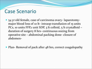
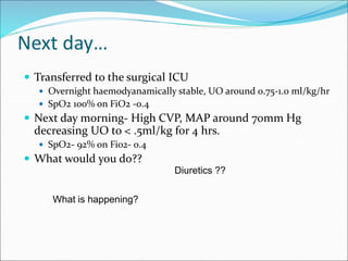
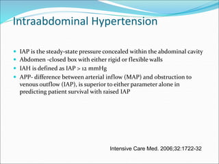
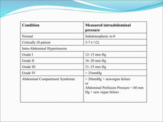
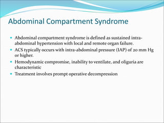
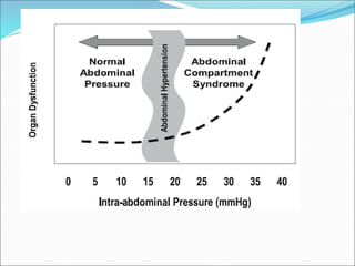
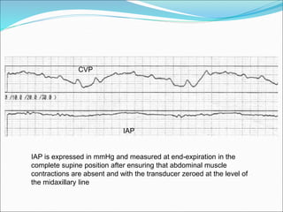
Dr Vijaya P.Patil
Professor,
Dept of Anaesthesia, Critical Care and Pain
Tata Memorial Hospital, Mumbai
Case Scenario
 54 yr old female, case of carcinoma ovary- laparotomy-
major blood loss of 10 lt- intraop transfusion of 15 units
PCs, 10 units FFP,1 unit SDP, 3 lt colloid, 5 lt crystalloid –
duration of surgery 8 hrs- continuous oozing from
operative site – abdominal packing done- closure of
abdomen-
 Plan- Removal of pack after 48 hrs, correct coagulopathy
Next day…
 Transferred to the surgical ICU
 Overnight haemodyanamically stable, UO around 0.75-1.0 ml/kg/hr
 SpO2 100% on FiO2 -0.4
 Next day morning- High CVP, MAP around 70mm Hg
decreasing UO to < .5ml/kg for 4 hrs.
 SpO2- 92% on Fi02- 0.4
 What would you do??
Diuretics ??
What is happening?
Intraabdominal Hypertension
 IAP is the steady-state pressure concealed within the abdominal cavity
 Abdomen -closed box with either rigid or flexible walls
 IAH is defined as IAP > 12 mmHg
 APP- difference between arterial inflow (MAP) and obstruction to
venous outflow (IAP), is superior to either parameter alone in
predicting patient survival with raised IAP
Intensive Care Med. 2006;32:1722-32
Condition Measured intraabdominal
pressure
Normal Subatmospheric to 0
Critically ill patient 5-7 (<12)
Intra-Abdominal Hypertension
Grade I 12–15 mm Hg
Grade II 16–20 mm Hg
Grade III 21–25 mm Hg
Grade IV > 25mmHg
Abdominal Compartment Syndrome > 20mmHg + neworgan failure
or
Abdominal Perfusion Pressure < 60 mm
Hg + new organ failure
Our Patient…
 By afternoon the patient developed worsening
abdominal distension, refractory hypotension,
hypoxia, oliguria, and elevated peak airway
pressures.
Abdominal Compartment Syndrome
 Abdominal compartment syndrome is defined as sustained intra-
abdominal hypertension with local and remote organ failure.
 ACS typically occurs with intra-abdominal pressure (IAP) of 20 mm Hg
or higher.
 Hemodynamic compromise, inability to ventilate, and oliguria are
characteristic
 Treatment involves prompt operative decompression
Risk factors for IAH/ACS
1. Diminished abdominal wall compliance
• ARF especially with elevated
intrathoracic pressure
• Abdominal surgery with primary fascial
closure
• Major trauma/burns
• Prone position
2. Increased intraluminal contents
• Gastroparesis
• Illeus
• Colonic pseudoobstruction
3.Increased abdominal contents
• Hemo/pneumo peritoneum
• Ascites/liver dysfunction
. 4.Capillary leak/ fluid rescuscitation
• Acidosis(pH<7.2)
• Hypotension
• Hypothermia(core temp<330C)
• Polytransfusion (>10 units blood/24hrs)
• Coagulopathy
• Massive fluid rescuscitation(>5lt/24hrs)
• Oligurea
• Sepsis
• Major trauma/ burns
• Damage control laparotomy
Raised IAP
Vascular compression Diaphragm elevation Organ compression
Decreased IVC flow
Venous stasis
/DVT
↑SVV/PVV
Decreased
cardiac preload
↓MAP
cardiac
compression
↑renin/Angiotensin
aldosterone
Increased SVR
↓pleural/
intrathoracic
pressure
↓TMP
↓EDV
↑CVP
↓cardiac contractility
↑PAOP
↓APP
↓ CO
Pulmonary sequelae
 Results from raised intra-abdominal pressure that splints the
diaphragm  reduced TLC, FRC, and RV.
 Intrathoracic and peak airway pressures rise precipitously.
 Decreased lung compliance and increased pulmonary vascular
resistance lead to hypoxia and hypercapnia.
 Increased alveolar dead space and VQ mismatch- hypoxia &
hypercapnea
Renal compromise
 Kidneys are particularly susceptible
 A drastic drop in urine output is a hallmark of ACS.
 Renal compromise can also be pre-renal.
 Increased IAP  compression of renal vein and renal
parenchyma increase in renal vascular resistance + low
CO  decreased RBF and decreased GFR  oliguria and
anuria.
Splanchnic perfusion
J Trauma 1992;33:45–49
CNS circulation
 At particular risk is the patient with concomitant head
trauma.
 The raised intra-abdominal and intrathoracic pressure 
increased CVP and impaired cerebral venous drainage 
raised ICP and intracerebral edema
Classification of IAH/ACS
 Primary ACS- Primary ACS is a condition associated with injury or
disease in the abdominopelvic region that frequently requires early
surgical intervention.
 Secondary ACS Secondary ACS refers to conditions that do not
originate from the abdominopelvic region
 Recurrent ACS (formerly termed tertiary ACS) represents a
redevelopment of ACS symptoms following resolution of an earlier
episode of either primary or secondary ACS and therefore represents a
“second-hit” phenomenon
Diagnose…
What does our patient have? primary
Blunt thoracoabdominal trauma with severe liver injury, hypotension primary
Septic shock related to a pneumonia secondary
Chronic liver failure complicated with a pneumonia primary
Severe burns to abdomen and chest develops
an IAP>20mmHg on day 7
secondary
Septic shock due to intestinal perforation and an IAP of 25mmHg primary
IAP Measurement
 Abdomen and its contents can be considered as relatively
noncompressive and primarily fluid in character, behaving
in accordance to Pascal’s law, the IAP measured at one
point may be assumed to represent the IAP throughout the
abdomen
Crit Care Med 2005 : 33;315-322
All new consecutive patients admitted in 14 intensive care units (ICUs) who
stayed 24 hrs, from six countries during a 4-wk period
Crit Care Med 2005 Vol. 33, No. 2
IAP Measurement
 Patients should be screened for IAH / ACS risk factors upon ICU
admission and in the presence of new or progressive organ failure
(GRADE 1B).
 If two or more risk factors for IAH / ACS are present, a baseline IAP
measurement should be obtained (GRADE 1B)
 If IAH is present, serial IAP measurements should be performed
throughout the patient’s critical illness (GRADE 1C).
IAP Measurement
 Direct-
 Intraperitoneal- during laparoscopy
 Indirect-
 Intragastric
 Transvesical
 IVC pressure via femoral route
Angiocatheter Technique
CVP
IAP
IAP is expressed in mmHg and measured at end-expiration in the
complete supine position after ensuring that abdominal muscle
contractions are absent and with the transducer zeroed at the level of
the midaxillary line
What other sites
 Intra-gastric pressure is a good alternative that correlates
well with bladder pressure.
 Infuse 50–100 ml of water through the NG tube into the stomach.
 The height of water column from the mid-axillary line equals intra-
abdominal pressure.
 A column > about 27 cm H2O is suspicious for ACS.
 Intra-gastric pressure is used when bladder pressure
cannot be measured in the patient post cystectomy or who
has a diseased or neurogenic bladder
Prompt diagnosis is key
 The clinical characteristics of ACS are:
 Abdominal distension
 Fall in urine output (less than 0.5 ml/kg/hr)
 Elevated peak airway pressure (> 40 cm H20)
 Reduced Oxygenation
 Serial monitoring of IAP
 Optimization of systemic perfusion and organ function in
the patient with elevated IAP (APP> 60)
Improve abdominal wall compliance
Evacuate abdominal fluid collection Percutaneous catheter decompression
(Grade IIC)
•Sedation and analgesia (Grade IIC)
•Neuromuscular blockade (Grade IIC)
•Body positioning (Grade IIC)
•Evacuate intraluminal contents
•Nasogastric/colonic decompression
•Prokinetic motility agents
Correct positive fluid balance •Optimal Fluid resuscitation and not
supranormal (Grade IB)
•Diuretics
•CVVHD/HF/ultrafiltration
Surgical decompression should be performed in patients with ACS
that is refractory to other treatment options (Grade 1B)
Medical treatment options
Further Progress
 IAP instituted
 Pressures- 24mm Hg
 Taken to OT- pack removal done
 UO improved, creat decreased, IAP 12
 On pressure support
 On 4th day –febrile, hypotensive, fluid rescuscitation, inotropes,
oliguria, hypoxia, increased RT aspirate
 IAP- 22
 CVVHF

Case Based teaching IAH.ppt

  • 1.
    Dr Vijaya P.Patil Professor, Deptof Anaesthesia, Critical Care and Pain Tata Memorial Hospital, Mumbai
  • 2.
    Case Scenario  54yr old female, case of carcinoma ovary- laparotomy- major blood loss of 10 lt- intraop transfusion of 15 units PCs, 10 units FFP,1 unit SDP, 3 lt colloid, 5 lt crystalloid – duration of surgery 8 hrs- continuous oozing from operative site – abdominal packing done- closure of abdomen-  Plan- Removal of pack after 48 hrs, correct coagulopathy
  • 3.
    Next day…  Transferredto the surgical ICU  Overnight haemodyanamically stable, UO around 0.75-1.0 ml/kg/hr  SpO2 100% on FiO2 -0.4  Next day morning- High CVP, MAP around 70mm Hg decreasing UO to < .5ml/kg for 4 hrs.  SpO2- 92% on Fi02- 0.4  What would you do?? Diuretics ?? What is happening?
  • 4.
    Intraabdominal Hypertension  IAPis the steady-state pressure concealed within the abdominal cavity  Abdomen -closed box with either rigid or flexible walls  IAH is defined as IAP > 12 mmHg  APP- difference between arterial inflow (MAP) and obstruction to venous outflow (IAP), is superior to either parameter alone in predicting patient survival with raised IAP Intensive Care Med. 2006;32:1722-32
  • 5.
    Condition Measured intraabdominal pressure NormalSubatmospheric to 0 Critically ill patient 5-7 (<12) Intra-Abdominal Hypertension Grade I 12–15 mm Hg Grade II 16–20 mm Hg Grade III 21–25 mm Hg Grade IV > 25mmHg Abdominal Compartment Syndrome > 20mmHg + neworgan failure or Abdominal Perfusion Pressure < 60 mm Hg + new organ failure
  • 6.
    Our Patient…  Byafternoon the patient developed worsening abdominal distension, refractory hypotension, hypoxia, oliguria, and elevated peak airway pressures.
  • 7.
    Abdominal Compartment Syndrome Abdominal compartment syndrome is defined as sustained intra- abdominal hypertension with local and remote organ failure.  ACS typically occurs with intra-abdominal pressure (IAP) of 20 mm Hg or higher.  Hemodynamic compromise, inability to ventilate, and oliguria are characteristic  Treatment involves prompt operative decompression
  • 9.
    Risk factors forIAH/ACS 1. Diminished abdominal wall compliance • ARF especially with elevated intrathoracic pressure • Abdominal surgery with primary fascial closure • Major trauma/burns • Prone position 2. Increased intraluminal contents • Gastroparesis • Illeus • Colonic pseudoobstruction 3.Increased abdominal contents • Hemo/pneumo peritoneum • Ascites/liver dysfunction . 4.Capillary leak/ fluid rescuscitation • Acidosis(pH<7.2) • Hypotension • Hypothermia(core temp<330C) • Polytransfusion (>10 units blood/24hrs) • Coagulopathy • Massive fluid rescuscitation(>5lt/24hrs) • Oligurea • Sepsis • Major trauma/ burns • Damage control laparotomy
  • 10.
    Raised IAP Vascular compressionDiaphragm elevation Organ compression Decreased IVC flow Venous stasis /DVT ↑SVV/PVV Decreased cardiac preload ↓MAP cardiac compression ↑renin/Angiotensin aldosterone Increased SVR ↓pleural/ intrathoracic pressure ↓TMP ↓EDV ↑CVP ↓cardiac contractility ↑PAOP ↓APP ↓ CO
  • 11.
    Pulmonary sequelae  Resultsfrom raised intra-abdominal pressure that splints the diaphragm  reduced TLC, FRC, and RV.  Intrathoracic and peak airway pressures rise precipitously.  Decreased lung compliance and increased pulmonary vascular resistance lead to hypoxia and hypercapnia.  Increased alveolar dead space and VQ mismatch- hypoxia & hypercapnea
  • 12.
    Renal compromise  Kidneysare particularly susceptible  A drastic drop in urine output is a hallmark of ACS.  Renal compromise can also be pre-renal.  Increased IAP  compression of renal vein and renal parenchyma increase in renal vascular resistance + low CO  decreased RBF and decreased GFR  oliguria and anuria.
  • 13.
  • 14.
    CNS circulation  Atparticular risk is the patient with concomitant head trauma.  The raised intra-abdominal and intrathoracic pressure  increased CVP and impaired cerebral venous drainage  raised ICP and intracerebral edema
  • 15.
    Classification of IAH/ACS Primary ACS- Primary ACS is a condition associated with injury or disease in the abdominopelvic region that frequently requires early surgical intervention.  Secondary ACS Secondary ACS refers to conditions that do not originate from the abdominopelvic region  Recurrent ACS (formerly termed tertiary ACS) represents a redevelopment of ACS symptoms following resolution of an earlier episode of either primary or secondary ACS and therefore represents a “second-hit” phenomenon
  • 16.
    Diagnose… What does ourpatient have? primary Blunt thoracoabdominal trauma with severe liver injury, hypotension primary Septic shock related to a pneumonia secondary Chronic liver failure complicated with a pneumonia primary Severe burns to abdomen and chest develops an IAP>20mmHg on day 7 secondary Septic shock due to intestinal perforation and an IAP of 25mmHg primary
  • 17.
    IAP Measurement  Abdomenand its contents can be considered as relatively noncompressive and primarily fluid in character, behaving in accordance to Pascal’s law, the IAP measured at one point may be assumed to represent the IAP throughout the abdomen
  • 18.
    Crit Care Med2005 : 33;315-322 All new consecutive patients admitted in 14 intensive care units (ICUs) who stayed 24 hrs, from six countries during a 4-wk period
  • 19.
    Crit Care Med2005 Vol. 33, No. 2
  • 20.
    IAP Measurement  Patientsshould be screened for IAH / ACS risk factors upon ICU admission and in the presence of new or progressive organ failure (GRADE 1B).  If two or more risk factors for IAH / ACS are present, a baseline IAP measurement should be obtained (GRADE 1B)  If IAH is present, serial IAP measurements should be performed throughout the patient’s critical illness (GRADE 1C).
  • 21.
    IAP Measurement  Direct- Intraperitoneal- during laparoscopy  Indirect-  Intragastric  Transvesical  IVC pressure via femoral route
  • 24.
  • 25.
    CVP IAP IAP is expressedin mmHg and measured at end-expiration in the complete supine position after ensuring that abdominal muscle contractions are absent and with the transducer zeroed at the level of the midaxillary line
  • 26.
    What other sites Intra-gastric pressure is a good alternative that correlates well with bladder pressure.  Infuse 50–100 ml of water through the NG tube into the stomach.  The height of water column from the mid-axillary line equals intra- abdominal pressure.  A column > about 27 cm H2O is suspicious for ACS.  Intra-gastric pressure is used when bladder pressure cannot be measured in the patient post cystectomy or who has a diseased or neurogenic bladder
  • 27.
    Prompt diagnosis iskey  The clinical characteristics of ACS are:  Abdominal distension  Fall in urine output (less than 0.5 ml/kg/hr)  Elevated peak airway pressure (> 40 cm H20)  Reduced Oxygenation  Serial monitoring of IAP  Optimization of systemic perfusion and organ function in the patient with elevated IAP (APP> 60)
  • 28.
    Improve abdominal wallcompliance Evacuate abdominal fluid collection Percutaneous catheter decompression (Grade IIC) •Sedation and analgesia (Grade IIC) •Neuromuscular blockade (Grade IIC) •Body positioning (Grade IIC) •Evacuate intraluminal contents •Nasogastric/colonic decompression •Prokinetic motility agents Correct positive fluid balance •Optimal Fluid resuscitation and not supranormal (Grade IB) •Diuretics •CVVHD/HF/ultrafiltration Surgical decompression should be performed in patients with ACS that is refractory to other treatment options (Grade 1B) Medical treatment options
  • 29.
    Further Progress  IAPinstituted  Pressures- 24mm Hg  Taken to OT- pack removal done  UO improved, creat decreased, IAP 12  On pressure support  On 4th day –febrile, hypotensive, fluid rescuscitation, inotropes, oliguria, hypoxia, increased RT aspirate  IAP- 22  CVVHF