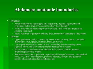
Trauma Conference January 9 th , 2006 Greg Feldman, MD PGY1, General Surgery Department Stanford Medical Center Blunt Abdominal Trauma: Evaluation
Outline Anatomic definition of abdomen Mechanisms of injury in blunt trauma Typical injury patterns Assessment of blunt abdominal trauma Diagnostic algorithms
Abdomen: anatomic boundaries External: Anterior abdomen: transnipple line superiorly, inguinal ligaments and symphasis pubis inferiorly, anterior axillary lines laterally. Flank: between anterior and posterior axillary lines from 6th intercostals space to iliac crest. Back: Posterior to posterior axillary lines, from tip of scapulae to iliac crests. Internal: Upper peritoneal cavity: covered by lower aspect of bony thorax.  Includes diaphragm, liver, spleen, stomach, transverse colon. Lower peritoneal cavity: small bowel, ascending and descending colon, sigmoid colon, and (in women) internal reproductive organs. Pelvic cavity: contains rectum, bladder, iliac vessels, and (in women) internal reproductive organs. Retroperitoneal space: posterior to peritoneal lining of abdomen.  Abdominal aorta, IVC, most of duodenum, pancreas kidneys, ureters, and posterior aspects of ascending and descending colon.
Mechanisms of injury Compression, crush, or sheer injury to abdominal viscera    deformation of solid or hollow organs, rupture (e.g. small bowel, gravid uterus) Deceleration injuries: differential movements of fixed and nonfixed structures (e.g. liver and spleen lacs at sites of supporting ligaments)
Common injury patterns In patients undergoing laparotomy for blunt trauma, most frequently injured organs are spleen (40-55%), liver (35-45%), and small bowel (5-10%).  (ATLS, 2001) Duodenum: Classically, frontal-impact MVC with unrestrained driver; or direct blow to abdomen. Bloody gastric aspirate, retroperitoneal air on XR or CT Confirmed with upper GI series or double contrast CT Small bowel injury: Generally from sudden deceleration with subsequent tearing near fixed points of attachment. Often associated with seat belt sign, lumbar distraction fracture (Chance fracture) DPL superior to FAST or CT for diagnosis.
Common injury patterns (2) Pancreas: Direct epigastric blow compressing pancreas against vertebral column. Early normal serum amylase does NOT exclude major pancreatic trauma. CT with PO/IV contrast – NOT particularly sensitive in immediate post-injury period. Diaphragm: Most commonly, 5-10 cm  rupture involving posterolateral hemidiaphragm. Noted on CXR: blurred or elevated hemidiaphragm, hemothorax, GT in chest Genitourinary: Anterior injuries (below UG diaphragm): usually from straddle impact. Posterior injuries (above UG diaphragm): in patient with multisystem injuries and pelvic fractures.
Common injury patterns (3) Solid organ injury Laceration to liver, spleen, or kidney Injury to one of these three + hemodynamic instability: considered indication for urgent laparotomy Isolated solid organ injury in hemodynamically stable patient: can often be managed nonoperatively. Pelvic fractures: Suggest major force applied to patient. Usually auto-ped, MVC, or motorcycle Significant association with intraperitoneal and retroperitoneal organs and vascular structures.
Restraining devices Lap seat belt Mesenteric tear or avulsion Rupture of small bowel or colon Iliac artery or abdominal aorta thrombosis Chance fracture of lumbar vertebrae (hyperflexion) Shoulder Harness Rupture of upper abdominal viscera Intimal tear or thrombosis in innominate, carotid, subclavian, or vertebral arteries Fracture or dislocation of C-spine Rib fractures Pulmonary contusion Air Bag Corneal abrasions, keratitis Abrasions of face, neck, chest Cardiac rupture C or T-spine fracture
Assessment: History Mechanism Symptoms, events, PMH, Meds, EtOH/drugs MVC: Speed Type of collision (frontal, lateral, sideswipe, rear, rollover) Vehicle intrusion into passenger compartment Types of restraints Deployment of air bag Patient's position in vehicle
Assessment: Physical Exam Inspection, auscultation, percussion, palpation Inspection: abrasions, contusions, lacerations, deformity Grey-Turner, Kehr, Balance, Cullen Auscultation: careful exam advised by ATLS.  (Controversial utility in trauma setting.) Percussion: subtle signs of peritonitis; tympany in gastric dilatation or free air; dullness with hemoperitoneum Palpation: elicit superficial, deep, or rebound tenderness; involuntary muscle guarding
Physical Exam: Eponyms Grey-Turner sign: Bluish discoloration of lower flanks, lower back; associated with retroperitoneal bleeding of pancreas, kidney, or pelvic fracture. Cullen sign: Bluish discoloration around umbilicus, indicates peritoneal bleeding, often pancreatic hemorrhage. Kehr sign: L shoulder pain while supine; caused by diaphragmatic irritation (splenic injury, free air, intra-abd bleeding) Balance sign: Dull percussion in LUQ.  Sign of splenic injury; blood accumulating in subcapsular or extracapsular spleen.
Diagnostic adjuncts Labs: BMP, CBC, coags, b-HCG, amy/lip, U/A, tox screen, T&C Plain films: CXR, pelvis; abd films generally lower priority DPL FAST CT
Diagnostic Peritoneal Lavage 98% sensitive for intraperitoneal bleeding (ATLS) Open or closed (Seldinger); usually infraumbilical, but may be supraumbilical in pelvic frxs or advanced pregnancy. Free aspiration of blood, GI contents, or bile in demodynamically abnormal pt: indication for laparotomy If gross blood (> 10 mL) or GI contents not aspirated, perform lavage with 1000 mL warmed LR.  Allow to mix, compress abdomen and logross paient, the sent to lab.  + test: >100,000 RBC/mm3, >500 WBC/mm3, Gram stain with bacteria. Alters subsequent examination of patient Has been somewhat superceded by FAST in common use; now generally performed in unstable patients with intermediate FAST exams, or with suspicion for small bowel injury.
FAST: Strengths and Limitations Strengths Rapid (~2 mins) Portable Inexpensive Technically simple, easy to train (studies show competence can be achieved after ~30 studies) Can be performed serially Useful for guiding triage decisions in trauma patients Limitations Does not typically identify source of bleeding, or detect injuries that do not cause hemoperitoneum Requires extensive training to assess parenchyma reliably Limited in detecting <250 cc intraperitoneal fluid Particularly poor at detecting bowel and mesentery damage (44% sensitivity) Difficult to assess retroperitoneum Limited by habitus in obese patients
FAST: Accuracy For identifying hemoperitoneum in blunt abdominal trauma: Sensitivity 76 - 90% Specificity 95 - 100% The larger the hemoperitoneum, the higher the sensitivity.  So sensitivity increases for  clinically significant  hemoperitoneum. How much fluid can FAST detect? 250 cc total 100 cc in Morison’s pouch
Does FAST replace CT? Only at the extremes. Unstable patient, (+) FAST    OR Stable patient, low force injury, (-) FAST    consider observing patient.  CT is far more sensitive than FAST for detecting and characterizing abdominal injury in trauma.  The gold standard for characterizing intraparenchymal injury. “ Death begins with a CT.”  Never send an unstable patient to CT.  FAST, however, can be performed during resuscitation.
CT EAST level I recommendations (2001): CT is recommended for evaluation of hemodynamically stable patients with equivocal findings on physical examination, associated neurologic injury, or multiple extra-abdominal injuries. CT is the diagnostic modality of choice for nonoperative management of solid visceral injuries.
EAST Algorithm: Unstable Eastern Association for the Surgery of Trauma, 2001
EAST Algorithm: Stable Eastern Association for the Surgery of Trauma, 2001
References Hoff et al.  EAST Practice Management Guidelines Work Group.  Practice Management Guidelines for the Evaluation of Blunt Abdominal Trauma, 2001.  www.east.org. American College of Surgeons Committee on Trauma.  Advanced Trauma Life Support for Doctors; Student Course Manual, 7 th  edition, 2004. Scalea TM, Rodriquez A, Chiu WC.  Focused Assessment with Sonography for Trauma (FAST): Results from an International Consensus Conference.  J. Trauma  1999;46:466-472. Yoshii H, Sato M, Yamamoto S.  Usefulness and Limitations of Ultrasonography in the Initial Evaluation of Blunt Abdominal Trauma.  J. Trauma  1998;45:45-51.
Acknowledgements Dr. Shelly Erford Dr. Denny Jenkins Carol Thomson Dr. Natalie Kirilchik Dr. Subarna Biswas Drs. Brundage, Spain, and Gregg Stanford Medical Center ACS/Trauma Service Noah Feinstein Dr. Gillian Lieberman Dr. Jason Tracy

01 blunt abdominal trauma

  • 1.
    Trauma Conference January9 th , 2006 Greg Feldman, MD PGY1, General Surgery Department Stanford Medical Center Blunt Abdominal Trauma: Evaluation
  • 2.
    Outline Anatomic definitionof abdomen Mechanisms of injury in blunt trauma Typical injury patterns Assessment of blunt abdominal trauma Diagnostic algorithms
  • 3.
    Abdomen: anatomic boundariesExternal: Anterior abdomen: transnipple line superiorly, inguinal ligaments and symphasis pubis inferiorly, anterior axillary lines laterally. Flank: between anterior and posterior axillary lines from 6th intercostals space to iliac crest. Back: Posterior to posterior axillary lines, from tip of scapulae to iliac crests. Internal: Upper peritoneal cavity: covered by lower aspect of bony thorax. Includes diaphragm, liver, spleen, stomach, transverse colon. Lower peritoneal cavity: small bowel, ascending and descending colon, sigmoid colon, and (in women) internal reproductive organs. Pelvic cavity: contains rectum, bladder, iliac vessels, and (in women) internal reproductive organs. Retroperitoneal space: posterior to peritoneal lining of abdomen. Abdominal aorta, IVC, most of duodenum, pancreas kidneys, ureters, and posterior aspects of ascending and descending colon.
  • 4.
    Mechanisms of injuryCompression, crush, or sheer injury to abdominal viscera  deformation of solid or hollow organs, rupture (e.g. small bowel, gravid uterus) Deceleration injuries: differential movements of fixed and nonfixed structures (e.g. liver and spleen lacs at sites of supporting ligaments)
  • 5.
    Common injury patternsIn patients undergoing laparotomy for blunt trauma, most frequently injured organs are spleen (40-55%), liver (35-45%), and small bowel (5-10%). (ATLS, 2001) Duodenum: Classically, frontal-impact MVC with unrestrained driver; or direct blow to abdomen. Bloody gastric aspirate, retroperitoneal air on XR or CT Confirmed with upper GI series or double contrast CT Small bowel injury: Generally from sudden deceleration with subsequent tearing near fixed points of attachment. Often associated with seat belt sign, lumbar distraction fracture (Chance fracture) DPL superior to FAST or CT for diagnosis.
  • 6.
    Common injury patterns(2) Pancreas: Direct epigastric blow compressing pancreas against vertebral column. Early normal serum amylase does NOT exclude major pancreatic trauma. CT with PO/IV contrast – NOT particularly sensitive in immediate post-injury period. Diaphragm: Most commonly, 5-10 cm rupture involving posterolateral hemidiaphragm. Noted on CXR: blurred or elevated hemidiaphragm, hemothorax, GT in chest Genitourinary: Anterior injuries (below UG diaphragm): usually from straddle impact. Posterior injuries (above UG diaphragm): in patient with multisystem injuries and pelvic fractures.
  • 7.
    Common injury patterns(3) Solid organ injury Laceration to liver, spleen, or kidney Injury to one of these three + hemodynamic instability: considered indication for urgent laparotomy Isolated solid organ injury in hemodynamically stable patient: can often be managed nonoperatively. Pelvic fractures: Suggest major force applied to patient. Usually auto-ped, MVC, or motorcycle Significant association with intraperitoneal and retroperitoneal organs and vascular structures.
  • 8.
    Restraining devices Lapseat belt Mesenteric tear or avulsion Rupture of small bowel or colon Iliac artery or abdominal aorta thrombosis Chance fracture of lumbar vertebrae (hyperflexion) Shoulder Harness Rupture of upper abdominal viscera Intimal tear or thrombosis in innominate, carotid, subclavian, or vertebral arteries Fracture or dislocation of C-spine Rib fractures Pulmonary contusion Air Bag Corneal abrasions, keratitis Abrasions of face, neck, chest Cardiac rupture C or T-spine fracture
  • 9.
    Assessment: History MechanismSymptoms, events, PMH, Meds, EtOH/drugs MVC: Speed Type of collision (frontal, lateral, sideswipe, rear, rollover) Vehicle intrusion into passenger compartment Types of restraints Deployment of air bag Patient's position in vehicle
  • 10.
    Assessment: Physical ExamInspection, auscultation, percussion, palpation Inspection: abrasions, contusions, lacerations, deformity Grey-Turner, Kehr, Balance, Cullen Auscultation: careful exam advised by ATLS. (Controversial utility in trauma setting.) Percussion: subtle signs of peritonitis; tympany in gastric dilatation or free air; dullness with hemoperitoneum Palpation: elicit superficial, deep, or rebound tenderness; involuntary muscle guarding
  • 11.
    Physical Exam: EponymsGrey-Turner sign: Bluish discoloration of lower flanks, lower back; associated with retroperitoneal bleeding of pancreas, kidney, or pelvic fracture. Cullen sign: Bluish discoloration around umbilicus, indicates peritoneal bleeding, often pancreatic hemorrhage. Kehr sign: L shoulder pain while supine; caused by diaphragmatic irritation (splenic injury, free air, intra-abd bleeding) Balance sign: Dull percussion in LUQ. Sign of splenic injury; blood accumulating in subcapsular or extracapsular spleen.
  • 12.
    Diagnostic adjuncts Labs:BMP, CBC, coags, b-HCG, amy/lip, U/A, tox screen, T&C Plain films: CXR, pelvis; abd films generally lower priority DPL FAST CT
  • 13.
    Diagnostic Peritoneal Lavage98% sensitive for intraperitoneal bleeding (ATLS) Open or closed (Seldinger); usually infraumbilical, but may be supraumbilical in pelvic frxs or advanced pregnancy. Free aspiration of blood, GI contents, or bile in demodynamically abnormal pt: indication for laparotomy If gross blood (> 10 mL) or GI contents not aspirated, perform lavage with 1000 mL warmed LR. Allow to mix, compress abdomen and logross paient, the sent to lab. + test: >100,000 RBC/mm3, >500 WBC/mm3, Gram stain with bacteria. Alters subsequent examination of patient Has been somewhat superceded by FAST in common use; now generally performed in unstable patients with intermediate FAST exams, or with suspicion for small bowel injury.
  • 14.
    FAST: Strengths andLimitations Strengths Rapid (~2 mins) Portable Inexpensive Technically simple, easy to train (studies show competence can be achieved after ~30 studies) Can be performed serially Useful for guiding triage decisions in trauma patients Limitations Does not typically identify source of bleeding, or detect injuries that do not cause hemoperitoneum Requires extensive training to assess parenchyma reliably Limited in detecting <250 cc intraperitoneal fluid Particularly poor at detecting bowel and mesentery damage (44% sensitivity) Difficult to assess retroperitoneum Limited by habitus in obese patients
  • 15.
    FAST: Accuracy Foridentifying hemoperitoneum in blunt abdominal trauma: Sensitivity 76 - 90% Specificity 95 - 100% The larger the hemoperitoneum, the higher the sensitivity. So sensitivity increases for clinically significant hemoperitoneum. How much fluid can FAST detect? 250 cc total 100 cc in Morison’s pouch
  • 16.
    Does FAST replaceCT? Only at the extremes. Unstable patient, (+) FAST  OR Stable patient, low force injury, (-) FAST  consider observing patient. CT is far more sensitive than FAST for detecting and characterizing abdominal injury in trauma. The gold standard for characterizing intraparenchymal injury. “ Death begins with a CT.” Never send an unstable patient to CT. FAST, however, can be performed during resuscitation.
  • 17.
    CT EAST levelI recommendations (2001): CT is recommended for evaluation of hemodynamically stable patients with equivocal findings on physical examination, associated neurologic injury, or multiple extra-abdominal injuries. CT is the diagnostic modality of choice for nonoperative management of solid visceral injuries.
  • 18.
    EAST Algorithm: UnstableEastern Association for the Surgery of Trauma, 2001
  • 19.
    EAST Algorithm: StableEastern Association for the Surgery of Trauma, 2001
  • 20.
    References Hoff etal. EAST Practice Management Guidelines Work Group. Practice Management Guidelines for the Evaluation of Blunt Abdominal Trauma, 2001. www.east.org. American College of Surgeons Committee on Trauma. Advanced Trauma Life Support for Doctors; Student Course Manual, 7 th edition, 2004. Scalea TM, Rodriquez A, Chiu WC. Focused Assessment with Sonography for Trauma (FAST): Results from an International Consensus Conference. J. Trauma 1999;46:466-472. Yoshii H, Sato M, Yamamoto S. Usefulness and Limitations of Ultrasonography in the Initial Evaluation of Blunt Abdominal Trauma. J. Trauma 1998;45:45-51.
  • 21.
    Acknowledgements Dr. ShellyErford Dr. Denny Jenkins Carol Thomson Dr. Natalie Kirilchik Dr. Subarna Biswas Drs. Brundage, Spain, and Gregg Stanford Medical Center ACS/Trauma Service Noah Feinstein Dr. Gillian Lieberman Dr. Jason Tracy